
Abstract
Garlic has been used as a food as well as a component of traditional medicine. Aged garlic extract (AGE) is claimed to promote human health through antioxidant/anti-inflammatory activities with neuroprotective effects. We evaluated the possible beneficial effect of AGE neurologically, pathologically, ultrastructurally, and biochemically in a spinal cord ischemia–reperfusion (I/R) model of rats. Twenty-four Sprague-Dawley rats were divided into three groups: sham (no I/R), I/R, and AGE (I/R+AGE); each group consisted of eight animals. Animals were evaluated neurologically with the Basso, Beattie, and Bresnahan (BBB) scoring system. The spinal cord tissue samples were harvested for pathological and ultrastructural examinations. Oxidative products (Malondialdehyde, nitric oxide), antioxidant enzymes (superoxide dismutase, catalase, glutathione peroxidase), inflammatory cytokines (tissue tumor necrosis factor alpha, interleukin-1), and caspase-3 activity were analyzed. The AGE group had significantly higher BBB scores than the I/R group. Pathologically, AGE group revealed reduced degree of ischemia and spinal cord edema. Ultrastructural results also showed preservation of tissue structure in the AGE group. Oxidative product levels of the I/R group were significantly higher than both the other groups, and antioxidant enzyme levels of AGE group were significantly higher than the I/R group. There was also significant difference between the sham and AGE groups in terms of total antioxidant enzyme levels. Furthermore, AGE treatment significantly reduced the inflammatory cytokines and caspase-3 activity than the I/R group. This study demonstrates the considerable neuroprotective effect of AGE on the neurological, pathological, ultrastructural, and biochemical status of rats with I/R-induced spinal cord injury.
Introduction
I
Garlic, Allium sativum, is a species of the onion family, Alliaceae. It has been extensively used throughout history for its prophylactic and therapeutic effects. Its immunomodulatory and antitumor effects have also been demonstrated by in vitro and in vivo experiments. 3 In addition, garlic is reported to attenuate various risk factors associated with cardiovascular diseases such as hyperlipidemia, high blood pressure, proinflammatory cytokine production, and platelet activation. 4 It is believed that aged garlic extract (AGE) has antioxidant potential for scavenging the reactive oxygen species (ROS). Furthermore, AGE acts as an enhancer for cellular antioxidant enzymes (superoxide dismutase, catalase, and glutathione peroxidase) and also increases glutathione levels in the cells. 5
Previously in the literature, neuroprotective effects of AGE have not been evaluated in a rat model of spinal cord I/R injury. The aim of this study was to assess the effects of AGE neurologically, biochemically, pathologically, and ultrastructurally in an I/R model of spinal cord ischemia induced by cross-clamping of the rat infrarenal abdominal aorta.
Material and Methods
Experimental groups
All the experimental procedures and protocols used in this investigation were reviewed and conducted with the prior approval of animal experimental ethics committee of Ankara Education and Research Hospital. Twenty-four male Sprague-Dawley rats weighing 350 to 450 g were randomly assigned into three groups: sham operated control group, I/R injury group, and AGE group. All rats were kept under environmentally controlled conditions at 23°C ± 2°C, with appropriate humidity and a 12-h light cycle and granted free access to food and water. The sham group (n = 8); laparotomy and infrarenal abdominal aorta dissection were completed, but occlusion was not performed. The I/R injury group (n = 8); rats administered 1 cc vehicle isotonic NaCl 0.9% orally by gavage for 15 days before I/R injury induction. The AGE group (n = 8); similar to I/R group, but all rats were given 250 mg/kg per day of AGE diluted in tap water orally by gavage for 15 days before the trauma. A commercially available AGE (KYOLIC®) was kindly provided by Wakunaga of America. 6
Surgical procedure
Spinal cord I/R injury was induced by occluding the abdominal aorta for 30 min, as previously described. 7 Rats were anesthetized with an intraperitoneal injection of 10 mg/kg xylazine (Rompun; Bayer) and 50 mg/kg ketamine (Ketalar; Parke Davis), and allowed to breathe spontaneously. A rectal probe was inserted, and the body temperature was maintained at 37°C ± 0.5°C with a heating pad. The abdominal aorta with its left renal artery division was exposed through an abdominal incision. The artery was occluded 0.5 cm below the left renal artery with an aneurysm clip for 30 min. The reperfusion had begun after the clip was removed. The wound was closed in layers after the operation. No surgical intervention was performed in the control group. The animals were kept alive for 24 h under appropriate conditions and veterinary control, after which decapitation took place after anesthetization using the same anesthetic agents. Spinal cord segments between L4 and L6 were obtained from the operated spinal cord area and divided into three (5 mm) equal parts. Cranial parts of the tissue samples were obtained for microscopy and electron microscopy evaluation; caudal parts were stored in a −20°C freezer for biochemical analysis.
Neurological evaluation
Rats were evaluated by an independent observer who was blinded to the protocols and group assignment for neurological examination at 24th hour after the I/R injury according to the Basso, Beattie, and Bresnahan (BBB) scoring system described by Basso et al. 8 The BBB scale ranges from 0 to 21. A score of “0” indicates that the animal exhibits complete hindlimb paralysis. A score of “21” denotes that the animal has completely normal locomotor function in the hindlimbs.
Pathological assessment
The specimens were immersed into 10% formaldehyde and stored at 4°C. The lumbar spinal cord specimens were then embedded in paraffin, cut into 5 μm thick sections and stained with hematoxylin–eosin (H&E). Sections were evaluated under a light microscope (Olympus BH-2, Japan; Olympus BH-2, Olympus Corp) by a pathologist blinded to the groups. Pathological changes were graded using Naslund's standard: grade I, neurons normal, or vacuole and granule denaturation of cytoplasm in neurons observed incidentally; grade II, normal neurons and ischemic neurons coexisting in very similar numbers, ischemic neurons identified by cytoplasmic eosinophilia with loss of Nissl substance and by the presence of pyknotic homogenous nuclei; grade III, many ischemic neurons, crimpled massive neurons, with nuclear dissolution and myelin swelling. 9
Ultrastructural examination
The specimens were fixed in 2.5% glutaraldehyde for 24 h, washed in phosphate buffer (pH 7.4), postfixed in 1% osmium tetroxide in phosphate buffer (pH 7.4) for 2 h, and dehydrated in increasing concentrations of alcohol. The tissues were washed with propylene oxide and embedded in epoxy resin embedding media. Using a glass knife on an LKB–Nova (LKB–Produkter AB) ultramicrotome, semithin sections about 2 μm in thickness and ultrathin sections about 60 nm in thickness were performed. The semithin sections were stained with methylene blue and examined by a light microscope. The tissue blocks were trimmed; the ultrathin sections were taken by the same ultramicrotome and stained with uranyl acetate and lead citrate. All the ultrathin sections were examined by Jeol JEM 1200 EX (Jeol Ltd.) transmission electron microscope. For each sample, 100 each of the large-diameter, medium-diameter, and small-diameter myelinated axons were counted, evaluated, and scored from 0 to 3 as described by Kaptanoglu et al. Scoring of myelinated axons is as follows: 0 = ultrastructurally normal myelinated axon, 1 = separation in myelin configuration, 2 = interruption in myelin configuration, and 3 = honey comb appearance in myelin configuration. 10 The scoring was performed to five samples of every group.
Biochemical analyses
Spinal cord tissues were homogenized in physiologic saline solution and centrifuged at 4000 g for 20 min. Then, upper clear supernatants were removed to be used in the analysis.
Oxidative products
Malondialdehyde (MDA) levels were measured by a method based on the reaction with thiobarbituric acid as described previously by Ohkawa et al. 11 and expressed as nmol/mg protein. Nitric oxide (NO) levels were determined by the method of Miranda et al. 12 and expressed as nmol/mg protein.
Antioxidant enzymes
The superoxide dismutase (SOD) activity was measured according to the nitroblue tetrazolium method described by Sun et al. 13 and expressed as U/mg protein. The glutathione peroxidase (GSH-Px) activity of the spinal cord was determined by photometric kinetic measurement using the GSH Assay Kit (Catalog No.: 703102; Cayman Chemical), following the oxidation of nicotinamide adenine dinucleotide phosphate spectrophotometrically (Bio-Tek ELx-800) at 340 nm. The GSH-Px activity was expressed as U/g protein. 14 The catalase (CAT) activity was measured by colorimetric method using the Catalase Assay Kit, Catalog No.: 707002 (Cayman Chemical), based on the determination of the decrease in the hydrogen peroxide decomposition in a medium through measuring the absorbance changes at 540 nm per minute. 15 The CAT activity was expressed as mmol/g protein.
Inflammatory cytokines
Tissue tumor necrosis factor alpha (TNF-α) and interleukin-1 (IL-1) levels were determined using the ELISA kit (Uscn Life Science, Inc., Wuhan.) and expressed as pg/mg.
Caspase-3 activity
The caspase-3 activity was measured using the ELISA kit (Uscn Life Science, Inc., Wuhan.) and expressed as ng/mg protein.
Statistical analyses
Analyses were performed using SPSS software 15.0 for Windows (SPSS). All values are presented as mean ± standard deviation. The significance of differences between histological grades was assessed using the χ 2 test. Comparisons were made using the Mann–Whitney U-test. Differences among the groups were assessed using the Kruskal–Wallis test. P < .05 was considered statistically significant.
Results
Neurological function
The BBB scores of the groups at 24 h after ischemia are shown in Table 1. Rats in the sham group had significantly better postoperative neurological outcomes than animals in the other groups (P < .05). The I/R group had the worst mean score. In addition, there was a statistically significant difference between the I/R group and the AGE group (P < .05).
Sham versus I/R (P < .05).
Sham versus AGE (P < .05).
I/R versus AGE (P < .05).
AGE, aged garlic extract; BBB, Basso, Beattie, and Bresnahan; I/R, ischemia-reperfusion.
Pathological results
Microscopic findings of the sham group were normal. The I/R group revealed increased edema, vascular proliferation, areas of severe ischemia, and the destruction of the neurons in the gray matter. In addition, mild inflammatory changes were observed in the sections. Also, the AGE group showed mild ischemia areas and markedly reduced edema (Fig. 1A–C). The pathological grades of the samples are shown in Table 2. There was a statistically significant difference among all groups (P < .05). In addition, cross-comparisons of the groups revealed statistically significant differences (P < .05).

Sections from all groups (H&E, original magnification, ×200):
Ultrastructural results
In the transmission electron microscopic examination of the tissue samples of the sham group, ultrastructural pathological changes were not observed in the gray and white matters of the spinal cord. In the I/R group, separations in myelin configuration and interruptions in myelin configuration were observed in the small-sized, medium–sized, and large-sized myelinated axons. However; the ultrastructural pathological changes were more severe in the large-sized myelinated axons. In the ultrastructural examination of the neuronal tissue; swollen mitochondria were present inside the cytoplasm of neurons. In addition, perineuronal edema was present around the neurons in the gray matter. In the AGE group, ultrastructural pathological changes were found in the white matter. In a few of the small-sized myelinated axons, separations were observed in the myelin configuration. In most of the medium-sized and large-sized myelinated axons, separations were observed in the myelin configuration. In addition, in a few of the large-sized myelinated axons, interruptions were observed in the myelin configuration. However, in this group, ultrastructurally normal medium-sized and large-sized myelinated axons were also present. In the ultrastructural examination of the neuronal tissue, the intracellular organelles, nuclei, and membranes of the neurons were found normal ultrastructurally. The perineuronal tissues did not show any pathological changes (Fig. 2A–C).
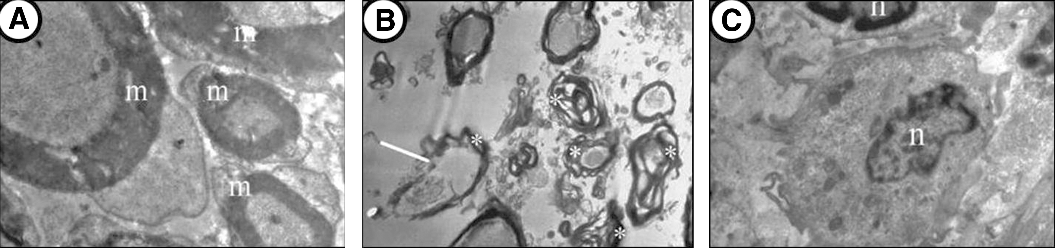
Ultrastructural findings of all groups (original magnification ×5000):
The I/R group had the worst mean score. Also, statistically significant difference was found among all groups. When compared with the I/R injury group, the AGE group showed statistically better scores on all sizes of myelinated axons (P < .05). In addition, there were statistically significant differences between the sham group and the other groups (P < .05) (Table 3).
Sham versus I/R (P < .05).
Sham versus AGE (P < .05).
I/R versus AGE (P < .05).
Biochemical results
Oxidative products (MDA, NO)
When MDA and NO levels had been evaluated, statistically significant differences were found among the groups (P < .05) (Table 4). I/R was found to produce significant elevation in MDA and NO levels (P < .05). For MDA and NO levels, the sham group was significantly different from the experimental groups (P < .05). The above results suggest that spinal cord I/R injury can lead to the increase of MDA and NO levels in spinal cord, which can be significantly attenuated by AGE treatment.
Sham versus I/R (P < .05).
Sham versus AGE (P < .05).
I/R versus AGE (P < .05).
Antioxidant enzymes (SOD, GSH-Px, catalase)
The results showed that spinal cord I/R damage significantly decreased the activities of SOD, GSH-Px, and CAT (P < .05) (Table 4). In addition, the sham group showed significantly the most elevated level from the others (P < .05). Compared with I/R group, AGE treatment significantly increased the activities of SOD, GSH-Px, and CAT (P < .05). The above results suggest that spinal cord I/R injury can cause the decrease of SOD, GSH-Px, and CAT activities in spinal cord, which can be significantly improved by AGE treatment.
Inflammatory cytokines (TNF, IL)
I/R animals had a significant increase of TNF-α and IL levels in spinal cord (P < .05) (Table 4). However, AGE treatment significantly reduced the levels of TNF-α and IL in spinal cord when compared with I/R group (P < .05). In addition, the sham group showed significantly the most decreased level from the others (P < .05). The results suggest that spinal cord I/R injury can lead to the increase of proinflammatory cytokines in spinal cord, which can be significantly attenuated by AGE treatment.
Caspase-3 activity
Statistically significant differences were found among the groups (P < .05) (Table 4), when the caspase-3 activity had been evaluated. I/R was found to produce significant elevation in the caspase-3 activity (P < .05). For the caspase-3 activity, the sham group was significantly different from the others (P < .05). The results suggest that spinal cord I/R damage can increase the neuron apoptosis of spinal cord, which can be significantly alleviated by AGE treatment.
Discussion
I/R injury of the spinal cord may be provoked by spinal dislocation, spinal fracture, spinal cord vascular malformations, spinal surgery, and aortic aneurysm repair. This event tends to cause paraplegia or even death. For instance, the incidence of paraplegia is up to 16% following thoracoabdominal aortic repair, 16 which seriously spoils life quality of the patients. 17
According to the pathophysiologic features, SCI is mainly categorized into primary injury and secondary injury. Primary injury mainly includes direct injury and ischemic injury, and it often occurs in a relatively short period of time after injury, with the irreversible nerve damage. 18 The perfusion after spinal cord ischemia may further aggravate the damage and cause spinal cord I/R injury. Spinal cord I/R injury is one of the most frequent types of secondary SCI and it aggravates the neurofunctional impairment of the limbs. 19 I/R not only insults immediate damage to neurons at the ischemic focus but also induces a multitude of pathological processes in the tissue surrounding the ischemic core, including acute inflammatory reactions, edema, lipid peroxidation, calcium overload, excitotoxicity, and apoptosis. 20 In I/R injury of the spinal cord, glutamate-mediated excitotoxicity, ROS production and ensuing oxidative stress, and inflammation are major contributors to neuronal cell death during the reperfusion period. 21
Garlic has long been used as a medicinal food. AGE is an odorless product resulting from prolonged extraction of fresh garlic at room temperature. AGE is sold in both dry form and as a liquid containing 10% ethanol. The process of aging gently modifies harsh and irritating compounds from the raw garlic and naturally generates unique and beneficial compounds through both enzymatic and natural chemical reactions.
22
AGE has been known to contain organosulfur compounds, mainly S-allylcysteine and allicin.
23
These compounds play an important role as antioxidants. These organosulfur compounds exert their antioxidant actions by scavenging ROS, enhancing cellular antioxidant enzymes, and increasing glutathione in the cells.
24,25
Chronic administration of AGE has been shown to prevent memory impairment in mice.
26
Mukherjee et al. reported that chronic administration of Lasuna, marketed formulation of crude garlic extract, inhibited AchE, while increasing GSH levels. Thus, long-term administration of crude garlic extract may improve learning and memory in mice, while the underlying mechanism of action may be attributed to the anti-AchE activity and antioxidant property of garlic.
26
Shi et al. showed that S-allyl-
Hereby, we found that the beneficial effects of AGE treatment against spinal cord I/R injury were associated with the decreased levels of oxidative products (MDA and NO) and proinflammatory cytokines (TNF-α and IL), increased activities of antioxidant enzymes (SOD, GPx, and CAT), as well as reduction of motor neuron apoptosis. The above results demonstrate that AGE treatment is beneficial to spinal cord I/R damage by reducing oxidative stress, inflammatory response, and apoptosis. In addition, we showed that AGE treatment induced after reperfusion significantly improved the pathologic features in spinal cord I/R-challenged rats. Also, the ultrastructural pathological changes of the AGE group were found statistically significantly better than the I/R group. Furthermore, AGE treatment also significantly attenuated spinal cord I/R-induced neurological dysfunction. These results suggest that AGE treatment can produce a beneficial effect against spinal cord I/R injury.
In whole literature, only one study has investigated the neuroprotective effects of the AGE in an experimental SCI model; in that study, AGE was evaluated functionally, pathologically, and biochemically. In that study, the AGE group demonstrated decreased MDA levels and increased SOD levels when compared with the SCI group. In addition, the AGE group showed better pathological findings than the SCI group. The result regarding the functional finding was similar. 29 In our study, we demonstrated neurological, biochemical, pathological, and ultrastructural protective effects of AGE for the first time in a rat model of spinal cord I/R injury. Measurements of this study were found compatible with this study. 29
In conclusion, the results of this study support that AGE may be a neuroprotective therapeutic agent for spinal cord I/R injury through reduction of oxidative stress, inflammatory cytokines, and apoptosis at 24 h after ischemic insult. In addition, neurological measurements, pathological, and/or ultrastructural evaluations also supported these results. Further studies are needed to show the therapeutic mechanisms and the correct dose of AGE necessary for maximal benefit.
Footnotes
Author Disclosure Statement
No competing financial interests exist.