
Abstract
The influence of metabolites of sulforaphane, natural compounds present in broccoli (Brassica oleracea var. botrytis italica) and in other cruciferous vegetables, on drug-metabolizing cytochrome P450 (CYP) enzymes in human liver microsomes and possible entry of sulforaphane into human hepatic cells were investigated. Metabolites studied are compounds derived from sulforaphane by the mercapturic acid pathway (conjugation with glutathione and by following reactions), namely sulforaphane glutathione and sulforaphane cysteine conjugates and sulforaphane-N-acetylcysteine. Their possible effect on four drug-metabolizing CYP enzymes, CYP3A4 (midazolam 1′-hydroxylation), CYP2D6 (bufuralol 1′-hydroxylation), CYP1A2 (7-ethoxyresorufin O-deethylation), and CYP2B6 (7-ethoxy-4-(trifluoromethyl)coumarin O-deethylation), was tested. Inhibition of four prototypical CYP activities by sulforaphane metabolites was studied in pooled human liver microsomes. Sulforaphane metabolites did not considerably affect biological function of drug-metabolizing CYPs in human liver microsomes except for CYP2D6, which was found to be inhibited down to 73–78% of the original activity. Analysis of the entry of sulforaphane into human hepatocytes was done by cell disruption by sonication, methylene chloride extraction, and modified high-performance liquid chromatography method. The results have shown penetration of sulforaphane into the human hepatic cells.
Introduction
S

Chemical structure of sulforaphane.
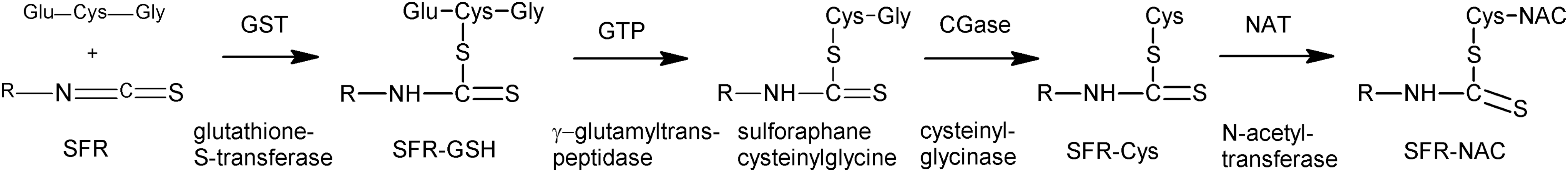
Metabolism of sulforaphane through the mercapturic acid pathway.
Sulforaphane is extensively investigated and is of interest in medicine for its health benefits. In the cited articles and in other works, sulforaphane has been identified as a chemoprotective agent potentially useful in clinical practice and as a substance beneficial to human health. In recent studies, it has been shown that sulforaphane may protect against various types of cancers, including pancreatic cancer, 3 leukemia, 4 colon cancer, 5 prostate cancer, 6 and may also decrease the risk of cardiovascular disease. 7 In addition, the effect of sulforaphane may also help in autism 8 and osteoporosis. 9 Sulforaphane is also known to be a potent Nrf2 activator and is capable of preventing toxicity of organic chemicals. 10,11 On the other hand, our recent experiments have shown that sulforaphane inhibits two major CYPs in human liver microsomes in vitro (3A4 and 2D6), 12 that is, enzymes that take part in a variety of important reactions of drug metabolism in humans. 13
In this work, we were interested also in possible inhibition of CYPs by the sulforaphane metabolites formed by the mercapturic acid pathway. To exhibit the biological activity, sulforaphane should be able to cross the cell wall and enter the respective cell. This is why we investigated the possible entry of sulforaphane into human hepatocytes. A reliable and robust method for determination of sulforaphane in the cells had to be developed.
For determination of sulforaphane in plant tissues, seeds, or in functional foods, many methods have been published based mostly on analysis by high-performance liquid chromatography (HPLC). 14 –16 Recently, an HPLC method was proposed involving heating the column at 60°C for determination of naturally occurring isothiocyanates 17 ; also an HPLC-electrospray ionization tandem mass spectrometry method for the simultaneous determination of glucosinolates and the corresponding isothiocyanates was published 18 ; and a ultra performance liquid chromatography method with a tandem mass spectrometry for determination of sulforaphane and its glucosinolate, glucoraphanin, in human urine was developed and validated. 19 As the methods cited above are rather elaborate, we have developed and validated a simple, reliable, and rapid HPLC method for determination of sulforaphane in the cells.
Materials and Methods
Chemicals and materials
Sulforaphane standard and sulforaphane metabolites (sulforaphane glutathione and sulforaphane cysteine conjugates and sulforaphane-N-acetylcysteine) were purchased from Santa Cruz Biotechnology, Inc. (Heidelberg, Germany). Dimethyl sulfoxide (DMSO) and potassium dihydrogen phosphate were obtained from Lach-Ner (Neratovice, Czech Republic); dichloromethane and methanol were from VWR Prolabo (Fontenay-sous-Bois, France).
For determination of CYP activities, ethoxyresorufin and 7-ethoxy-4-(trifluoromethyl)coumarin were obtained from Fluka (Buchs, Switzerland). Other specific substrates and metabolites of respective CYP enzymes (resorufin, bufuralol, midazolam, 7-hydroxy-4-(trifluoromethyl)coumarin, hydroxybufuralol, hydroxymidazolam) and all other chemicals were supplied by Sigma-Aldrich (Prague, Czech Republic).
Primary human hepatocytes used in this study were obtained from multiorgan donors; the use of liver cells of donors was approved by the Ethics committee at the Faculty Hospital Olomouc, in accordance with Czech Transplantation Law No. 285/2002 Sb. Human liver microsomes were obtained from Xenotech (Lenexa, KS).
Methods
Cytochrome P450 activities
Determination of cytochrome P450 activities
All activities of individual CYP forms were determined according to established published protocols. The following microsomal CYP activities were assayed: CYP1A2, 7-ethoxyresorufin O-deethylation (EROD) 20 ; CYP2B6, 7-ethoxy-4-(trifluoromethyl)coumarin 7-deethylation 21 ; CYP2D6, bufuralol 1′-hydroxylation 22 ; and CYP3A4 midazolam 1′-hydroxylation. 23 HPLC analyses of respective metabolites [resorufin for CYP1A2, 7-ethoxy-4-(trifluoromethyl)coumarin for CYP2B6, 1′-hydroxybufuralol CYP2D6, and 1-hydroxymidazolam for CYP3A4] were performed using the Prominence system (Shimadzu, Kyoto, Japan).
Inhibition of cytochrome P450 enzyme activities in microsomal fraction by metabolites of sulforaphane
For each enzyme assay, the Michaelis constant (Km) and maximum velocity (Vmax) values were determined to get the substrate concentration suitable for the inhibition experiments (substrate concentration was chosen in the range corresponding to the value of the Km).
Inhibition experiments were routinely performed with five concentration levels of individual sulforaphane metabolites (5; 10; 50; 100; and 200 μmol/L) and inhibitor-free control (buffer control). The stock solution of sulforaphane-N-acetylcysteine was 400 mmol/L in 100% methanol; concentration levels of sulforaphane glutathione and sulforaphane cysteine were obtained by diluting a 200 mmol/L solution in water. In all cases, controls with solvent (without tested compounds) were included because dimethyl sulfoxide, like other organic solvents, can inhibit CYP enzyme activity. 24
The amounts of human liver microsomes were as follows: 35 pmol of cytochrome P450 for CYP1A2 and CYP2B6; 13 pmol of cytochrome P450 for CYP3A4; and 70 pmol of cytochrome P450 for CYP2D6. Incubation mixtures contained 5 mmol/L Mg2+; 0.5 mmol/L NADP+; and an isocitrate/isocitric dehydrogenase NADPH-generating system and were buffered by 100 mmol/L K/PO4 (pH 7.4).
Concentrations of individual substrates in reaction mixtures were as follows: ethoxyresorufin (2.6 μmol/L); bufuralol (14.3 μmol/L); 7-ethoxy-4-(trifluoromethyl)coumarin (15 μmol/L); and midazolam (2.2 μmol/L).
Influence of sulforaphane metabolites on activities of individual CYPs was evaluated by plotting respective remaining activity against inhibitor concentration. All determinations of inhibition were done in triplicates and the results are expressed as means of three to five independent determinations with the difference between triplicates being lower than 10%. For evaluation of inhibition degree, scientific graphic software GraphPad Prism 5 (GraphPad Software, San Diego, CA) was used.
HPLC method for determination of sulforaphane in human liver cells
HPLC instrumentation and condition of analysis
Determination of sulforaphane was performed using the Prominence system (Shimadzu) equipped with a DGU-20A5R degassing unit, SIL-20ACXR autosampler, communication bus module CBM-20A, SPD-M20A diode array detector, CTO-20AC column oven, and LC-20ACXR pump. Chromatographic separation was performed on the LiChroCART® RP-18 (25 cm × 4 mm, 5 μm) reversed-phase column. The column was thermostated to 30°C and the injection volume was 50 μL. Analysis was carried out with isocratic elution by mobile phase (54% methanol in water) with flow rate of 0.6 mL/min. Under these conditions, sulforaphane was detected by absorbance at 245 nm with retention time 7 min.
Method validation
Linearity and calibration curve
Linearity of this HPLC method was evaluated by analysis of eight different concentration levels of standard solution of sulforaphane (0.9; 1.8; 2.7; 3.5; 4.4; 7.1; 10.3; and 17.7 μg/ml). Calibration samples were prepared as described below, each to be injected in volume 50 μL using autosampler, detected at 245 nm, and measured independently five times. For obtaining the calibration curve, means of peak height responses were plotted as a function of concentration with least-square linear regression analysis.
For calibration, samples containing various concentration levels of sulforaphane were obtained by diluting a 400 mmol/L stock solution in 100% methanol (calibration standards contained 10 μL of sonicated drug-free human liver cells). The final concentrations of tested compounds were in the range of 0.9–17.7 μg/mL in volume of 100 μL. Each sample of standard concentration levels was performed in five individual replicates.
Limits of detection and quantification
Limit of detection (LOD) and limit of quantification (LOQ) have been determined at a signal-to-noise ratio of 3.3 and 10, respectively, from the equations LOD = 3.3 × (σ/S), LOQ = 10 × (σ/S); where σ is the standard deviation of the response and S is the slope of the calibration curve.
Precision, accuracy, and recovery
Precision of this method was determined by the intraday and interday repeatability responses of analyzed working solutions of sulforaphane for three concentration (3.5; 7.1; and 17.7 μg/mL) levels and was expressed as % relative standard deviation (RSD). The intraday variation was determined by analyzing each concentration level for four repetitions on the same day.
The interday variation was determined by analyzing each concentration level for four repetitions on three different days. In this study, overall recovery of the method was evaluated by the analysis of aliquots of standard sulforaphane and sample with known concentration of determined compound, including the liquid–liquid extraction process used for cleaning up of samples. Percentage value of recovery was calculated by the following equation: % recovery = (recovered concentration/injected concentration) × 100 and expressed as mean ± RSD.
Preparation of samples of sulforaphane incubated with human hepatic cells
Sulforaphane in concentrations, 65 and 130 μmol/L, was incubated with human hepatocyte cultures (concentration of the cells 1.27 × 10⁵/cm2) for 24 h at 37°C in culture medium solution. Thereafter, human liver cells were washed by 100 mmol/L K/PO4 buffer (pH 7.4). The washed cells were briefly centrifuged (10 min; 1100 g), subsequently transferred into Eppendorf tubes, and frozen at −80°C and stored for determination of sulforaphane.
Then, the human liver cells with sulforaphane were thawed at laboratory temperature and diluted in 100 μL of 50 mmol/L phosphate buffer, and the cells were subsequently disrupted by sonication. The solution of disrupted cells was centrifugated (10 min; 14,000 g) and after transferring the supernatant into glass test-tubes, the mixture was extracted with 2 mL of dichloromethane by vortexing for 15 sec and again centrifuged (10 min; 1600 g; 4°C). After extraction, 1 mL of organic phase was taken and transferred into the new glass test-tubes and dried at 40°C under the stream of nitrogen. The residue was redissolved with 100 μL of 54% methanol in water (mobile phase). The resulting solution was gently vortexed for 15 sec and 50 μL of this sample was injected into the HPLC.
The conditions of HPLC analysis were described above. All determinations of sulforaphane concentration levels in prepared cell samples were done in triplicates and the results are expressed as means of three independent determinations ± SDs.
Results
Effects of sulforaphane metabolites on catalytic activities of drug-metabolizing cytochrome P450 in human liver microsomes
The effect of three sulforaphane metabolites (sulforaphane glutathione, sulforaphane cysteine conjugates, and sulforaphane-N-acetylcysteine) on activities of four forms of CYP enzymes present in human liver microsomes (CYPs 1A2, 3A4, 2B6, and 2D6) was studied.
The influence of sulforaphane metabolites on activities of four drug-metabolizing cytochrome P450 enzymes is shown in Figure 3. Contrary to the results obtained with the parent compound (sulforaphane) alone, where inhibition of CYP2D6 and CYP3A4 activities was found in human liver microsomes (and also of the CYP2A6 in human hepatocytes), the metabolites inhibit only CYP2D6 activity.

Effect of sulforaphane metabolites on specific activities of drug-metabolizing cytochrome P450 in human liver microsomes; concentrations of tested compounds (sulforaphane-glutathion, sulforaphane-cysteine, and sulforaphane-N-acetylcysteine) in reaction mixture were 0, 5, 10, 50, 100, and 200 μmol/L. For experimental details, see the Methods section.
In this study, the bufuralol 1′-hydroxylation (prototypical activity of CYP2D6 enzyme) was inhibited by sulforaphane glutathione and sulforaphane cysteine down to 78% and 73% of the original activity (in the absence of tested compounds) (Fig. 3), both at the concentration of 200 μmol/L. In the presence of the highest concentration (200 μmol/L) of all tested compounds (sulforaphane glutathione and sulforaphane cysteine conjugates and sulforaphane-N-acetylcysteine), the enzymatic activities of CYP1A2, CYP3A4, and 2B6 were decreased only to a low extent (to 97–87% of the initial activity, i.e., without tested compound added).
HPLC method for determination of sulforaphane in human liver cells
As the aim of this study was also to prove whether sulforaphane enters the cells, namely the human liver cells, it was necessary to develop a reliable method of determination of sulforaphane in the cell interior. The methods used in the literature were modified (based on a reversed-phase chromatography with isocratic elution by mobile phase) and validated and a simple and rapid HPLC method with UV detection was developed. The parameters of validation of the method described (see the Methods section) are presented in Table 1.
LOD, limit of detection; LOQ, limit of quantification; RSD, relative standard deviation.
Presence of sulforaphane in human hepatocytes
The experiment has demonstrated that sulforaphane has penetrated into the human liver cells. Figure 4 shows a typical chromatogram of sulforaphane standard and sulforaphane sample obtained after incubation with human liver cells. The data obtained indicate clearly a penetration of sulforaphane inside the hepatocytes. The sulforaphane levels 4.2 ± 0.3 μg/mL (i.e., 24 ± 2 μmol/L) for incubation with 11.5 μg/mL (i.e., 65 μmol/L) sulforaphane and 5.3 ± 0.3 μg/mL (i.e., 30 ± 2 μmol/L) for incubation with 23 μg/mL (i.e., 130 μmol/L) document the entry of sulforaphane inside the human hepatocyte.

Typical HPLC chromatogram of sulforaphane standard, including human liver cells (the black line), and sample of sulforaphane incubated with human hepatic cells (the gray line). HPLC, high-performance liquid chromatography.
Discussion
In this article, the influence of sulforaphane metabolites on drug-metabolizing cytochrome P450 enzymes in human liver microsomes is reported. The data obtained here revealed that sulforaphane metabolites had no prominent effects on the CYP1A2, CYP3A4, and 2B6 activities; however, two sulforaphane metabolites (sulforaphane glutathione and sulforaphane cysteine conjugates) weakly interacted with CYP2D6 form (Fig. 3); moreover, the inhibition occurred at rather high concentration of sulforaphane-glutathione and sulforaphane-cysteine conjugates (up to 200 μmol/L). Nevertheless, these interactions are unlikely to be clinically important because the enzymatic activities were not significantly affected at physiologically relevant concentrations of the potential inhibitor (plasma concentration of sulforaphane and its metabolites does not exceed 1 μmol/L). 25
In addition, in this study, a simple and rapid HPLC method has been used for determination of sulforaphane in human liver cells treated with this compound. Many HPLC methods have been already published for determination of sulforaphane (using a multitude of different detectors) mostly in plant tissues, 14 –19,26 in human plasma, 25 or in mouse organs. 27 The validation parameters of the method described here are in principle similar to methods published earlier 14 –16 ; on the other hand, the method presented here allows to get information on the sulforaphane level also inside the cells with very good precision and at a relatively low level. The differences in validation parameters of other methods are in general related to the treatment and preparation of samples. The method presented was finally successfully applied to the evaluation of sulforaphane intake by human hepatic cells.
The result related to penetration of sulforaphane into human liver cells was rather expected as it was reported earlier that sulforaphane can influence the activities of CYPs (namely of the forms, CYP3A4 and CYP2D6) not only in the human liver microsomes but also in human hepatocytes. 12 These obtained levels of sulforaphane in the cells (see the Results section) should be compared with typical plasma levels of these compounds, that is, about 1 μmol/L. 25 It can be concluded here that the sulforaphane enters the human hepatocytes to a rather significant extent. Hence, the drug-metabolizing enzymes, located inside the cells, can be influenced by action of this compound. Nevertheless, it has been found 27 that the free sulforaphane is not a major compound present in the organs of mice; instead, rather the glutathione, cysteinyl, and N-acetylcysteine conjugates are present at even higher levels.
Footnotes
Acknowledgments
Our laboratories are supported by a grant GACR 303/12/G163 from the Grant Agency of the Czech Republic and by students' grant of Palacky University IGA UPOL_LF_2016_006.
Author Disclosure Statement
No competing financial interests exist.