




Abstract
White sesame seed oil (WSSO) has been used in cooking and food preparations for centuries. It has many purported health benefits and may be a promising nutraceutical. The primary purpose of this study was to examine the effects of WSSO on fasting blood glucose (GLU) and insulin (INS) in male Sprague-Dawley rats with chemically induced diabetes. A secondary aim was to explore other hematological biomarkers of hepatic, cardiac, and renal function. Sixty-three male Sprague-Dawley rats were randomized into standard diet groups, normal control (NCON) (n = 21) and diabetic control (DCON) (n = 21), and a diabetic sesame oil (DSO) (n = 21) group, which were fed a diet containing 12% WSSO. Blood samples were analyzed at 0, 30, and 60 days. Differences between groups and across days were assessed with two-way repeated measures analysis of variance. At baseline, GLU and INS were similar in both diabetic groups, mean 248.4 ± 2.8 mg/dL and mean 23.4 ± 0.4 μU/mL, respectively. At 60 days, GLU was significantly (P < .05) higher in DCON (298.0 ± 2.3 mg/dL) compared with DSO (202.1 ± 1.0 mg/dL). INS showed similar favorable trends after WSSO supplementation. Consumption of WSSO significantly improved glucose control and other biomarkers of hepatic stress, as well as cardiac and renal health. WSSO may be a viable functional food to help reduce the detrimental effects of diabetes.
Introduction
T
In recent years, there has been increasing interest in developing and using natural plant products as nutraceuticals to treat diseases such as T2DM. Consumers have begun to examine foods not only for their basic nutrients but also for their potential health benefits. 2 The upsurge in using commonly obtained foods as functional foods has also prompted a critical need to scientifically study the purported health claims involved. Specifically, much attention is being given to the usage of plant species that have antidiabetic activity. 3 Scientific investigation of traditional or natural remedies for T2DM may improve the options for physicians and their patients, who are struggling with diabetes and its complications.
White sesame seeds (Sesamum indicum) are known as the queen of oil seeds because of their high oil (50–60%) content. 4 White sesame seed oil (WSSO) is an excellent source of unsaturated fatty acids with 37% from oleic and 46% from linoleic fatty acids, 5 which have been shown to improve insulin sensitivity and thus glucose regulation through increasing the presence of unsaturated fatty acids in the sarcolemma. 6
Sesame seeds also contain appreciable amounts of bioactive components, including tocopherols, polyphenols, phytosterols, and flavonoids. 7 Additionally, sesame oil has a concentration of 1% to 2% of phenolic lignans, sesamin, and sesamolin. All of these bioactive components are considered protective 8,9 and likely act synergistically to contribute to the reported antioxidant, antihypertensive, antimutagenic, anti-inflammatory, antithrombotic, and cardioprotective properties for which sesame seed oil is known. 9,10
In an earlier study by Ramesh et al., 11 the consumption of 6% sesame seed oil added to normal rat diet was shown to significantly reduce blood glucose levels, lipid peroxidation, and antioxidant status in normal and diabetic (streptozotocin [STZ]-induced) female Wistar rats. However, this previous investigation did not assess insulin, specific hepatic enzyme activities, liver, cardiac, or renal markers, electrolyte/mineral balance, or hematological components. Additionally, Ramesh and colleagues 11 performed their experiments on female Wistar rats and used commercially purchased sesame oil and did not describe the purity of the oil.
Our study is unique, in that we have greatly extended the study published by Ramesh et al. 11 by examining additional biomarkers and a full hematological profile in response to pure virgin sesame seed oil that we extracted directly from the seeds in the laboratory. Furthermore, we examined younger male rats of a different strain (Sprague-Dawley) and used a higher dose of STZ (65 mg/kg vs. 40 mg/kg). Finally, in contrast to the previously mentioned study, 11 we addressed changes between three time points over a longer time period. Thus, this study is unique as it is a highly controlled examination of the effects of WSSO on glucose regulation and markers of hepatic, cardiac, and renal functions in STZ-induced diabetes in adolescent, male Sprague-Dawley rats.
The primary aim of this study was to examine the effects of 12% WSSO on fasting blood glucose (GLU) and insulin (INS) in male Sprague-Dawley rats with chemically induced diabetes at baseline, 30, and 60 days. A secondary aim was to explore other hematological biomarkers of hepatic, cardiac, and renal function. We hypothesized that diabetic rats supplemented with WSSO will have lower blood glucose and improved glucose regulation than diabetic rats on a normal diet. In addition, we expected that the WSSO would show positive indications of enhancing liver and kidney function as well as improvements in cardioprotective properties.
Materials and Methods
Materials
White sesame seeds (PB-Till 90) were purchased from Ayub Agriculture Research Institute Faisalabad, Pakistan. Oil extracted from white sesame seeds through solvent extraction method 4 was used for further studies.
Chemicals
All chemicals used were of analytical grade. Analytical grade STZ (Cat. No. 41910012-4 (714992) (bioWORLD-Dublin, Dublin, OH) of the highest purity was used (Supplementary Data; Supplementary Data are available online at
Animals
Sixty-three adolescent, male Sprague-Dawley rats (age: six weeks, weight: 125–175 gm) were purchased from National Institute of Health Islamabad, Pakistan, and housed at the animal center of the University of Veterinary and Animal Sciences Lahore, Pakistan. Upon arrival, rats were randomly distributed into three groups and were acclimatized to the laboratory for seven days. Rats were housed in separate cages, with 4 rats/cage (cage size: L 61 cm × W 46 × H 46). Rats were kept in a temperature-controlled (25°C) room with 12-h light/12-h dark cycles. Rats were given free access to water and food. However, each day, intake was carefully measured to determine how much was consumed.
All food consumed was measured, including any that dropped on the cage floor. Each rat consumed 18–25 gm per day of food. Additionally, individual rats were weighed monthly to determine if there were any discrepancies in food intake of any rat within a cage. Last, rats were not provided exercise wheels, but were allowed to roam freely in their cages.
Induction of diabetes
Forty-two rats were injected with STZ to induce diabetes. The STZ was dissolved in citrate buffer (0.05 M, pH 4.5) and then was injected intraperitoneally at a dose of 65 mg/kg of the body weight. 12 By day 6, blood sugar levels were checked and treated rats had become diabetic (defined as blood sugar level more than 220 mg/dL). Diabetic rats were grouped as either diabetic control (DCON) (n = 21) and fed a standard diet containing soybean oil or diabetic sesame oil (DSO) (n = 21) and fed the standard diet containing WSSO.
Preparation of rat chow
The diets were prepared in the laboratory following the composition and ingredients of the standard AIN-93G purified diet 13 (Harlan, Madison, WI). The two control diets (NCON and DCON) and the DSO diet were prepared identically. Control diets contained soybean oil (12%) and DSO diet contained WSSO (12%). After mixing all the ingredients, the final batter was formed into pellets, air-dried, and stored at 4°C. All rat food contained 18% protein, 12% fat, and 60% carbohydrates.
Experimental design
A total of 63 male Sprague-Dawley rats were randomized into three groups: DCON, DSO, and normal control (NCON). Blood samples were analyzed at 0, 30, and 60 days. Differences between and within groups were assessed with a two-way repeated measures analysis of variance (ANOVA).
Sample collection
Blood samples were taken from each rat under mild chloroform anesthesia (inhalant) at three time periods: (baseline, day 30, and day 60). Blood was collected in different tubes for whole blood samples (EDTA as anticoagulant), for plasma (Heparin tubes), and for serum (serum separator tubes). Serum/plasma was separated by centrifugation at 3000 rpm for 15 min. Samples were stored at −40°C and were analyzed within 7–10 days.
Methods and biochemical assays
Serum glucose and insulin levels
Serum glucose concentrations were measured following the glucose oxidase-phenol amino phenazone (GOD-PAP) method by enzymatic colorimetric method described by Thomas and Labor. 14 Glucose assay kit GAGO-20 from Sigma-Aldrich, UK was used. Changes in serum insulin were measured using the RAB0817 kit (Sigma-Aldrich, UK) according to the manufacturer's protocol as previously described by Besch et al. 15
Cardiac enzymes
Levels of cardiac enzymes were also determined by following the procedure of Pagana and Pagana 16 and using diagnostic kits (CK: MAK116 and CK-Mb: ERMAD455) from Sigma-Aldrich.
Serum biochemistry
Serum levels of alanine aminotransferase (ALT) and aspartate aminotransferase (AST) were measured using the dinitrophenylhydrazine method from diagnostic kits (ALT: Cat. No. MAK052; and AST: Cat. No. MAK055 Sigma-Aldrich). Alkaline phosphatase (ALP) was measured through alkaline phosphatase-DGKC (German Society of Clinical Chemistry) method, 17 and using diagnostic kit (Cat. No. AP0100) from Sigma-Aldrich, UK, while urea, creatinine, and uric acid concentrations were analyzed by following the procedure of Jacob 18 and using diagnostic kits (MAK006; MAK080; and MAK077; Sigma-Aldrich).
Electrolyte balance and mineral contents
Electrolytes (Na and K) were measured using commercially available kits according to the manufacturer's protocols (Na: K 391-100 BioVision and K: 37202-1EA Sigma-Aldrich) by adopting the method described by Marshall, 19 and minerals (Ca, Fe, and Zn) were estimated using commercially available kits according to the manufacturer's protocols (Ca; MAK022; Fe: MAK025; and Zn: MAK032 Sigma-Aldrich) by adopting the procedures as described by Kazi et al. 20
Hematological components
Red blood cell counts, white blood cell counts, packed cell volume, and hemoglobin were determined by adopting the methods described by Widmann. 21 Total proteins and the serum protein profile (albumin, globulin, and A/G ratio) were determined using diagnostic kits (Total protein: TP0300; Albumin: MAK-124; and Globulin: G9894; Sigma-Aldrich), following the method described by Bradford. 22
Statistical approach
All data are expressed as mean ± standard error of mean. Between- and within-subject variations were analyzed by two-way RM ANOVA (SigmaPlot 10.0; Systat Software, San Jose, CA). Where significant effects occurred, Tukey's post hoc analyses were used. A P-value of <.05 was considered statistically significant.
Results
Morphometrics
As shown in Table 1, mean weight of the two diabetic groups dropped significantly (P < .05), while the body weight of the NCON group increased significantly (P < .05), as would be expected with normal growth with ad libitum feeding. However, between 30 and 60 days, the mean weight in the DCON group continued to drop, while the weight of the DSO group seemed to stabilize across time.
Rat 1 to Rat 21 are the body weights in grams of DCON group rats, Rat 22 to Rat 42 are the body weights in grams of DSO group rats, and from Rat 43 to Rat 63 are the body weights in grams of NCON group rats.
Significant difference P < .05 between baseline and 60 days.
DCON, diabetic control; DSO, diabetic sesame oil; NCON, normal control.
Glucose and insulin
Changes in blood glucose and insulin level for each group across time periods are shown in Figure 1. NCON had normal glucose levels from baseline to day 60 (range: 92.5 ± 0.7 mg/dL to 101.5 ± 1.6 mg/dL). In addition, in the NCON group, insulin did not change significantly across time (baseline: 29.9 ± 0.50 μU/mL to 60 days: 29.4 ± 0.2 μU/mL) and it was significantly (P < .05) higher than the DCON. At baseline, there were minimal differences in glucose between the two diabetic groups. However, there was a significant group by time interaction in glucose and insulin in the two diabetic groups.

Effects of WSSO on glucose and insulin. *Significantly different, P < .05 between groups at 60 days. WSSO, white sesame seed oil.
In the DSO group, fasting blood glucose significantly (P < .05) decreased across time (baseline: 253.9 ± 3.3 mg/dL to 60 days: 202.1 ± 1.0 mg/dL), whereas in the DCON group, glucose levels significantly (P < .05) increased from baseline (242.9 ± 2.3 mg/dL) to 60 days (298.0 ± 2.3 mg/dL). Serum insulin showed a similar trend. In the DSO group, insulin increased from 22.9 ± 0.5 μU/mL to 33.3 ± 0.7 μU/mL at 60 days, which was similar (P > .05) to the NCON group, whereas in the DCON group, insulin level significantly (P < .05) decreased from 23.8 ± 0.2 μU/mL to 16.8 ± 0.8 μU/mL across time.
Cardiac enzymes
Figure 2 shows the changes in the activities of cardiac enzymes; creatine kinase and creatine kinase-myoglobin (CK and CK-Mb) over time for each group. In the DSO group, CK decreased significantly (P < .05) from baseline (291.1 ± 0.9 U/L) to 60 days (245.5 ± 7.2 U/L) in both the control groups, while CK-Mb decreased significantly (P < .05) from baseline (550.5 ± 3.9 U/L) to 60 days (510.8 ± 6.8 U/L) in the NCON group, but was not significantly different in the DCON group. NCON and DCON groups were significantly different for CK-Mb.
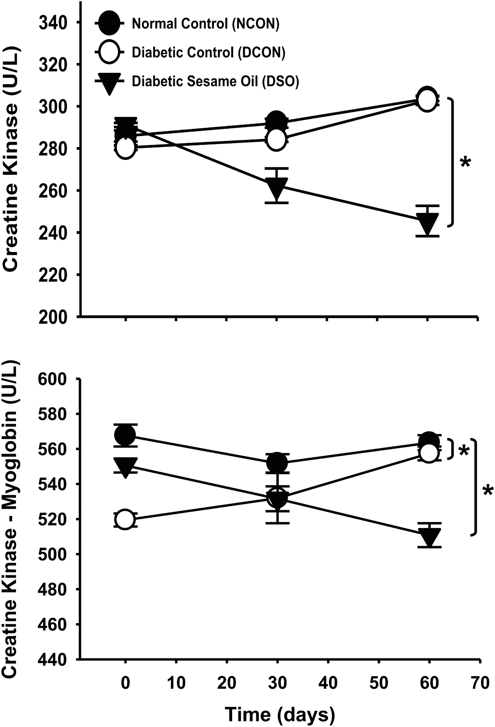
Effects of WSSO on cardiac enzymes. *Significantly different, P < .05 between groups at 60 days.
Serum biochemistry
Figure 3 indicates changes across time in various liver function values (ALP, ALT, and AST). The results indicated that ALP increased over time in both diabetic groups (i.e., in DSO group, from baseline to 60 days, it was raised from 246.7 ± 3.3 U/L to 277.7 ± 2.8 U/L) and, at 60 days, was significantly higher (P < .05) than NCON in both groups, but values were not significantly different from each other. In contrast, ALT from baseline (81.5 ± 3.7 U/L) to 60 days (67.4 ± 2.7 U/L) and AST from baseline (148.7 ± 3.5 U/L) to 60 days (118.3 ± 1.2 U/L) significantly decreased (P < .05) in the DSO group compared with DCON or NCON, resulting in significantly lower values than both control groups by 60 days. The overall results indicated that by 60 days, liver function values were lower in the DSO group compared with rats in the control groups.
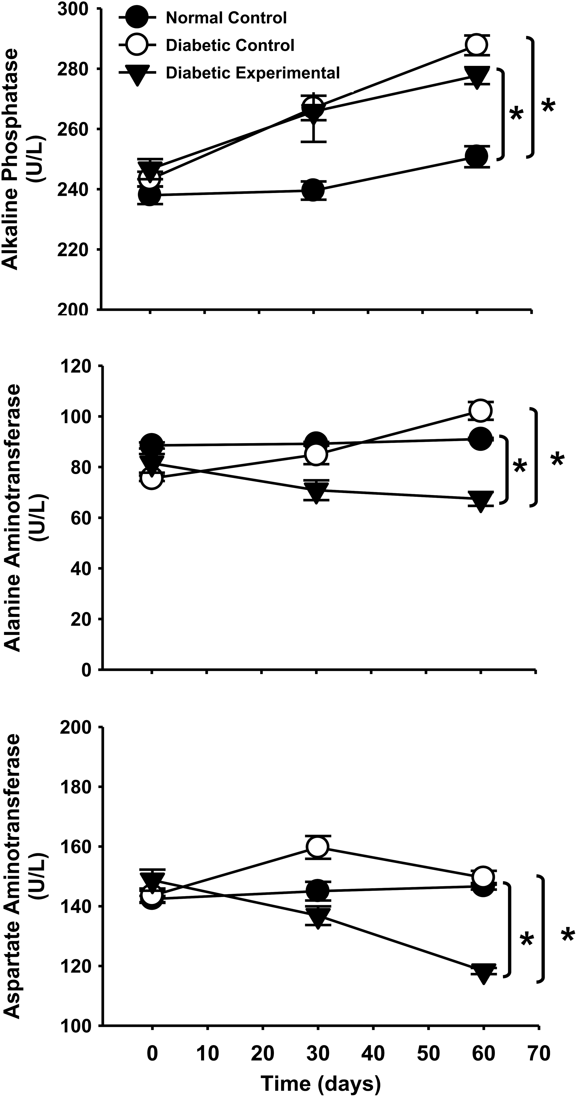
Effects of WSSO on serum biochemistry (liver function tests). *Significantly different, P < .05 between groups at 60 days.
Figure 4 indicates changes in the basic kidney function values (urea, creatinine, and uric acid). Urea was not statistically significant between groups at baseline and at 30 days. At 60 days, urea in the DSO group decreased from baseline (38.5 ± 2.3 to 30.9 ± 1.1) such that it was significantly lower (P < .05) than both control groups. From baseline to 60 days, creatinine significantly increased (P < .05) in the two diabetic groups; in the DSO group at baseline, creatinine was 0.3 ± 0.0 mg/dL and increased up to 0.4 ± 0.1 after 60 days, whereas it remained fairly stable in the NCON group. At 60 days, creatinine was significantly higher in both the diabetic groups compared with NCON. For uric acid, the results indicated that in both of the control groups, uric acid increased, whereas in the DSO group, uric acid remained fairly stable across time.
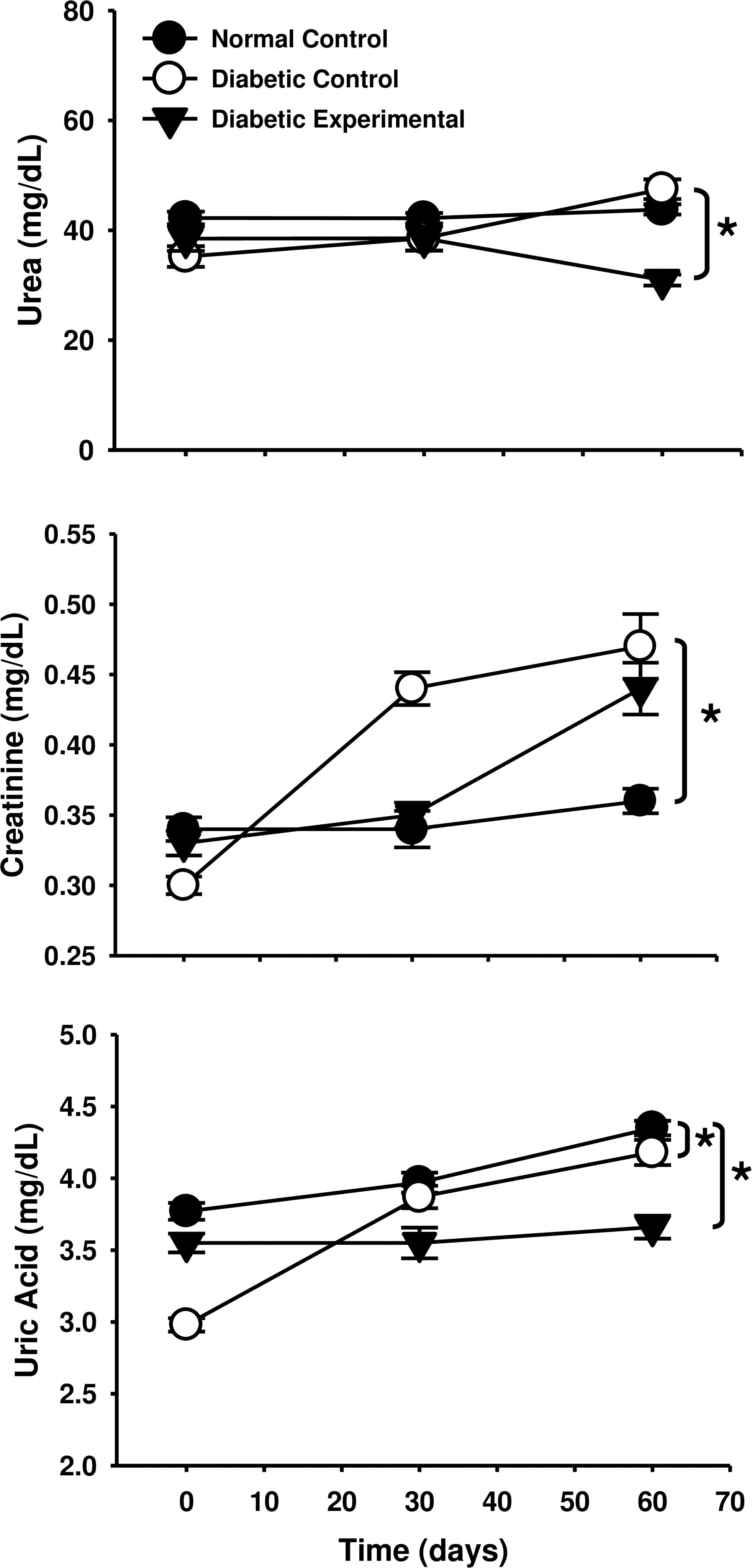
Effects of WSSO on serum biochemistry (renal function tests). *Significantly different, P < .05 between groups at 60 days.
Electrolyte balance, mineral contents, and hematological components
Table 2 indicates the changes in electrolytes and minerals across time. When comparing differences between the two diabetic groups (DCON and DSO); calcium was significantly increased and iron was significantly decreased in DSO compared with DCON. In fact, while the values of sodium, potassium, and zinc varied between groups, there was no significant group by time differences. Table 3 reports the hematological values for each group at each time period. The results indicated that there were no significant differences between the DSO or DCON groups in any of the hematological values.
A/G, albumin to globulin; Hb, hemoglobin; RBC, red blood cell; WBC, white blood cell.
Discussion
In this study, we demonstrated that consumption of WSSO controls blood glucose levels and improves the insulin level in rats with chemically induced diabetes. We used sesame seed oil in this study, as oil comprises the major portion of the sesame seed. It also contains the greatest quantity of bioactive components (i.e., fat-soluble lignans, sesamin, sesamolin, and γ-tocopherol) that may have antidiabetic activity. The results of this study suggest that ingesting a small amount of sesame seed oil over time may improve health in those with diabetes. This study agrees with, and greatly extends, the results presented by Ramesh et al. 11 by describing the glucoregulatory effects of WSSO in chemically induced adolescent male diabetic rats as well as illuminating changes in the characteristic biomarkers of liver, kidney, and heart function in response to WSSO.
Blood glucose and insulin
The process of inducing diabetes through STZ results in the development of high levels of reactive oxygen and nitrogen species, which function to destroy pancreatic β-cells. 23 Since STZ enters tissues through GLUT2 glucose transporters in the plasma membrane, it can also affect other tissues with GLUT2 transporters such as liver and kidney. 24 When the pancreatic β-cells are destroyed, insulin secretion is reduced, resulting in an increase in glucose and an overall decrease in glucose regulation. Interestingly, since STZ-induced diabetes is in essence caused by oxidative stress, it is reasonable that the process of improving pancreatic β-cell function with WSSO, and hence increased insulin levels, may have occurred through increased free radical scavenging. 25,26
While much of the pancreatic β-cell function may have been destroyed by the oxidative stress caused by STZ in the DSO group, the function of any remaining β-cells may have increased such that insulin secretion was restored, thereby improving glucose control. STZ works by inhibiting the activities of free radical scavenging enzymes that in turn increase superoxide radical and nitric oxide generation. These activities may generate reactive oxygen species (ROS) or oxidative stress that may cause oxidative damage that in turn causes β-cell cytotoxicity, 27 resulting in decreased insulin production and increased blood sugar. In the case of diabetes, cell dysfunction leads to increased production of ROS, which generates oxidative stress. 28 Increased production of ROS/reactive nitrogen species (RNS) as a result of oxidative metabolism can damage valuable biomolecules and cell components (lipids, DNA, protein) and trigger the activation of signaling pathways, as well as disrupt normal repair mechanisms. These effects may lead to the development of chronic diseases such as diabetes. 29,30
Another potent regulator of glucose homeostasis is liver glucokinase (GCK) activity. Iynedjian and colleagues 31 showed that in rats with STZ-induced diabetes, the gene expression of GCK was reduced. Furthermore, they noted that insulin restores GCK expression. In this study, GCK was not measured, but it is possible that the increase in insulin resulted from improving β-cell function subsequent to lowering oxidative stress with WSSO, which may have increased GCK activity and stimulated an increase in glucose uptake and lower blood sugar levels. Indeed, the results of this study indicate that in the DSO rats, WSSO improved both β-cell function and glucose regulation. β-Cells are more sensitive to ROS because they have low free radical scavenging enzymes. 32
Thus, ROS may have increased oxidative stress by upregulating production of p21 and decreasing insulin messenger RNA cytosolic adenosine triphosphate, as well as calcium flux in the cytosol and mitochondria. 33 In rat liver, sesamin is metabolized by cytochrome P450 that converts methylenedioxyphenyl to dihydrophenyl (catechol) moiety. This conversion to catechol is reported to have strong free radical scavenging activities 34 that reduce the generation of free radicals and play a protective role against STZ-induced oxidative stress, β-cell damage, and ultimately diabetes. 26,27,35 The process of improving pancreatic β-cell function with WSSO, and hence restoring insulin levels, may have occurred through increased free radical scavenging activity. In addition to improved insulin bioavailability, which has also been observed by Ibrahiem, 36 glucose regulation was likely also improved by augmenting insulin sensitivity through increased presence of unsaturated fatty acids in the sarcolemma as has been shown following supplementation with polyunsaturated fatty acids. 6 The glucose-lowering effects of sesame seed oil following STZ-induced diabetes are consistent with findings from several other recent studies. 11,37,38
Finally, monounsaturated-rich diets are known to be capable of reducing blood glucose levels by delaying glucose absorption. 24 Sesame oil is highly unsaturated oil, thus the very nature of the oil itself may be able to regulate glucose levels. For example, Takeuchi et al. 38 discovered that if diabetic mice were fed the defatted portions of sesame seeds, then glucose uptake was reduced, thereby establishing that the oil in the sesame seeds is able to delay glucose absorption and aid in glucose regulation. Similarly, Ramesh 39 also showed that consumption of sesame oil helped to improve blood glucose regulation in diabetic rats. A limitation of the current study is that oxidative stress markers were not reported.
Cardiac enzymes
Oxidative stress that develops in response to prolonged exposure to hyperglycemia also leads to diabetic cardiomyopathy. 40 This oxidative stress causes cardiomyocyte damage, resulting in increased activity of the cardiac enzymes, such as CK and CK-Mb. In this study, the CK activity and CK-Mb levels were significantly decreased in the DSO group compared with DCON. This provides evidence that WSSO may have protected the heart from hyperglycemia. Last, with diabetes, both glucose uptake and utilization by the heart muscles are inhibited. This imbalance in glucose uptake and utilization in heart muscles is known to play a significant role in the development of heart disease. 41,42 Thus, WSSO intake may be a good strategy to decrease the risk for developing heart disease in those with chronic hyperglycemia.
Serum biochemistry
As noted previously, hyperglycemia and oxidative stress caused by both reactive nitrogen species and ROS result in systematic cellular damage. It is known that in STZ-induced hyperglycemia, liver cell function is disrupted. In fact, the free radicals generated from exposure to hyperglycemia disturb normal defense mechanisms, which in turn put increased stress on the plasma membranes of liver cells and increase the chance of peroxidation. 26,35 In the liver, this oxidative stress destroys cell membranes, 43 resulting in the release of ALP that is located in the cytoplasm.
In this study, ALP in both diabetes groups was significantly increased beyond that of the normal control group, providing further evidence of the liver damage characteristic of this induced diabetic state. The two groups with induced diabetes seemed to respond similarly. However, although not significantly different, at 60 days, the level of ALP was trending to be lower in the DSO group compared with DCON. This may suggest that with a longer treatment period, WSSO may have protected the liver cell membranes. Giannini and colleagues indicated that when the mitochondria of the cell are damaged or destroyed, then both ALT and AST are released. 44 In this study, both ALT and AST steadily increased over time in the DCON group, but remained stable in the NCON group.
However, in the DSO group, ALT and AST levels significantly decreased such that by 60 days, they were lower than the NCON group. This suggests that WSSO may have protected liver mitochondria.
In the kidneys, elevated values of serum urea, creatinine, and uric acid often indicate renal dysfunction. 45 The findings of the current study indicate that in the two diabetic groups, creatinine was significantly increased in comparison with NCON. This finding suggests that STZ may have promoted oxidative stress. However, by day 60, urea and uric acid were significantly lower in the DSO group than both control groups. Since urea and uric acid are derived primarily from protein catabolism, it may be that these lower values are reflective of less overall protein breakdown in the DSO group. Thus, these findings support a protective role for WSSO in the kidney as well.
Electrolyte balance, minerals, and hematological values
In diabetes, changes in electrolytes and minerals are often a good indication of disease complications and progression. Electrolyte balance becomes disturbed in hyperglycemia due to reduced bioavailability and release of insulin, which reduces the absorption of these electrolytes. 46,47 In this study, sodium and potassium electrolyte balance was not significantly different between groups. This could be because insulin was stable in the NCON group and began to stabilize in the DSO group. However, insulin was still disturbed in DCON, possibly creating some variability in electrolyte balance.
Minerals are essential for biochemical reactions in the body. Oxidative stress and less free radical scavenging activity reduce the availability of minerals for various biochemical reactions. In addition, the presence of increased antioxidant activity and reduced free radical activity can change the intestinal absorption of minerals such as iron and zinc. 20 However, high amounts of some minerals, such as iron, may actually serve as a booster for lipid and protein oxidation and generate an increase in free ROS. 48 Thus, in diabetes, metabolic availability of several trace elements/minerals may be altered in response to prolonged hyperglycemia. 20
In this study, there were no clear patterns in the changes in the mineral values between groups over time. Over time, calcium was significantly elevated in DSO compared with DCON, while iron was highest in the DCON group at 60 days. The values of sodium, potassium, and zinc varied between groups, but there were no significant group by time differences.
The results indicated that there were no significant differences in any of the hematological values between DSO and DCON. As previously noted, in the presence of hyperglycemia, liver and kidney tissues can become damaged or necrotic, which could impact hematological variables. 49 In this study, it is likely that the degree of the exposure to STZ was not of sufficient intensity or duration to damage the liver and kidney tissues enough to impact the hematological values.
Conclusion
This study provides evidence that the consumption of WSSO by adolescent, male diabetic rats may reverse much of the STZ-induced glucose dysregulation through improved insulin bioavailability. Although not measured directly in the current study, it is possible that some of the protective effects were due to changes in liver enzymes in response to WSSO. Additionally, it is likely that WSSO further helped myocardial, liver, and kidney functions. The results of this study indicate that consumption of WSSO positively influences blood glucose, cardiac, liver, and kidney functions in adolescent, male Sprague-Dawley rats with STZ-induced diabetes.
Footnotes
Acknowledgments
This work was financially supported under an Indigenous PhD Fellowship for 5000 Scholars (Phase-II) from the Higher Education Commission of Pakistan. The authors thank Dr. Muhammad Rafique Asi, Dr. Mateen Abbas, Mr. Matee-ur-Rehman, Mr. Rizwan Razzaq, Mr. Aamir Shahzad, and Mr. Amir Rasheed for their continuous support and help during this project.
Author Disclosure Statement
No competing financial interests exist.
References
Supplementary Material
Please find the following supplemental material available below.
For Open Access articles published under a Creative Commons License, all supplemental material carries the same license as the article it is associated with.
For non-Open Access articles published, all supplemental material carries a non-exclusive license, and permission requests for re-use of supplemental material or any part of supplemental material shall be sent directly to the copyright owner as specified in the copyright notice associated with the article.
