




Abstract
One of the useful properties of probiotic bacteria is their capacity to bind different targets, thus eliminating them through feces. It is supposed that one of these targets could be cadmium, a widespread environmental toxicant that causes various disturbances in biological systems. This study examined the protective effects of probiotic supplementation against cadmium-induced toxicity in the rat. The experiment was conducted in the course of 5 weeks. Animals were divided into four groups: (1) controls, (2) probiotics treated, (3) cadmium treated, and (4) probiotics + cadmium treated. The cadmium concentration was measured in the blood, liver, kidney, and feces, as well as the blood alanine aminotransferase (ALT) and aspartate aminotransferase (AST) as biomarkers of the liver function. Histomorphological changes in the liver and kidney were also determined. Our results revealed that probiotics combined with cadmium increase this metal concentration in feces. As a result, blood, liver, and kidney Cd levels, as well as blood ALT and AST activities were lessened compared to the rat group treated with cadmium only. Besides, probiotics consumed simultaneously with cadmium attenuated histomorphological changes in the liver and kidney caused by cadmium. The rise in lactobacilli number in feces of rats treated simultaneously with cadmium and probiotics results in strong correlation with the increase of Cd concentration in their feces and the decrease of Cd concentration in their blood. We speculate that probiotics actively contribute to cadmium excretion through feces, probably, by its binding to their bacterial cell wall.
Introduction
C
Extreme cadmium toxicity for mammals has been verified in many experimental models 4 and its harmfulness to humans in accidental toxicity is also well known. 5 Cadmium is toxic to almost every organ in the body, with the liver and kidneys as main targets. 6 In the liver, it causes various histological changes, including damage to hepatic parenchyma and increased infiltration of inflammatory cells. 7 These disturbances lead to elevation of plasma alanine aminotransferase (ALT), aspartate aminotransferase (AST), lactate dehydrogenase, and alkaline phosphatase. 7,8
One of the useful properties of probiotic bacteria is their capacity to adhere to different targets. 9 –11 For example, some probiotic strains, such as Lactobacillus rhamnosus GG (ATCC 53103) and L. rhamnosus LC-705 (DSM 7061), may remove aflatoxin B from aqueous solution, 10 as well as mutagen pollutants from foodstuffs. 11 Also, Bifidobacterium longum 46, L. fermentum ME3, and B. lactis Bb12 bind to cadmium and lead in the water. 9
However, all those studies were conducted in vitro. However, recently, the Cd binding capacity of probiotics was also demonstrated in vivo 12 : only cadmium given orally could be removed through feces, but not that injected intraperitoneally. 13
The results of our trial examining whether probiotics may exert similar effects in rats are presented in this study. We have selected rat as a model given that study results can be better translated to humans since it is widely believed that rats are physiologically more similar to humans than mice. 14 Besides, rat model is believed to be superior to mouse models for the study of toxicity, because of similarity in the number and type of detoxifying enzymes to those in humans. 15
Materials and Methods
Animals and treatments
This study was carried out in strict accordance with the recommendations of Animal Welfare Act of Serbia. The protocols were approved by the Committee on the Ethics of Animal Experiments of the University of Belgrade, Faculty of Biology.
Male rats of Wistar strain (Rattus norvegicus), weighing 130 ± 10 g, were used for the experiments. The animals were acclimated to 22°C ± 2°C, exposed to a 12-h light/12-h dark regime with the light on at 6 a.m. and off at 6 p.m., with free access to commercial rat food (Veterinary Institute, Subotica, Serbia) and tap water. Rats were randomly divided into four groups, each consisting of six animals. Animals were individually housed, with cages randomly distributed on shelves close to each other to allow social interactions among animals. Animals were treated as follows:
Control group: rats given tap water to drink ad libitum and fed with commercial food;
Probiotic group: rats given tap water to drink ad libitum and fed with commercial food supplemented with probiotic bacteria (commercial preparation PROBIOTIC®; Ivančić i sinovi d.o.o.). The capsules were declared to contain 5 × 10⁹ lyophilized cells of Lactobacillus rhamnosus Rosell-11, Lactobacillus acidophilus Rosell-52, and Bifidobacterium longum Rosell-175 strains. However, after identifying the number of bacteria present in the capsules, we found that the number of viable cells was around 5 × 10⁸. One capsule was mixed with one gram of food to achieve a final probiotic concentration of 5 × 10⁸ cfu/g food. Based on the rat food consumption during 5 weeks, the total probiotic supplementation was on average around 7.5 × 10⁹ cfu/100 g body mass/day;
Cadmium group: rats given cadmium chloride (CdCl2, analytical grade; Fisher Scientific) in a volume concentration of 70 ppm in tap water to drink ad libitum and fed with commercial food. Based on the rat water consumption during 5 weeks, the total cadmium intake was on average around 1.25 mg of CdCl2/100 g body mass/day;
Probiotic + cadmium group: rats fed with probiotics and given to drink CdCl2. Based on the rat food and water consumption during 5 weeks, the total probiotic supplementation was on average around 6.9 × 10⁹ cfu/100 g body mass/day and 1.09 mg of CdCl2/100 g body mass/day.
The experiment was conducted for 5 weeks. The animal body mass and food and water consumption were measured daily. At the end of the treatment, the feces were collected and stored at −80°C until analysis. Animals were decapitated with a guillotine (Harward-Apparatus) without anesthesia. The blood, collected from the trunk, was divided into two sets of tubes, with EDTA added to get plasma.
Biochemical analyses
Serum, plasma and feces were frozen at −80°C for subsequent determination of AST, ALT, and Cd concentrations.
ALT and AST were measured in serum by a Randox kit, where the products of ALT and AST action (pyruvate and oxaloacetate, respectively), in the presence of NADH, and the enzymes lactate and malate dehydrogenase, respectively, oxidize NADH to NAD. This reaction is followed by a decrement in absorbance at 340 nm. ALT and AST activities were calculated as U/L = 1746 × ΔA(340nm/min), where ΔA(340nm/min) is the mean absorbance decrease between the first and second, and second and third minutes.
Cadmium was determined in the whole blood, liver, and kidney of individual animals, as well as in feces collected during the last week of the experiment. Before spectrophotochemical determination of the Cd concentration, the samples were digested with 1 mL of H2O2 and 7 mL of concentrated HNO3. Digestion was performed according to the US EPA SW-846 Method 3052 with a Milestone ETHOS One microwave digestion system at 200°C. The feces digests were filtered to remove undestroyed silicate salts, although the procedure extracts the total bioavailable cadmium amount. The metal concentration was determined according to the US EPA SW-846 7000B method for flame atomic absorption spectroscopy analysis with a Varian, SpectraAA atomic absorption spectrophotometer. A Merck 1000 mg/L cadmium solution was used for the standard curve.
Histopathological analysis
Samples of the left kidney (sagittal sectioned halves) and liver (left lobe) were removed, thoroughly rinsed in ice-cold physiological saline (0.9% NaCl), and fixed in 10% neutral-buffered formalin (AnalaR NORMAPUR®; VWR). After dehydration in a rising series of ethanol solutions (50%, 70%, 95%, 100%), tissue samples were cleared in xylene and set in paraffin. Five-micrometer-thick tissue sections were subjected to the periodic acid-Schiff reaction using a commercial detection kit (Bio Optica). Specimens were photographed using a Leica DMLB light microscope and the histological structure was examined.
Statistical analysis and graphic presentation
Data were evaluated by two-way analysis of variance (ANOVA) and the Spearman Rank-Order correlation using SigmaStat (Version 3.10) software. Holm–Sidak comparisons were performed when ANOVA was significant. Results are presented as mean ± SEM of six animals using P < .05 as the minimum level of significance.
Results
Animals from each experimental group consumed very similar amounts of water and food throughout the experiment (Figs. 1 and 2). During the same period, a 45% decrease was found in the body weight gain in the group of animals treated with cadmium, compared to the controls (Fig. 3). In contrast, no decrease in the body weight gain was present either in the group of animals treated with probiotics or in that given probiotics + cadmium.
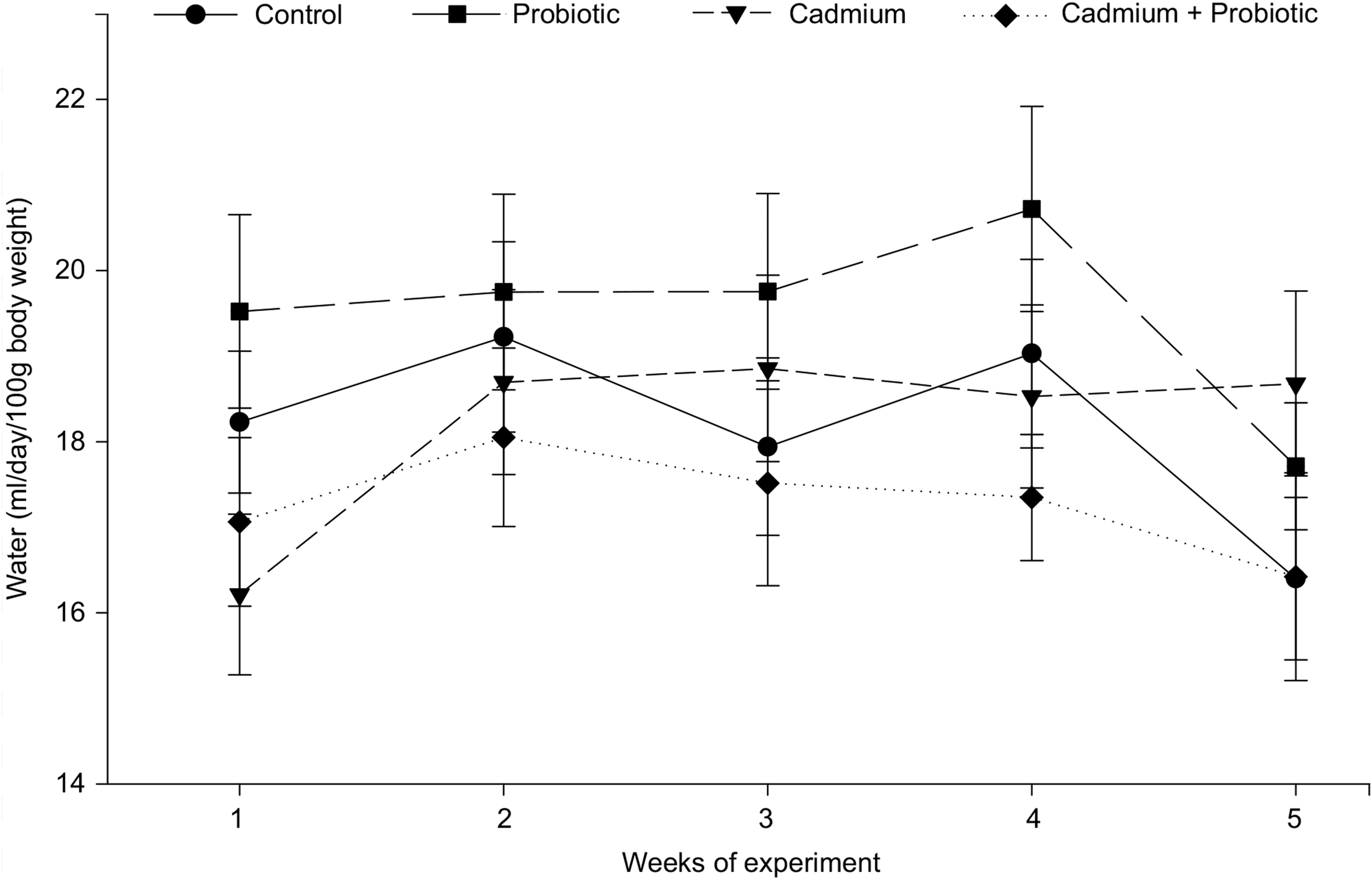
Water consumption (mL/day/100 g of body weight) of controls, and rats treated with probiotics, cadmium, or their combination, over a period of treatment (5 weeks), presented as mean ± SEM.
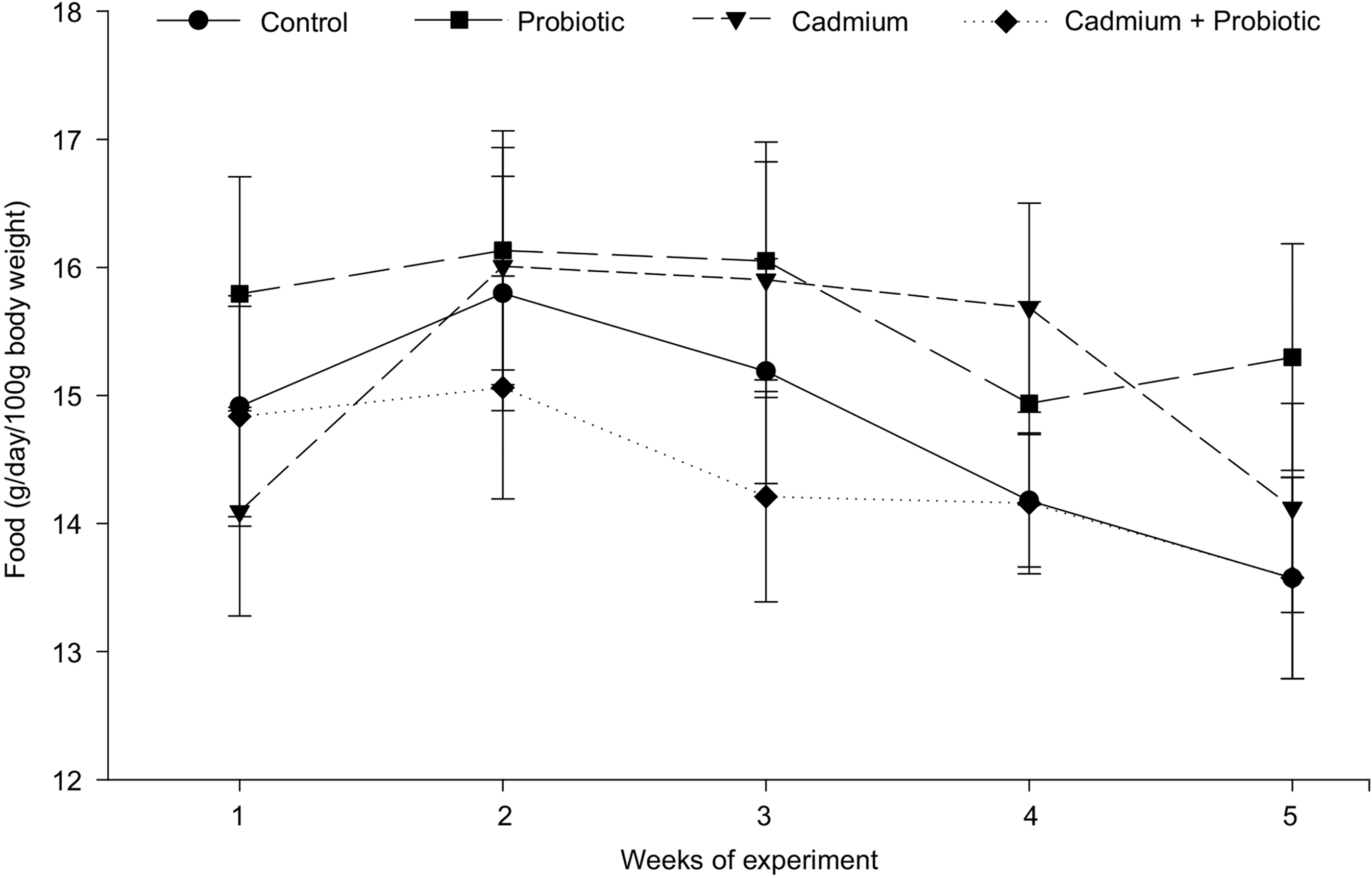
Food consumption (g/day/100 g of body weight) of controls, and rats treated with probiotics, cadmium, or their combination, over a period of treatment (5 weeks), presented as mean ± SEM.
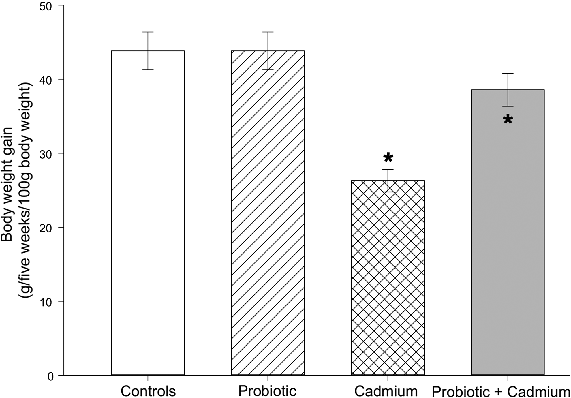
Body weight gain (g/5 weeks/100 g of body weight) of controls, and rats treated with probiotics, cadmium, or their combination, over a period of treatment (5 weeks), presented as mean ± SEM. An asterisk above the bars marks a significant difference against control group of animals, whereas an asterisk inside the bars indicates a significant difference between cadmium-treated versus probiotic + cadmium-treated group of animals.
Exposure of animals to CdCl2 increased cadmium concentration in both the blood and feces, compared with the control group of animals (Fig. 4). Adding probiotics to cadmium increased feces Cd concentration, but decreased blood Cd concentration compared to the CdCl2-treated group.
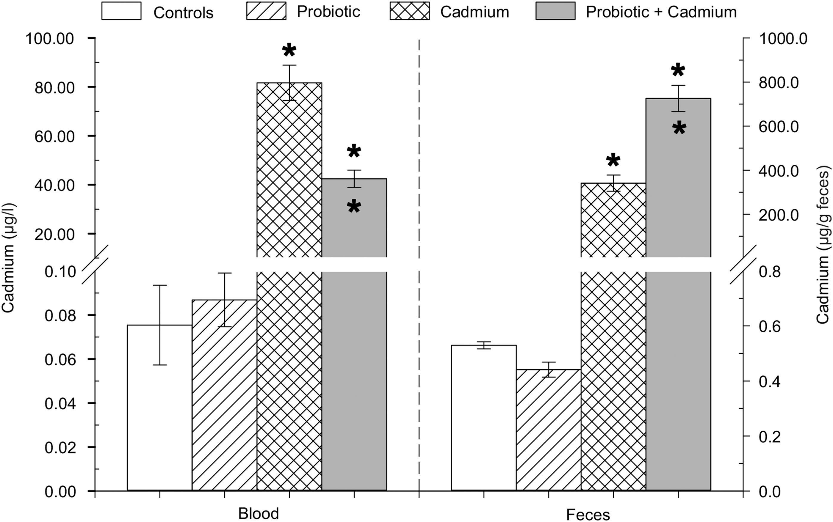
Cadmium concentration (ppm) in the feces and blood of controls, and rats treated with probiotics, cadmium, or their combination, presented as mean ± SEM. An asterisk above the bars marks a significant difference against control group of animals, whereas an asterisk inside the bars indicates a significant difference between cadmium-treated versus probiotic + cadmium-treated group of animals.
Alterations in the liver and kidney cadmium accumulation show that administration of CdCl2 increases this metal concentration in both tissues (Fig. 5). However, it should be noted that the maximum Cd accumulation in the liver is twice as high as that in the kidney. When animals were treated with probiotics and cadmium simultaneously, both liver and kidney Cd contents decreased significantly in respect to the group treated with cadmium only.
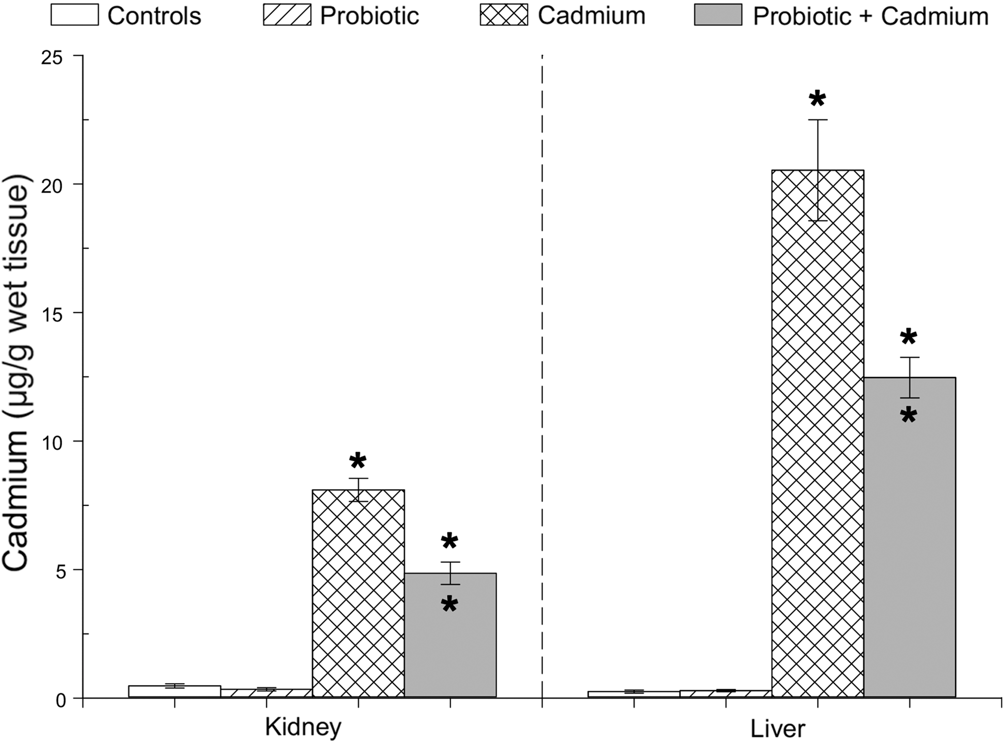
Cadmium concentration (ppm) in the liver and kidneys of controls, and rats treated with probiotics, cadmium, or their combination, presented as mean ± SEM. An asterisk above the bars marks a significant difference against control group of animals, whereas an asterisk inside the bars indicates a significant difference between cadmium-treated versus probiotic + cadmium-treated group of animals.
As regard to the activity of liver enzymes, administration of cadmium chloride alone resulted in an increased activity of serum ALT and AST in comparison to both control and probiotic-treated animals (Fig. 6). At the same time, probiotics in combination with CdCl2 significantly decreased both ALT and AST cadmium-induced increase.
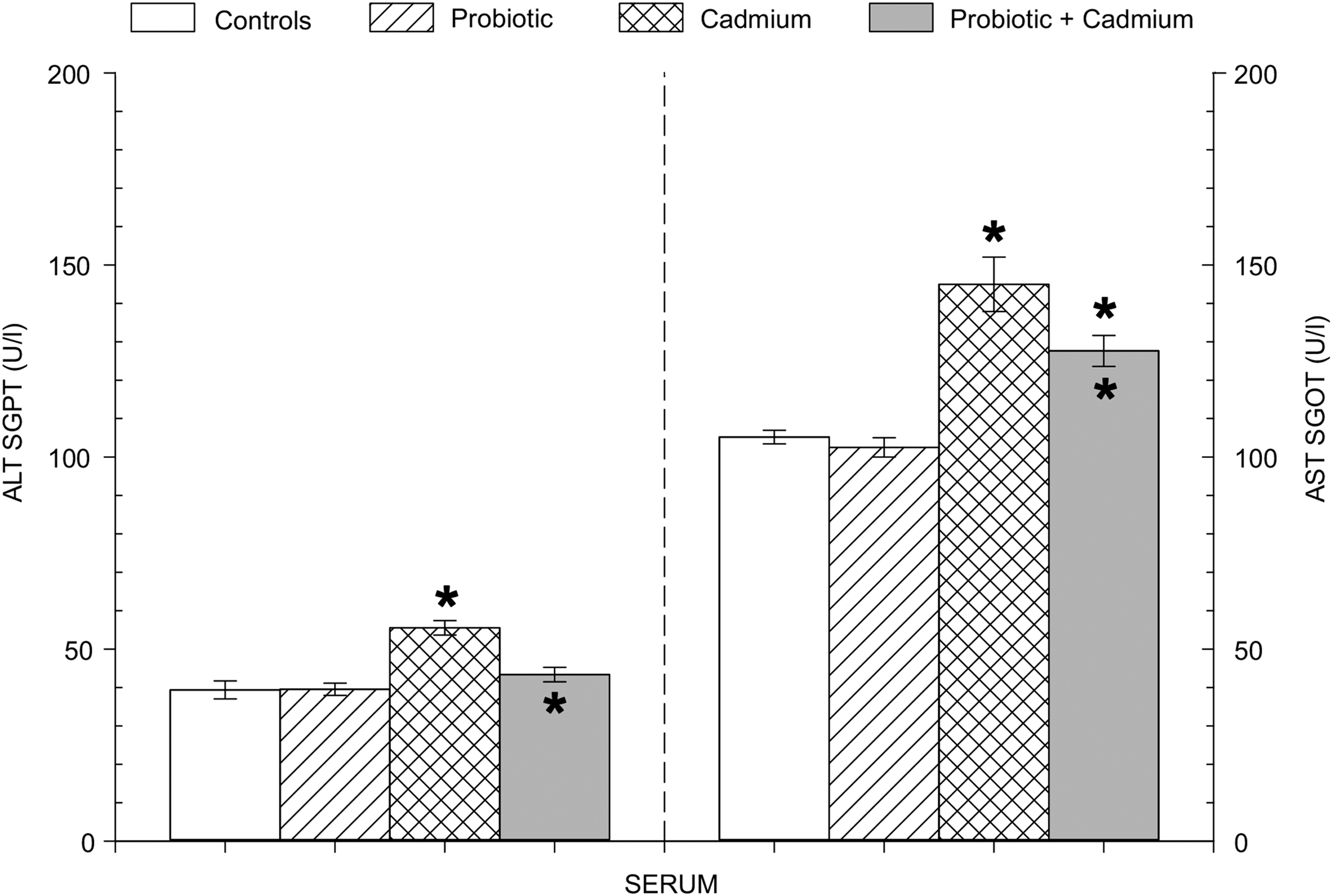
Serum ALT and AST activity (U/L) in controls, and rats treated with probiotics, cadmium, or their combination, presented as mean ± SEM. An asterisk above the bars marks a significant difference against control group of animals, whereas an asterisk inside the bars indicates a significant difference between cadmium-treated versus probiotic + cadmium-treated group of animals. ALT, alanine aminotransferase; AST, aspartate aminotransferase.
The liver and kidney, as main target organs of cadmium accumulation, were subjected to histomorphological analysis (Figs. 7 and 8 respectively, light microscopy, magnification 100 × ). Organs taken from control rats (Figs. 7A and 8A), as well from those that consumed probiotics (Figs. 7B and 8B), had normal macroscopic appearance without any sign of injury.
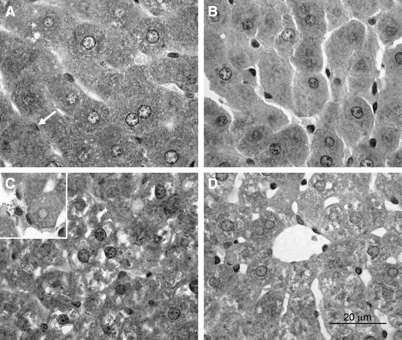
Light microscopy of the liver of control
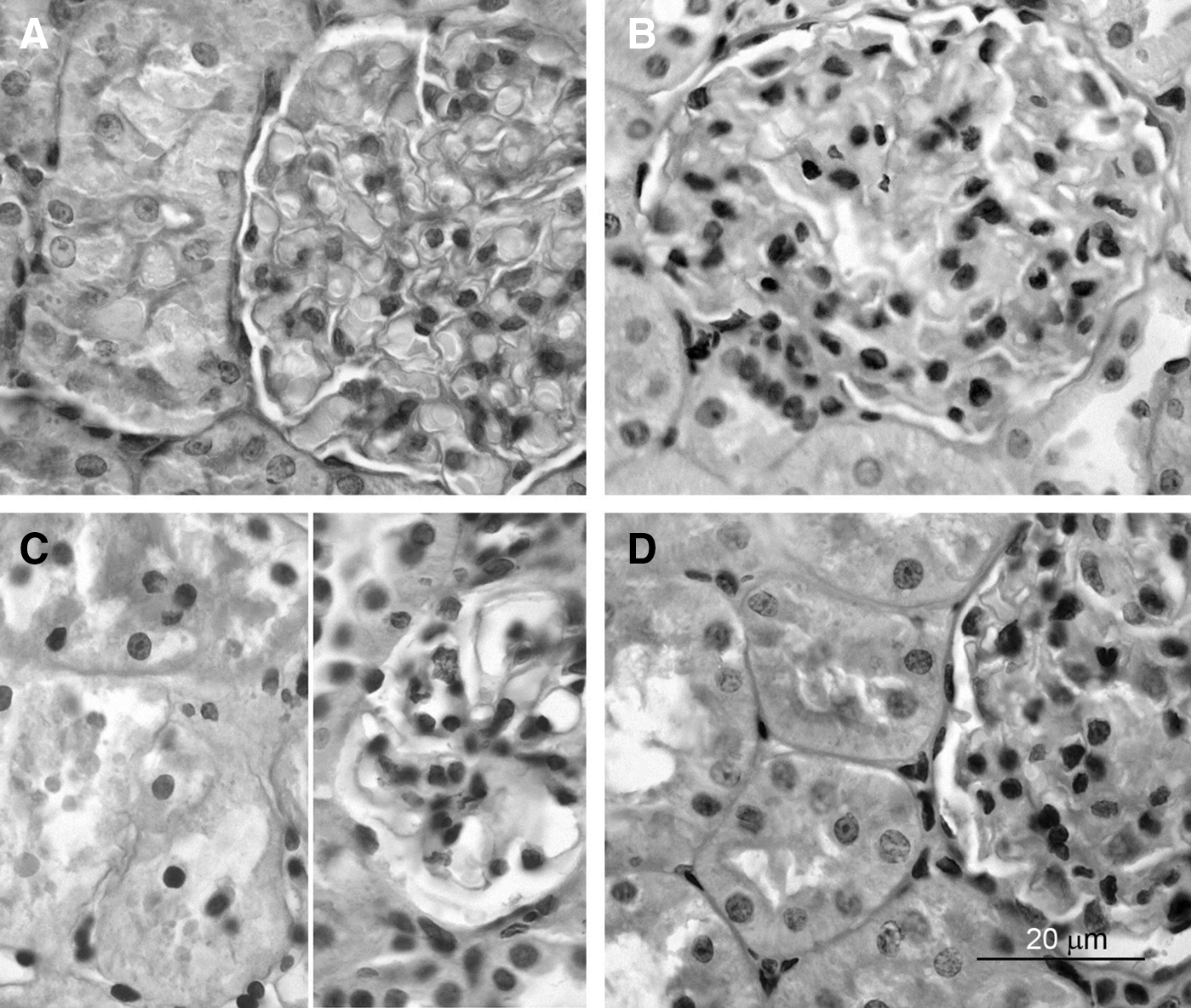
Light microscopy of the kidney of control
In CdCl2-treated rats, liver sinusoids, especially those which are closer to the central vein, became enlarged (Fig. 7C). Kupffer cells were activated, with many cytoplasmic protrusions at the surface and residual bodies inside the cell (inset of Fig. 7C). Clusters of hepatocytes containing large, irregularly shaped, poorly delineated intracytoplasmic vacuoles and small pyknotic nucleus were scattered throughout the parenchyma. In these areas, the structure of hepatic cords was blurred. Small foci of hepatocytes in different stages of cell death were also seen in some sites. It should be noted that despite rich vascularization of the liver, there was almost no bleeding during isolation and prefixation procedures.
In the kidney, renal corpuscles and proximal tubular epithelium showed clear signs of injury after CdCl2 treatment (Fig. 8C). Proximal nephrocytes were typically vacuolated, with nucleus displaced from the cell center, while brush border was occasionally degenerated (Fig. 8C left). Desquamated dead nephrocytes and nephrocyte nuclei might be seen inside the tubular lumen. Sporadic occurrence of tubular necrosis was also noted. Glomeruli showed gradual signs of injury, from hypercellularity, capillary enlargement, and “tuft segmentation” to glomerular shrinkage and widening of urinary space (Fig. 8C right).
However, probiotics consumed simultaneously with CdCl2 extenuated described changes in both organs examined, although they remained obvious (Figs. 7D and 8D).
The Spearman Rank-Order correlation among feces lactobacilli number and feces and blood Cd concentration in cadmium- and cadmium + probiotic-treated rats is shown in Figure 9. We combined multiple results obtained from the same experiment: previously published data on lactobacilli number in feces, 16 with the current data on the cadmium concentration in blood and feces (Fig. 4). The rise in lactobacilli number in feces of rats treated simultaneously with cadmium and probiotic results is in strong positive correlation with the increase of Cd concentration in their feces (r = 0.769 and P = .00228). The increase of Cd concentration in feces leads to the decrease of Cd concentration in blood (r = −0.783 and P = .00139).
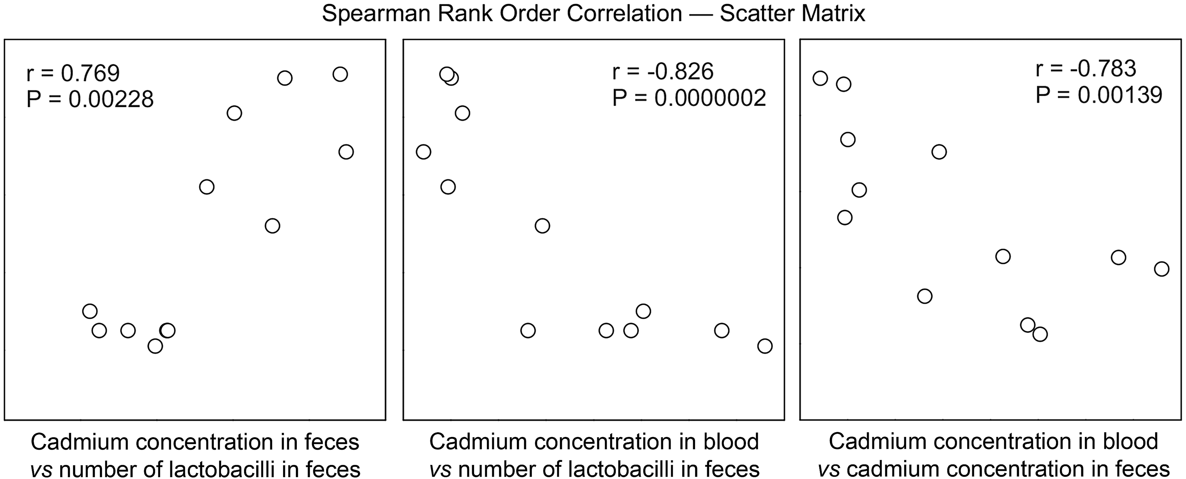
The scatter matrix of Spearman Rank-Order correlation among feces lactobacilli number and cadmium concentration in feces and blood of cadmium-treated and probiotic + cadmium-treated rats.
Discussion
Administration of cadmium or probiotics (alone or in combination) did not influence either water or food consumption over the period of treatment (Figs. 1 and 2). Accordingly, it was intriguing to find out that cadmium decreased the body weight gain in animals treated with CdCl2 only (Fig. 3). The same has already been reported for various species, 17 –21 suggesting that Cd could affect enzymes involved in glycogenesis, causing depletion of liver and muscular glycogen and changes in the energetic metabolism. 17,22
The most of orally given cadmium is excreted through feces, 23 while the absorbed part is transported through blood, with 50–70% of its accumulation in the kidneys and liver. 24 This is probably due to these organs' high concentration of metallothionein, which is known to bind a wide range of metals, including cadmium. 25
In line with above-mentioned studies, our results show that in rats administered with cadmium, its concentration is markedly elevated in the blood, feces, liver, and kidney (Figs. 4 and 5). In contrast, the exposure of rats to the combination of probiotics and cadmium increases Cd content in the feces, but decreases it in the blood, liver, and kidneys (Figs. 4 and 5). Our results suggest that probiotics accelerate cadmium elimination through feces, which causes an accompanying fall of its accumulation in blood and tissues.
Administration of cadmium causes a significant elevation in AST and ALT activity (Fig. 6). These findings are in line with previously published reports, which indicated that all these parameters were increased during cadmium toxicity. 6,8,13 Both ALT and AST are synthesized by the hepatic cells and have been widely used as indicators of liver function. When the hepatocytes are damaged (as we confirmed by histomorphological analyses), these enzymes are released into the extracellular fluid, which results in the rise of their activity in circulation. 26 However, probiotic supplementation together with the cadmium diminishes the cadmium-induced rise in AST and ALT (Fig. 6).
Histopathological analyses show that Cd alone provokes serious lesions in both the liver and kidneys (Figs. 7C and 8C). Patterns of these changes are typical for cadmium toxicity and were previously reported and explained by many authors. 5,13,27 –32 In general, liver injury is based mainly on initial endothelial and hepatocellular damage, which lead to activation of Kupffer cells and further inflammatory response. In the kidneys, tubular damage precedes progressive and irreversible glomerular changes and may explain renal impairment. However, coadministration of probiotics with cadmium leads to a considerable decrease of described changes, including necrosis (Figs. 7D and 8D).
As we showed previously, exposure to Cd leads to the significant decrease in lactobacilli counts in the feces, while adding probiotics to cadmium causes their subsequent increase. 16 By combining those data with these data on the cadmium concentration in blood and feces (Fig. 4), we found strong correlations between feces lactobacilli increase and feces Cd concentration increase and blood Cd concentration decrease (Fig. 9).
We speculate that probiotics actively contribute to cadmium elimination through feces probably by binding it to their bacterial cell wall. The surface of lactic acid bacteria comprised a thick layer of peptidoglycan, teichoic acid, proteins, and polysaccharides. All these structures contain different kinds of negatively charged groups, like carboxyl, hydroxyl, and phosphate groups, which are able to bind cations such as cadmium. 9,33
In conclusion, our results showed protective effects of probiotics in cadmium-exposed animals, which leads open the possibility of their therapeutic applications when oral cadmium toxicity is involved.
Footnotes
Acknowledgments
This work was supported by the Serbian Ministry of Education, Science and Technological Development [project nos. 173023, 176006, and 172058].
Author Disclosure Statement
We wish to confirm that the material in the article has not been, nor will be, submitted or published elsewhere and there are no known conflicts of interest associated with this publication.
We confirm that the article has been read and approved by all named authors and that there are no other persons who satisfied the criteria for authorship, but are not listed. We further confirm that all of us have approved the order of authors listed in the article.
We further confirm that any aspect of the work covered in this article that has involved experimental animals has been conducted with the ethical approval of all relevant bodies and such approvals are acknowledged within the article.
We understand that the Corresponding Author is the sole contact for the Editorial process (including Editorial Manager and direct communications with the office). He is responsible for communicating with the other authors about progress, submissions of revisions, and final approval of proofs.