
Abstract
Taraxacum officinale, the common dandelion, is a plant of the Asteraceae family, which is used as a food and medical herb. Various secondary plant metabolites such as sesquiterpene lactones, triterpenoids, flavonoids, phenolic acids, coumarins, and steroids have been described to be present in T. officinale. Dandelion may exhibit various health benefits, including antioxidant, anti-inflammatory, and anticarcinogenic properties. We analyzed the leaves and roots of the common dandelion (T. officinale) using high-performance liquid chromatography/mass spectrometry to determine its sesquiterpene lactone composition. The main compound of the leaf extract taraxinic acid β-
Introduction
T

Taraxacum officinale with inflorescence
Moreover, in its entirety, that is, inflorescences, leaves, and roots, dandelion is an edible weed. 1 It is consumed raw (salad), cooked, or roasted (used as coffee substitute). Furthermore, as flavoring it is used in, for example, alcoholic drinks, candy, and cheese. 1,2,5
Different dandelion tissues contain sesquiterpene lactones, triterpenoids, steroids, flavonoids, phenolic acids, and coumarins in various amounts.
6
Sesquiterpenes are probably responsible for the bitter taste of the dandelion.
7
T. officinale roots contain various sesquiterpene lactones such as eudesmanolides, that is, tetrahydroridentin B and taraxacolide-O-β-glucopyranoside, guaianolides, that is, 11β,13-dihydrolactucin and ixerin D, and esterified germacranolide acids, that is, taraxinic acid β-glucopyranosyl ester, its 11,13-dihydroderivative and ainslioside.
8,9
Also, the presence of taraxacoside (acylated γ-butyrolactone glycoside),
10
triterpenes, phytosterols,
3
and various phenolic acids, for example, chicoric acid, monocaffeoyltartaric, chlorogenic, and caffeic acids,
11
is known. Moreover, beside the secondary plant metabolites, roots contain considerable amounts of inulin.
12
The leaves contain the bitter sesquiterpene lactones taraxinic acid β-
In the past decades, the perennial weed was used as a medicinal plant for the treatment of various diseases, for example, anorexia and hepatitis. Various health benefits have been described so far, such as antioxidative, anti-inflammatory, anticarcinogenic, antihyperglycemic, anticoagulatory, analgesic, diuretic, choleretic, and prebiotic properties.
1
However, a search with the term “Taraxacum” or “dandelion” in the medical database PubMed under the category clinical trials (
The Keap1 (Kelch-like ECH-associated protein 1)–Nrf2 (nuclear factor erythroid 2-related factor 2) pathway plays a central role in cellular stress response and antioxidant defense mechanisms. 14,15 Nrf2 orchestrates the expression of genes encoding antioxidant and phase II enzymes such as heme oxygenase 1 (HO-1 or Hmox1), glutathione S-transferase (Gst), NAD(P)H quinone dehydrogenase 1 (Nqo1). 15,16 Under basal conditions, Nrf2 is bound to its repressor Keap1 in the cytoplasm. 17 Oxidants and electrophiles are able to inactivate Keap1 and stop the ubiquitination of Nrf2, leading to the release of Nrf2 from Keap1 and activation of Nrf2. 14,17 Thus, Nrf2 translocates into the cell nucleus, where it binds to the so-called antioxidant response element (ARE), thereby inducing the expression of Nrf2 target genes. 14,17 One important Nrf2 target gene is Hmox1, which catalyzes the degradation of heme to iron, biliverdin, and carbon monoxide. 18
In the current study, the sesquiterpene composition of T. officinale leaves and roots was analyzed by high-performance liquid chromatography/mass spectrometry (HPLC/MS). It is known that T. officinale may counteract oxidative stress.
19
Therefore, the objective of our cell culture experiments was to determine Nrf2 transcription factor activity in response to a leaf and root extract treatment. The leaf sesquiterpenes induced Nrf2 and its target gene Hmox1. The most active Nrf2 inducer of the leaves, taraxinic acid β-
Experimental
Plant material
T. officinale leaves and roots were collected in spring 2012 in the Fulda valley (Germany).
Chemicals
If not indicated otherwise, chemicals were obtained from Carl Roth (Karlsruhe, Germany), Sigma-Aldrich (Seelze, Germany), or Merck Millipore (Darmstadt, Germany). All chemicals were used as received from the supplier. Chemicals for cell culture such as
Extraction and isolation of 1 from T. officinale
Leaves and roots were extracted according to the same procedure. About 350 g of T. officinale leaves and about 100 g roots were freeze-dried to yield 41.6 or 21.8 g of dry powder, respectively. The freeze-dried leaves or roots were extracted twice with 350 mL of dry methanol for 60 min. Sodium sulfate was added and the mixture was stirred for 40 min, filtered, and evaporated. One milliliter of the residue was applied onto a silica gel flash chromatography (Carl Roth). Acetone was used as an elution solvent. The fractions were collected in test tubes by a fraction collector. After analysis of the eluate by thin-layer chromatography (TLC; see Thin-layer chromatography), test tubes were combined. Two leaf extracts (combined fractions 7–13 and 15–22) were collected. About 100 mg of the fraction 15–22 was obtained, which was enriched with compound
Thin-layer chromatography
Silica gel 60G F254 applied on aluminum (Merck Millipore) was used as stationary phase. A mixture of acetone:methanol (80:20, v/v) was applied as mobile phase. After development, the detection was conducted by UV detection and derivatization with the sodium permanganate solution.
High-performance liquid chromatography/mass spectrometry (HPLC/ESI-MSn)
An HPLC-MS instrument equipped with a PE 200 pump, PE 200 autosampler (PerkinElmer, Rodgau, Germany), a G1315B PDA detector (Agilent Technologies, Waldbronn, Germany), and a light scattering detector (Sedex 75; Sedere, Alfortville Cedex, France) controlled by Analyst 1.3 Software (AB Sciex, Darmstadt, Germany) was used. An API150ex mass spectrometer (AB Sciex) with electrospray ionization in negative and positive fast switching mode was used for analysis.
LC analyses were performed on a LiChrospher® 60 RP-Select B column (250 × 4 mm, 5 μm; Merck Millipore) equipped with a precolumn of the same material at 23°C. Mobile phases consisted of 5 mM ammonium formiate and 0.1% formic acid (A) and acetonitrile:methanol (50:50, v/v) with 5 mM ammonium formiate and 0.1% formic acid (B). The following gradient program was used: 0 min 15% B, 30 min 100% B, 40 min 100% B, and 45 min 15% B. The flow rate was set at 1.0 mL/min. All compounds were dissolved in methanol, and 20 μL was injected into the system. Analyst 1.3 software was used for data analyses.
Isolation of taraxinic acid β-d -glucopyranosyl ester by semipreparative HPLC
The semipreparative isolation was performed using an HPLC system from Hitachi (Düsseldorf, Germany) equipped with HPLC Pump l7150, autosampler L7250, UV detector L-4000A, and a semipreparative HPLC column (LiChrospher 60 RP-select B, 250 × 16 mm, 10 μm; Merck Millipore). HSM software (Dießen am Ammersee, Germany) was used for data analyses. Water (A) and methanol:acetonitrile (50:50, v/v) (B) were used as mobile phases. The linear gradient starting with 22% B up to 48% B in 57 min was used. The flow rate was set at 15 mL/min. The sample was dissolved in methanol, and 1 mL of the sample was injected. The chromatograms were recorded at λ 250 nm. Fractions were collected every 30 sec from 7.7 to 57.7 min. Fractions 29 to 31 were combined, evaporated, and analyzed by HPLC/MS and NMR.
Nuclear magnetic resonance spectroscopy
A DRX500 (500 MHz; Bruker Biospin, Rheinstetten, Germany) was used to measure 1D-
1
H and
13
C and 2D-NMR (HSQC, HMBC, HH-COSY) spectra of the sesquiterpene lactone glycoside
Cell culture
Huh7 human hepatoma cells were obtained from the Institute of Applied Cell Culture, Munich, Germany. Huh7 cells were maintained in high glucose (4 g/L) Dulbecco's Modified Eagle's Medium supplemented with 10% (v/v) fetal bovine serum, 4 mM
Neutral red assay for cell viability determination
Huh7 cells were seeded at a density of 0.15 × 10⁶ cells/well in a 24-well plate for 24 h and treated with 1–100 μg/mL B7-13, B15-22, or W33-47 extracts or 1–100 μM compound
Transient transfection and luciferase reporter gene analysis for Nrf2 transactivation
Transient transfection of Huh7 cells (24-well plate at a density of 0.15 × 10⁶ cells for 24 h) and luciferase reporter gene analysis for Nrf2 were conducted according to Wagner et al. (2011).
20
Huh7 cells were treated with 1 and 50 μg/mL B7-13, B15-22, or W33-47 and in case of compound
RNA isolation and quantitative reverse transcription-polymerase chain reaction
Huh7 cells were seeded at a density of 0.9 × 10⁶ cells/well in a six-well plate for 24 h and treated with 1 and 50 μg/mL B7-13 for 6 h. Resveratrol (25 μM) was used as positive control. Subsequently, cells were harvested with TriFAST (VWR International GmbH, Erlangen, Germany), and RNA was isolated according to the manufacturer's instructions. mRNA expression levels of human Hmox1 were determined. Primer3 Input software version 0.4.0 was used for primer design, and primers were ordered from Eurofins MWG (Ebersberg, Germany). Primer sequence was as follows: forward 5′-CCAGGCAGAGAATGCTGAGT-3′ and reverse 5′-GTAGACAGGGGCGAAGACTG-3′. Real-time PCR was performed with a one-step procedure using the SensiFAST™ SYBR No-ROX One-Step Kit (Bioline, Luckenwalde, Germany) with SYBR Green detection on a Rotorgene 6000 cycler (Corbett Life Science, Sydney, Australia). Quantitation was done by an external standard curve. Results were normalized to glyceraldehyde 3-phosphate dehydrogenase (Gapdh) (forward 5′-CAATGACCCCTTCATTGACC-3′ and reverse 5′-GATCTCGCTCCTGGAAGATG-3′), which served as a housekeeping gene.
Statistical analysis
Statistical analysis was performed using SPSS software version 23.0 (SPSS, Inc., Munich, Germany). Normal distribution of data was tested by Kolmogorov–Smirnov and Shapiro–Wilk tests. Student's t-test or one-way analysis of variance with a Dunnett-T post hoc test was applied. Results are presented as mean + SEM. Significance was accepted at P-values <.05.
Results and Discussion
HPLC/MS analysis of the leaf and root extracts of T. officinale
The freeze-dried leaves and roots of T. officinale were extracted with dry methanol. These evaporated extracts were applied on a silica gel flash chromatography and eluted with acetone. All fractions were analyzed by TLC. Two leaf extracts B7-13 and B15-22 and a root extract W33-47 were obtained and analyzed by LC/MS. HPLC chromatograms of the leaf and root extracts of T. officinale are displayed in Figure 2.
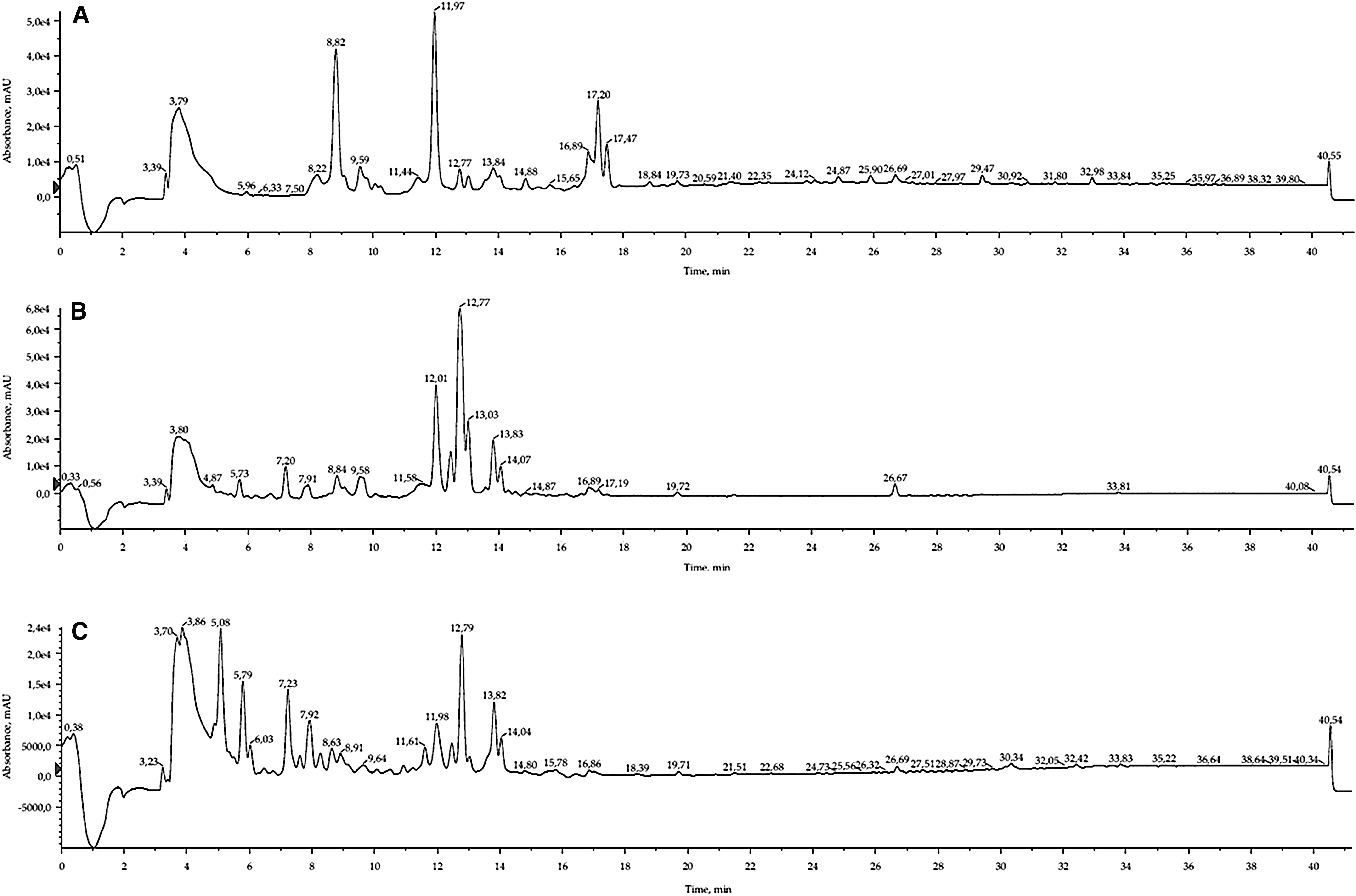
HPLC chromatograms of
About 3 mg of pure compound
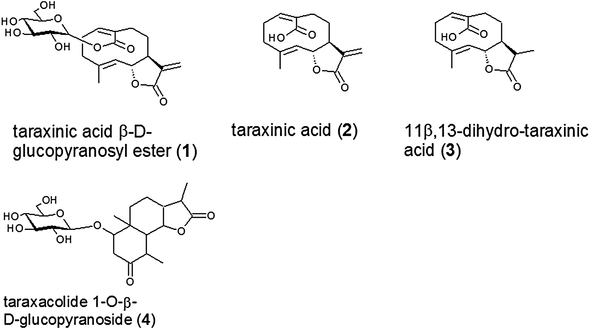
Chemical structures of sesquiterpenes from T. officinale.
In fraction B7-13, the inositol derivative 1,5-bis(4-hydroxyphenylacetyl)-
In a recent study, differences between Taraxacum species in terms of their chemical composition and antioxidant activity have been described.
2
In the present study, the inositol 1,5-bis(4-hydroxyphenylacetyl)-
Cytotoxicity of the leaf and root extracts of T. officinale
The neutral red assay was applied to determine the cytotoxicity after incubation with B7-13, B15-22, or W33-47 extracts and the pure compound
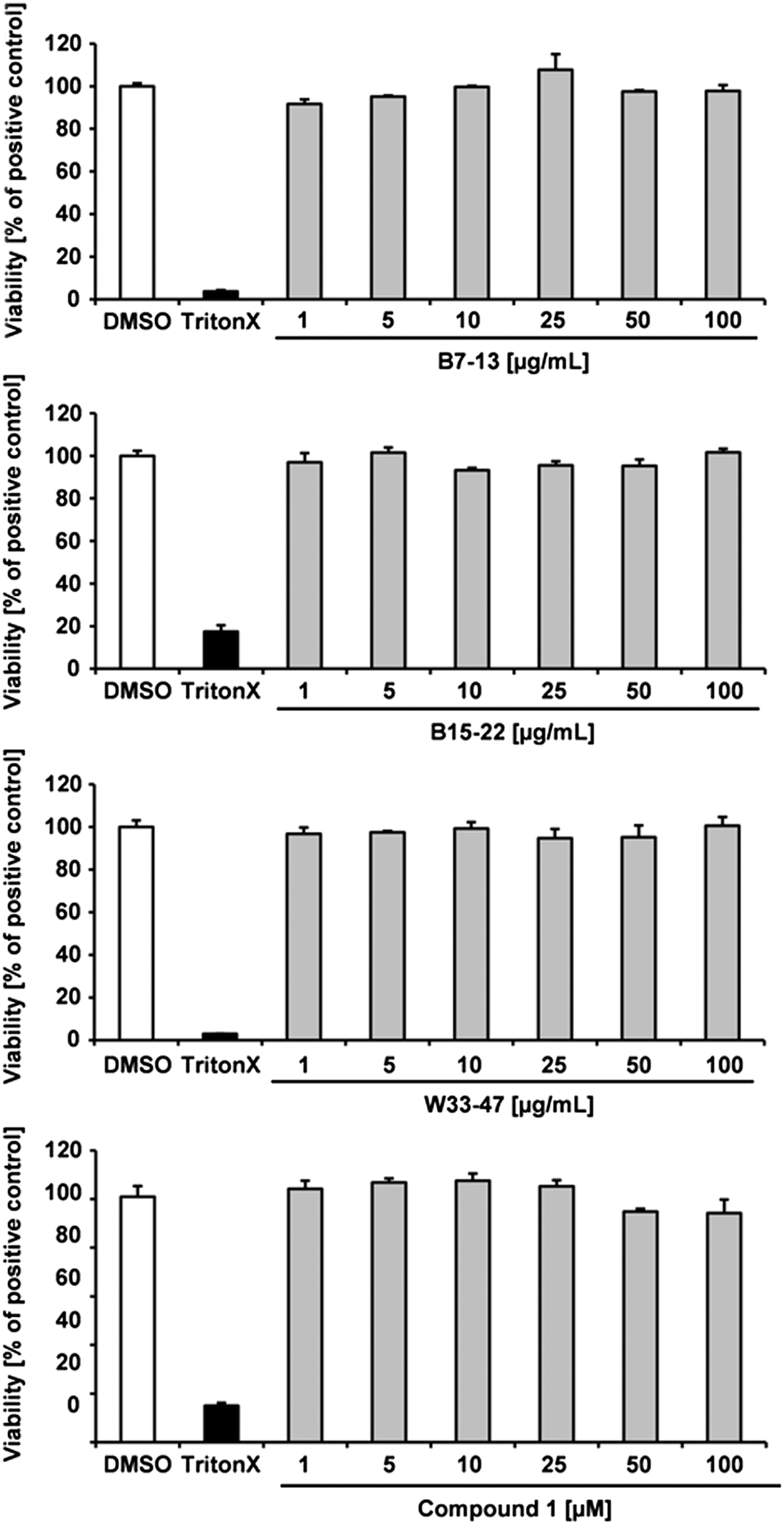
Cytotoxicity of the leaf and root extracts and compound
Induction of the transcription factor Nrf2 and its target gene heme oxygenase 1
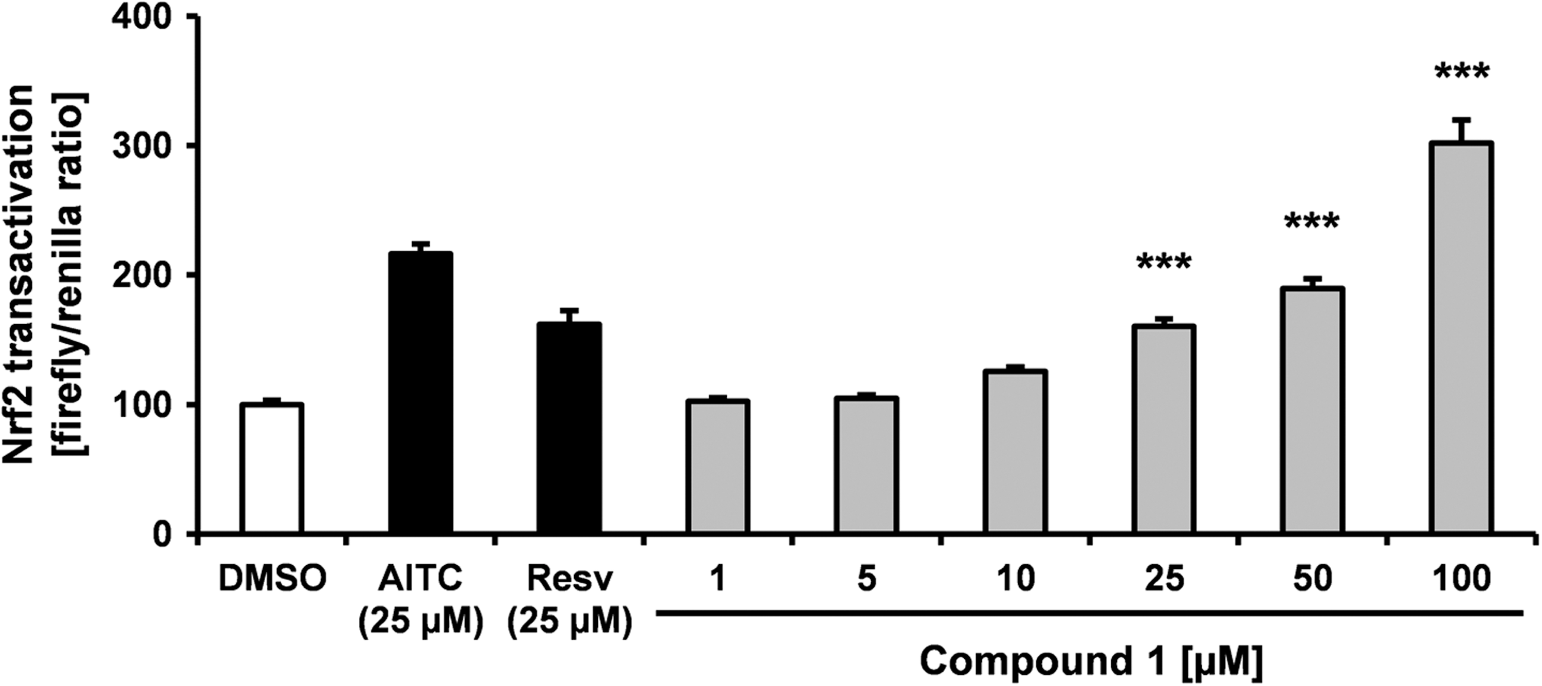
The Nrf2 transactivation activity of the B7-13, B15-22, and W33-47 extracts was determined as shown in Figure 5. The leaf extracts exhibited a more potent Nrf2 transactivation activity compared to the root extract. Supplementation of Huh7 cells with 50 μg/mL B7-13 extract resulted in a significant increase in Hmox1 mRNA levels (Fig. 6).
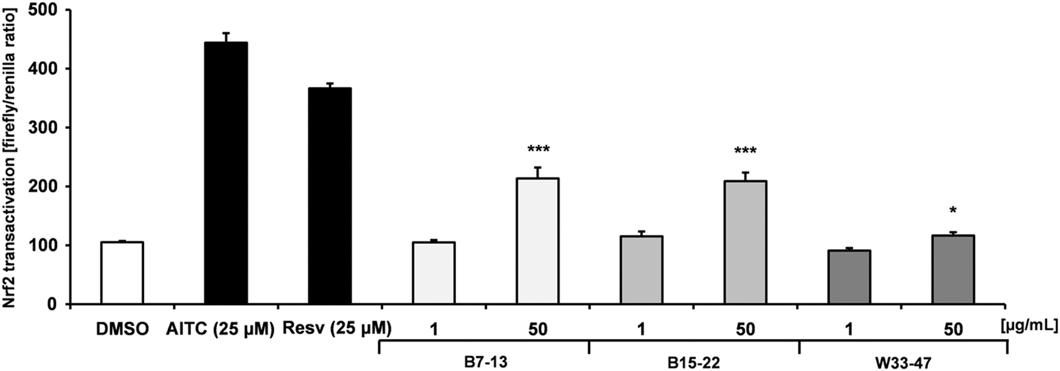
Effects of the leaf and root extracts of T. officinale on Nrf2 transactivation in human Huh7 cells. Human liver Huh7 cells (0.15 × 10⁶ cells/well) were supplemented after transient transfection with 1 and 50 μg/mL B7-13, B15-22, or W33-47 extracts for further 24 h. Resveratrol (Resv; 25 μM) and allyl isothiocyanate (AITC; 25 μM) served as positive controls. Data are mean + SEM of at least three experiments performed in triplicate (n = 3). * and *** indicate statistically significant differences between DMSO control cells and supplemented cells with extracts P < .05 and P < .001, respectively; Student's t-test. DMSO, dimethyl sulfoxide.
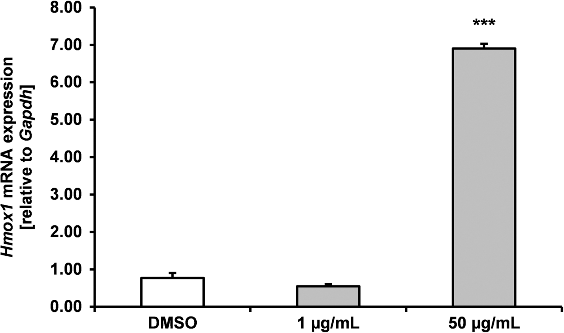
mRNA levels of heme oxygenase 1 (Hmox1) in human hepatocytes (Huh7). Huh7 cells (0.9 × 10⁶ cells/well) were supplemented with 1 and 50 μg/mL B7-13 extract after 24 h incubation for 6 h. Resveratrol (Resv; 25 μM) was used as positive control. The data are expressed as the mean + SEM (n = 3). *** significant differences in B7-13-treated cells compared with DMSO control cells P < .001, Student's t-test. SEM, standard error of the mean.
The main compound of the B15-22 extract, compound
Effects of compound
Some Taraxacum species may decrease cellular levels of reactive oxygen species and may also exhibit free radical scavenging activities.
2
Furthermore, T. officinale leaf extracts have been reported to decrease lipid peroxidation in mice.
21
Our results indicate that the leaf extracts and the compound
Structure elucidation
Compound
Conclusions
The sesquiterpene lactone composition in the root and leaf extracts of T. officinale was determined by HPLC/MS. T. officinale leaf and root extracts were potent inducers of Nrf2. Moreover, the leaves of T. officinale induced the Nrf2 target gene Hmox1. The pure compound
Footnotes
Acknowledgments
We thank Vivien Schmuck, Gaby Steinkamp, and Inga Richter for excellent technical help. We are thankful for the financial support of the “Interne Forschungsförderung” from the University of Applied Sciences Fulda.
Author Disclosure Statement
No competing financial interests exist.