
Abstract
Treating infections in pregnant patients is potentially dangerous even when herbal medicines are used. Many herbal medicines, among them extracts from plants of Rhodiola genus, have antimicrobial, anti-inflammatory, and immunostimulatory properties owing to their polyphenol content; they may, however, affect fetal development due to their antiangiogenic properties. The aim of this study was to explain whether daily feeding pregnant and lactating mice with 20 mg/kg Rhodiola kirilowii aqueous (RKW) or 50% hydro-alcoholic (RKW-A) extracts, or 0.2 mg/kg epigallocatechin (EGC, antiangiogenic compound of Rhodiola extracts), may lead to abnormalities in morphology and function of the kidneys of adult progeny. Such abnormalities were not observed in the kidneys of 6-week-old offspring, neither in RKW nor in the control group. However, the progeny of RKW-A- or EGC-fed mothers presented morphometric abnormalities in the kidney structure, with a significantly higher number of glomeruli/mm2 and a lower diameter of glomeruli (RKW-A group) or a significantly higher glomeruli diameter (EGC), than in the control and RKW groups. Abnormalities in serum vascular endothelial growth factor, tumor necrosis factor (TNF)-alpha, urea, creatinine, and cystatin C levels were also found. We recommend caution in long-term use of RKW-A extract and EGC-rich foods during pregnancy and lactation.
Introduction
T
Our earlier studies revealed that extracts prepared from Radix and Rhizoma of Rhodiola rosea, Rhodiola quadrifida, and Rhodiola kirilowii and their compounds rosavin and salidroside had antiangiogenic properties in the case of tumor angiogenesis. 6 –9
Previously, we also observed undesirable effects of some angiogenesis inhibitors (theobromine, cocoa catechins); when administered to pregnant mice, they affected embryonic angiogenesis, morphology, and function of some of the progeny organs, among them of kidneys, and lymphatic system. 10 –13
In our previous research, 400 mg of dark chocolate given daily to a pregnant mouse for 18 days suppressed angiogenic activity of embryonic tissue measured with in vivo cutaneous angiogenesis assay (embryo-induced angiogenesis). A significant negative correlation was found between epigallocatechin (EGC) content of homogenates and their angiogenic activity. 14
An antiangiogenic effect was also obtained after feeding pregnant mice a mixture of the main groups of cocoa catechins present in chocolate. The same mixture of cocoa catechins exerted the antiangiogenic effect in tumor-induced angiogenesis assay. Moreover, L-1 sarcoma tumors in the group of catechin-fed mice appeared later and during all observation periods were significantly smaller than in the control group. 15
Finally, morphometric and functional abnormalities were observed in kidneys of adult progeny of the chocolate-fed mothers. 13
Therefore, the aim of the present study was to evaluate whether regular administration of R. kirilowii extracts or EGC, to pregnant and lactating mice, might affect kidney morphology and function in the progeny.
Materials and Methods
Preparation of materials
R. kirilowii roots and rhizomes were cultivated, collected, and identified by the Department of Botany, Breeding and Agriculture of Institute of Natural Fibres and Medicinal Plants, Poznań, Poland, where the voucher specimen is kept. Roots and rhizomes were dried and powdered; extracts were prepared as previously described, 16 lyophilized, and stored at −70°C until used.
EGC was purchased from Sigma Aldrich (cat no: E3768-5 MG), dissolved in distilled water, and stored at −70°C until used.
Animals
Experiments were performed on adult inbred females of Balb/c strain, 8–9 weeks old, mated with adult males from the same strain. Animals were handled according to the Polish law on the protection of animals and NIH standards. All experiments were accepted and conducted according to the ethical guidance of Local Bioethical Committee (permission 73/2011). Mothers, from the time the copulatory plug was noted up to the 28th day after delivery, were fed Rhodiola kirilowii aqueous (RKW) or 50% hydro-alcoholic (RKW-A) extract (20 mg/kg b.m.) or EGC (0.2 mg/kg b.m.) daily. 17 The control group received distilled water. Mothers were housed separately. For avoiding stress connected with gavage and handling that can lead to miscarriage, 20 μL of tested substance dissolved in distilled water was placed on one corn crisp and served to the mouse in a Petri dish. On the 28th day after delivery, mothers were anesthetized, bled (sera for high-performance liquid chromatography [HPLC] analysis), and euthanized.
Six weeks after birth, the progenies of control mothers (105 mice), RKW-fed mothers (64 mice), RKW-A-fed mothers (78 mice), and EGC mothers (41 mice) were weighed, anesthetized, bled, and euthanized.
Serum
Mice were bled in anesthesia (intraperitoneal injection of ketamine 120 mg/kg of b.w. and xylazine 12 mg/kg of b.w. solution) from retroorbital plexus. Sera were separated by 1 h clotting (room temperature [RT]), centrifuged at 2000 g for 20 min, and stored at −70°C until analysis.
HPLC analysis (mothers' sera)
Total serum polyphenol/flavonoid concentration from the sera of mothers was assayed by applying the HPLC system (Dionex) equipped with the CoulArray electrochemical detector (ESA, Inc.). The extraction procedure of polyphenols/flavonoids was performed as described by Zdanowski et al. 18
Biochemical analysis (progeny sera)
Creatinine and urea concentrations were evaluated by the enzymatic colorimetric method, according to the manufacturer`s protocol (Biosystems). Results are presented as median mg% with range (min.—max., creatinine) and mean mg% ± SEM (urea). Cystatin C level was evaluated from serum by the Mouse/Rat Cystatin C Quantikine ELISA kit (R&D Systems) and presented as mean pg/mL ± SEM.
The level of vascular endothelial growth factor (VEGF) was determined by ELISA test (R&D Systems) according to the producer's protocols.
Determination of tumor necrosis factor (TNF)-alpha concentration was evaluated using flow cytometry (FACS Calibur; BD Bioscience) and the Cytometric Bead Array Mouse Th1/Th2/Th17 Cytokine Kit (cat. no. 560485; BD Biosciences), according to the manufacturer's protocol.
All analyses were performed in duplicate and the data entered to the statistical analysis as one mean value.
Kidneys
After blood sampling, the mice were euthanized (pentobarbital 400 mg/kg), and the kidneys were excised and weighed. Relative kidney weight was calculated as a ratio of the weight of kidney (mg) to body mass (g). Kidneys were fixed in 4% buffered formalin and processed for light microscopy. Histological evaluation and quantitative analyses were performed on the hematoxylin–eosin-stained paraffin sections of the kidneys obtained from 6-week-old progeny. Histotechnical criteria applied for quantitative analysis of kidney sections were as follows: thickness 3–5 μm, no evidence of traumatic artifacts within the sample (e.g., fragmentation hemorrhages), the complete frontal section of the kidney, including the cortex, medulla, and renal pelvis structures.
The light microscopic examination was based on the standard morphological criteria. The examination was conducted using the optical microscope Delta Optical Evolution 100 TRINO at 400 × magnification. The device was connected to a photometric color CCD camera UCMOS05100KPA. All photographs obtained were processed using ToupView 3.7 software.
The entire dissected surface of the frontal section of the kidney was analyzed in respect to the following: the total cortical area and total number of glomeruli with the results expressed as a number/1 mm2 and the glomeruli diameters measured in consecutive glomeruli of each sample.
Statistical analysis of data
The Shapiro–Wilk normality test (all data). Creatinine: chi-square analysis and Kruskal–Wallis test. Urea: one-way analysis of variance (ANOVA) and Tukey's multiple comparison test. Cystatin C: one-way ANOVA and unpaired t-test. VEGF and TNF-alpha: one-way ANOVA and unpaired t-test. Morphometry and HPLC results: unpaired t-test. The results are presented as mean ± SEM or median with minimum–maximum range. A value of P < .05 was considered significant (GraphPad Prism version 5).
Results
Body weight
Animals in all groups were weighed 6 weeks after birth. Mean weight of a control group mouse was 18.22 g. RKW extract did not affect mean body mass of the progeny, however, the mice from mothers fed RKW-A extract or EGC during pregnancy and lactation were significantly lighter in comparison to the control or RKW group (Table 1).
Unpaired t-test.
EGC, epigallocatechin; n, number of animals; P, statistical level in comparison to control group; RKW, Rhodiola kirilowii water extract; RKW-A, Rhodiola kirilowii hydro-alcoholic extract.
HPLC analysis
In serum of all mice-mothers (regardless of the group studied), we did not notice the following polyphenols present in both Rhodiola extracts: naringenin, luteolin, p-coumaric, and ferulic acid. Salidroside and kaempferol were found only in the serum of mice from RKW and RKW-A groups, where the concentrations of both compounds were significantly higher in the RKW-A group (2 and 1.5 times, respectively, P < .05). Total concentration of flavonols (fisetin, kaempferol, and quercetin) had the highest value in R. kirilowii groups (RKW-A, P < .05) and the lowest in EGC group (P < .05) in comparison to control. On the contrary, the total concentration of catechins (catechin, epicatechin, and EGC) was significantly higher in EGC group (P < .05) in comparison with control and R. kirilowii groups. In EGC group, the total concentration of phenolic acids was found to be more than 10 times lower than in the control group (P < .05). In sera of the progeny, polyphenolic compounds were not detected. The results are presented in Table 2.
n = 20 mice (5 mice/group).
Significant differences (P < .05) in comparison to control group (a) or RKW-A group (b), level of significance α < 0.05.
n.d., values below detection limit.
Biochemical analysis of progeny sera
Analysis of progeny sera revealed significantly more creatinine results above the upper value of the confidence limit (0.65 mg%) in RKW and RKW-A sera than in the sera collected from the controls (Fig. 1A). However, median values did not differ significantly (Fig. 1B).

Creatinine in progeny sera presented as
Urea level was increased only in EGC group (Fig. 2). There were no significant differences between control and Rhodiola groups as well as Rhodiola groups and EGC. Concentration of cystatin C was slightly lower in the sera of RKW-A group (P = .0555, Fig. 3).
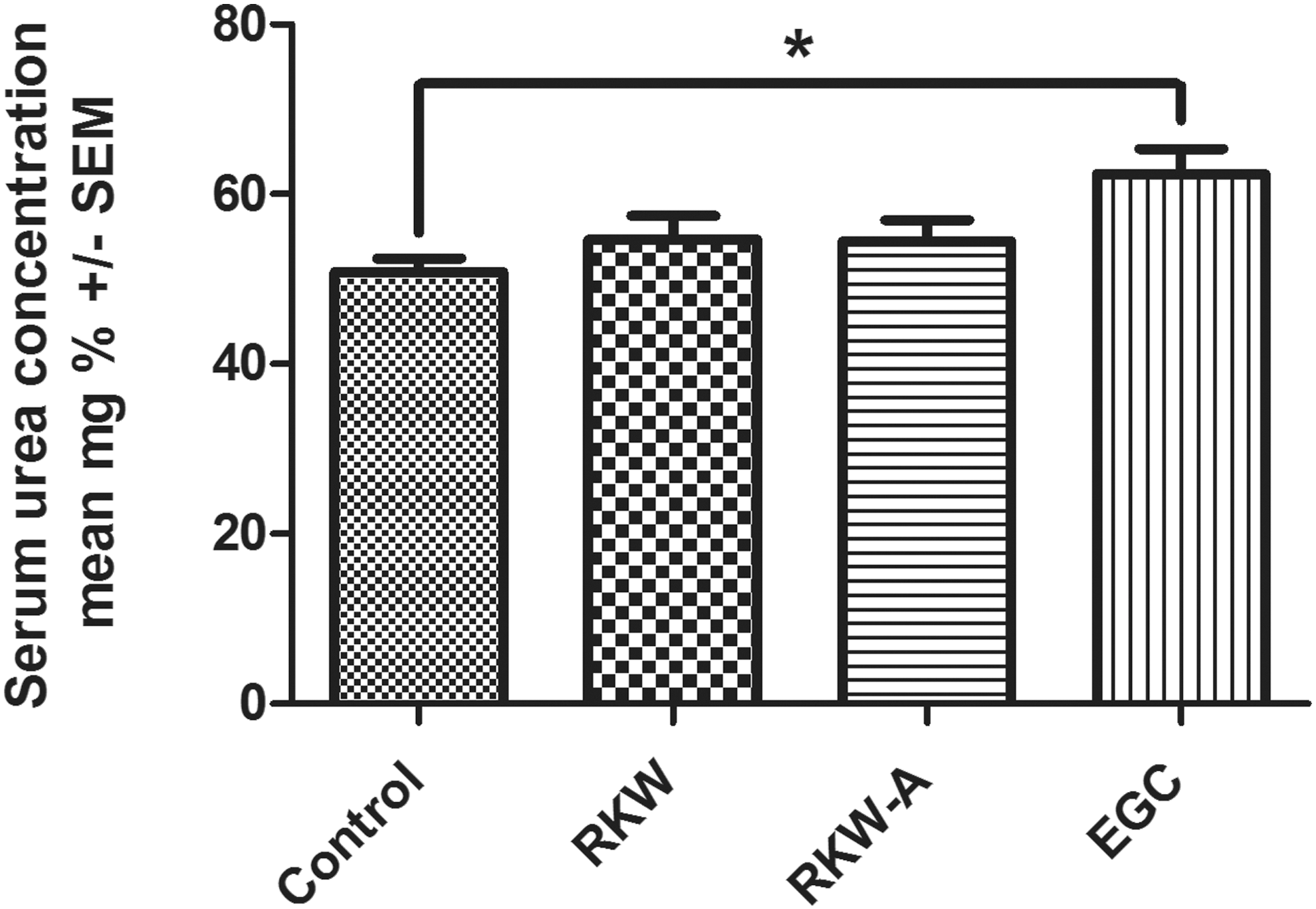
Mean urea concentration (mg%) in progeny sera. n = 89 mice (34 control, 21 RKW, 24 RKW-A, 10 EGC). One-way ANOVA and Tukey's test, *P = .0410. ANOVA, analysis of variance.

Mean cystatin C concentration (pg/mL) in progeny sera. n = 32 mice (8 mice/group). One-way ANOVA and unpaired t-test.
VEGF concentration was significantly lower in sera of progeny mice belonging to RKW-A group than in the controls (P = .031), and slightly higher in sera of EGC group (P = .085, Fig. 4). No other differences between control and studied groups were found.

Mean VEGF concentration (pg/mL) in progeny sera. n = 57 mice (18 control, 16 RKW, 16 RKW-A, 7 EGC). One-way ANOVA: P = .0054. Unpaired t-test. *P = .028, @ P = .085, # P = .016. VEGF, vascular endothelial growth factor.
Concentration of TNF-alpha was significantly lower in sera of progeny belonging to EGC group and significantly higher in sera of progeny belonging to RKW-A group, than in the controls (Fig. 5). No other differences between control and studied groups were found.
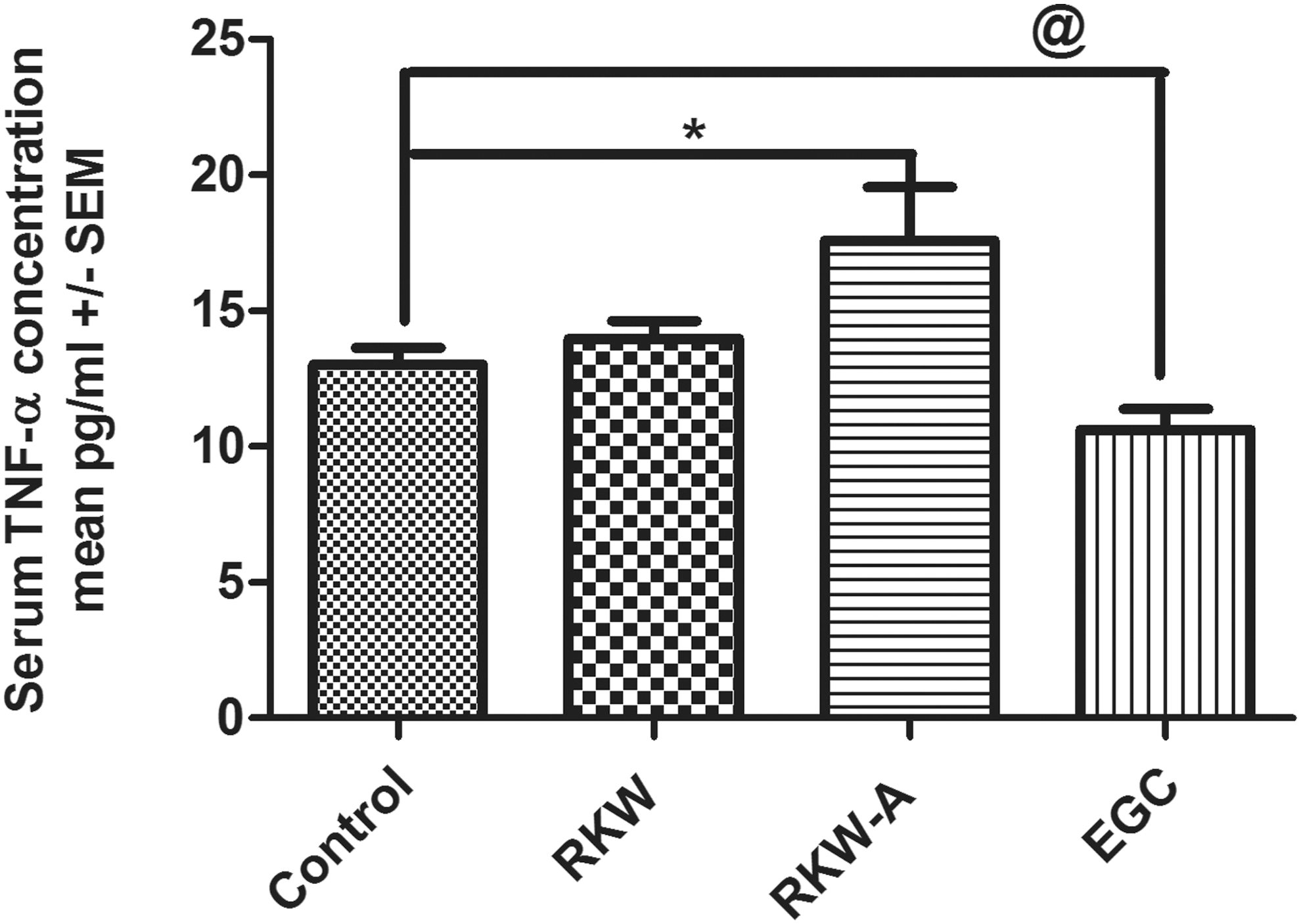
TNF-alpha concentration (pg/mL) in progeny sera. n = 72 mice (25 control, 14 RKW, 25 RKW-A, 8 EGC). One-way ANOVA P = .014 and unpaired t-test: *P = .031, @ P = .049. TNF, tumor necrosis factor alpha.
Kidneys
No macroscopic abnormalities were noticed in the anatomy of kidneys and no differences were found between control and experimental groups in the kidney relative weight (mean ± SEM 7.3 ± 0.1 in the control, 7.5 ± 0.2 in RKW group, 7.0 ± 0.1 in RKW-A group, and 7.4 ± 0.1 in EGC group). Morphologically, in the experimental groups, no changes in the structure of the renal tissue, compared with the control group, were observed. However, morphometric analysis did reveal differences in the number of glomeruli per microscopic field between RKW-A kidneys (more) and kidneys from the control, EGC, and RKW groups (less). Moreover, slight but statistically significant glomerular atrophy was found, manifested as a smaller diameter of glomeruli in the experimental RKW-A than in the control and other groups. In contrast to this, morphometric analysis of kidney sections from animals belonging to EGC group of progeny mice revealed a significantly higher size of renal glomeruli than that observed in the controls (Table 3).
Mice mothers were fed RKW or RKW-A extract as well as EGC during pregnancy and lactation. n = 40 mice (10 mice/group).
Level of significance (α < 0.05) in comparison to control group.* P < .05, *** P < .001. Unpaired t-test.
Discussion
The confirmation of kidney cell damage and functionality impairment caused by external factors in the developmental period is not straightforward; the damage may vary depending on the “grip point” and period of administration of an injurious compound to the mother and also on the ability of a young kidney to compensate for the function of damaged nephrons. It has been noted that in physiological processes, not all nephrons are engaged at the same time. Acute kidney damage should lead to increasing, in a short period of time, serum creatinine concentration and may be associated with oliguria. 19 Unfortunately, in acute kidney damage in young organisms, serum creatinine concentration rises relatively late, and the proper concentration of this parameter does not mean that kidney function is normal. 20,21 In the present work, we observed increased percent of progeny with creatinine concentration above 0.65 mg/dL. The increase of creatinine concentration from 0.3 mg/dL to the value of 0.6 mg/dL may be clinically significant despite both parameters being within the normal range. We are aware of the fact that creatinine should be also measured in urine (daily urine collection) and creatinine clearance should be calculated. 22
In mice, however properly conducted daily urine collection, it is almost “mission impossible.” Therefore, we used another kidney damage indicator—cystatin C. 23 Cystatin C is a compound whose production is maintained at a constant level, independent of age, muscle mass, sex, and diet. Ninety-nine percent of it is filtered by the glomerulus, then resorbed, and degraded in the proximal tubules. Increasing concentration of cystatin C is observed after 12 h of nephrotoxicity activation. It is believed that cystatin C is a better predictor of glomeruli function than creatinine. A special role is assigned to cystatin C as an early indicator of deterioration of glomerular filtration rate in the early stages of renal failure. 24 The decrease of cystatin C level in serum of RKW-A progeny noted in this work may be the effect of increased levels of glomerular filtration rate which, in turn, can signify early stages of kidney cell damage. It is associated with an elevated proportion of progeny with the creatinine concentration in serum above 0.65 mg/dL. However, to further clarify the relationships between these markers in the early stage of kidney damage, other markers should be used. Currently, it is a common opinion that there is no possibility that a single marker can differentiate etiology and monitor the severity and course of acute kidney damage. Clinically, the chances of acute kidney injury diagnosis are greatly increased with the detection of cystatin C and lipocalin gelatinase-associated neutrophils, which can be determined in the serum or urine, or interleukin-18, enzyme N-acetyl-β-D-glucosaminidase and kidney injury molecule-1 protein identified only in the urine. 25,26
To confirm the results obtained from serum analysis, we conducted a morphometric evaluation of progeny kidneys. Glomerular abnormalities include many conditions with a variety of genetic and environmental causes, but they fall into two major categories—glomerulonephritis and glomerulosclerosis. Although glomerulosclerosis and glomerulonephritis have different etiologies, they can both lead to kidney failure. In our research, we found significantly smaller size glomeruli in kidneys in the progeny of RKW-A group and significantly higher size glomeruli in the progeny of EGC group, than in kidneys from the progeny of control and RKW-fed mothers.
Possible mechanism by which R. kirilowii extracts or EGC affects kidney development and/or functionality may be associated with the presence of biologically active polyphenols in serum (and afterward in milk). Both R. kirilowii extracts contained varied concentrations of biological active compounds, however, there were no such big differences in the serum. 18 Both extracts differed in, among others, salidroside and catechin concentrations. It was shown in human microvascular endothelial cells (HMVEC) that green tea catechins inhibit VEGF-induced angiogenesis. 27 Also, Leong et al. demonstrated that on HMVEC and breast cancer cells, MB231 reduced angiogenesis after green tea catechin supplementation. 28 On the contrary, Negrão et al. revealed that catechins did not change angiogenesis and inflammation in skin wound-healing model and substantially decreased these processes in Matrigel plug assay. 29 Moreover, catechin in vitro decreased the level of TNF-alpha in endothelial cells (EC) and vascular smooth muscle cells, as well as in lipopolysaccharide (LPS)-stimulated macrophages. 29,30 Currently presented observations are in accordance with the previously published results. 31 In that study, the highest level of catechins (especially EGC and epicatechin) was found in the serum of mice-mothers fed EGC, which was associated with a lack of inhibition of endothelial cell proliferation in mice (HECa10 cell line). In contrast, sera from mothers fed RKW-A extracts inhibited proliferation of HECa10 cell line and exhibited lower concentration of catechins than sera from EGC and RKW group.
The data described in the present work may suggest that the disruption caused by RKW-A extracts (glomerular atrophy) is not connected with catechin action. Probably the other polyphenol—salidroside, is responsible for this. In this study, we noted almost twice as high concentration of salidroside in the sera of mothers fed RKW-A extracts as RKW, and an absence of this polyphenol in the control and EGC groups. Moreover, salidroside exhibits antiangiogenic character that we reported previously. 8 This finding was congruent with the results of Sun et al. who showed SW1116 cells downregulated concentration of VEGF and VEGF receptor after salidroside application in colonic carcinoma. 32 Those relationships were also observed in this work. Contrary to the results obtained for sera of EGC mothers and their offspring, a different situation in the RKW-A group was noted. The progeny from mothers fed RKW-A extract had lower serum concentration of VEGF and higher concentration of TNF-alpha, than controls, what suggested the inflammatory process in these mice. We suppose that inflammation may be one of the major factors responsible for the development of glomerular atrophy. Larger number of glomeruli/mm2 in this group of mice could be the result of some compensatory mechanism.
It would be more difficult to explain hypertrophic changes in renal glomeruli from progeny of the EGC group. During glomerulogenesis, the glomerular basement membrane (GBM) is formed from two components, one synthesized by epithelial cell (podocyte) and the other by endothelial cell. The GBM contains unique, specific isoforms of laminin, type IV collagen, nidogen, and heparin sulfate proteoglycan. The VEGF is an essential molecule for the development of kidney glomeruli, mediating communication between Bowman's capsule and capillary endothelial cells. 33 VEGF produced by epithelial cells appears to be an important regulator of glomerular endothelial cell functionality and permeability, and both its deficiency and overproduction may lead to glomerulopathies. 34 Podocytes express VEGF-A during glomerular development. Endothelial cells express VEGF receptors (fetal liver kinase 1 and fms-like tyrosine kinase 1). Correct regulation of VEGF-A signaling is critical for proper development and function of kidney glomeruli. 35
Mean concentrations of VEGF and TNF-alpha in serum of EGC mothers did not differ from the control group. 31 In contrast, in this work, VEGF concentration was higher and TNF-alpha was lower in the sera of progeny from mothers fed EGC during pregnancy and lactation, than in progeny of the control group. These changes in the sera of progeny from mothers fed EGC, and glomerular hypertrophy, were associated with higher levels of urea in serum.
In conclusion, it might be assumed that varying levels of VEGF production, as observed in this study, played some role in the observed morphometrical abnormalities in kidneys obtained from the offspring of mothers fed RKW-A extract or EGC, since this cytokine had been proven to mediate glomerulo- and nephrogenesis, both during pre- and postnatal periods. The described aforementioned differences in VEGF and TNF-alpha concentration in progeny serum may confirm the existence of different mechanisms, which lead to glomerular atrophy or hypertrophy in the examined groups of mice.
Footnotes
Acknowledgment
The study was supported by the National Centre of Science, grant number: 2012/05/B/NZ 7/03219.
Author Disclosure Statement
No competing financial interests exist.