
Abstract
Different polysaccharides were isolated from the leaves of Aloe arborescens using the gradient power of hydrogen followed by antitumor and immunomodulatory assay. The total polysaccharide content of different fractions, water-soluble polysaccharide (WAP), acid-soluble polysaccharide (ACP), and alkaline-soluble polysaccharide (ALP), was estimated using a phenol-sulfuric acid spectrophotometric method. WAP possessed a higher content of mannose and glucose than either ACP or ALP. In vitro antitumor activity was investigated in three different cancer cell lines, and in vitro immunomodulatory potential was assessed through phagocytosis and lymphocyte transformation assay. The results showed that WAP and ALP exhibited the most significant cytotoxicity against HepG2 human liver cancer cells, with IC50 values of 26.14 and 21.46 μg/mL, respectively. In contrast, ALP was able to enhance lymphocyte transformation, whereas WAP had the most potent phagocytic activity. Molecular weight, total sugar and uronic acid content, Fourier transform-infrared analysis, and linkage type of bioactive polysaccharides were investigated. These findings revealed that the potential antitumor activity of the natural agents WAP and ALP was through an immunomodulation mechanism, which verifies the use of the plant as adjuvant supplement for cancer patients suffering immunosuppression during chemotherapy.
Introduction
R
Cancer is the leading cause of death in economically developing countries and the second highest death cause in developing ones. 4 The chemotherapy is the most frequently used therapy for the treatment of cancer. However, if it is used alone no satisfactory outcome is achieved in terms of prevention of metastasis and complete tumor remission. 5 Since an increase in the use of synthetic drugs in cancer therapy has many side effects, there is a worldwide trend to natural resources, where plants serve as novel or additional treatment methods for cancer therapy. 6 Discovering and evaluating antitumor properties of polysaccharides has emerged as an important field in chemistry and biology. 7 Natural polysaccharides have been investigated for their capability in inhibiting tumor growth and inducing apoptosis in cancer cells using different animal models and human cancer patients, which showed improvement in cancer treatment. 8 –11 Most plant polysaccharides are relatively safe compared to other immunomodulators. Hence, plant polysaccharides are ideal candidates for immunomodulator and antitumor therapeutics that possess low toxicity. 12
Aloe arborescens Miller is a member of genus Aloe (Family Asphodelaceae). It is a traditionally valued herbal medicine for gastrointestinal complaints, skin injuries, and burns. 13 Recent reports showed that it has immunostimulating activity in animal models 14 in addition to other important phytotherapeutic and anticancer properties. 15,16 Different studies investigated the effect of different plant extracts on the inhibition of various cancer types, such as intestinal tumor, 17 colon carcinogenesis, 18,19 and duodenal tumorigenesis. 20 Furthermore, evaluation of the in vivo effect of its preparations as tumor angiogenesis inhibitors was investigated. 21 Considerable clinical investigations have been carried out to verify the therapeutic effects of chemotherapy plus leaves extract in patients with metastatic cancer and revealed significant results. 15
Despite various reports about anticancer therapeutic activities of A. arborescens extracts, there is a paucity of information concerning the antitumor effect of Aloe at the molecular level. Aloes are characterized by the presence of a variety of polysaccharide composition. According to previous literature, the antitumor activity of polysaccharides was suggested to be related mainly to its activation of the immune system. Therefore, the current study aimed to investigate the response to polysaccharide treatment through a cellular approach. To fulfill this aim, different polysaccharides were separated from A. arborescens using a pH gradient followed by standardization and characterization of monosaccharide chemical composition through GC analysis, molecular weight, uronic acid content, Fourier transform-infrared (FT-IR) analysis, and Linkage type of bioactive polysaccharides. Then to explore the mechanism underlying the antitumor effect, the polysaccharide activities were evaluated in vitro through cytotoxic assay. In addition, the cell proliferation responses and phagocytosis activity were assessed using human peripheral blood mononuclear cells (PBMCs).
Materials and Chemicals
Plant material
Samples of A. arborescens were collected from East deserts of Egypt. A voucher specimen (No. 32012) was identified by a botanist senior researcher, at the flora and taxonomy research department, Agricultural Museum, Giza, Egypt. The specimen was deposited at the Department of Pharmacognosy, Faculty of Pharmacy, Ain Shams University.
Chemicals and reagents
Phenol and standards of glucose and mannose were purchased from Sigma Chemical Co. Dialysis tubing was purchased from the Serva Co (D-69115 Heidelberg). PC3 human prostate cancer cells, HepG2 human hepatocellular carcinoma cells, and MCF-7 human breast cancer cells were purchased from VACSERA and then maintained in the tissue culture facility (Faculty of Pharmacy, Ain Shams University, Cairo, Egypt). Trypsin-EDTA and Sulforhodamine B (SRB) were obtained from Sigma-Aldrich. The remaining tissue culture reagents, including D-Hanks solution, fetal calf serum (FBS), RPMI-1640 medium, penicillin/streptomycin, and phosphate-buffered saline (PBS), were obtained from Lonza. Ficoll-Paque was obtained from GE Healthcare (Biosciences AB) and phytohemagglutinin (PHA) from Gibco. Dimethyl sulfoxide (DMSO), Dimethylformamide (DMF), and all other reagents were purchased from Sigma-Aldrich and were of the highest grade.
Extraction of different polysaccharides
Thirty kg of Aloe leaves yielded 12 kg gel that was homogenized in Ultraturrex followed by percolation with 60% alcohol containing10% acetic acid to extract phenolics. The marc leaves filet was air-dried and grinded to precisely weighed powder (146 gm); several steps were taken to extract different polysaccharides as shown in Fig. 1.

Schematic diagram for extraction and fractionation of different polysaccharides from Aloe arborescens.
Removal of lipids and coloring matter
Marc was treated under reflux (90°C) with EtOH: CHCl3 (1:1) (2 L) to remove lipids, followed by acetone (1 L) to remove the coloring matter.
Extraction of water soluble polysaccharide
Marc was air-dried followed by extraction with 1500 mL distilled water under a reflux at 90°C for 3 h; this extraction process was repeated four times. The supernatants obtained were pooled and filtered through a Buchner funnel to separate the insoluble material. The clear supernatant concentrated to a small volume by a rotary vacuum evaporator at 65°C, then water-soluble polysaccharide (WAP) was precipitated by absolute ethanol (four volumes), stirred vigorously, and left overnight at −4°C. The precipitate was separated using a Buchner funnel, redissolved in distilled water, and reprecipitated again by addition of absolute ethanol. The precipitate formed was resuspended in distilled water and left in refrigerator for further purification.
Extraction of acid-soluble polysaccharide
The marc residues were immersed in 2 L of 1 M HCl at 40°C under reflux and left overnight. After filtration, the supernatant was collected, and the marc was subjected to the abovementioned extraction four times. The supernatants obtained were combined and adjusted to neutrality with 0.008 g/mL NaOH till pH 7 to precipitate the polysaccharides. The precipitate formed was centrifuged at 4000 g for 15 min; the decant was discarded and the residue redissolved in distilled water, followed by precipitating a second time by adding four volumes of absolute ethanol, left in the refrigerator overnight at −4°C, followed by decantation. The precipitate was dried by Buchner funnel to yield (acid-soluble polysaccharide, ACP) crude acid ACPs.
Extraction of alkali-soluble polysaccharides
The marc residue was extracted four times using (2 L) 1% NaOH at 40°C under reflux. The supernatant was adjusted to neutrality with 1 M HCl, and the alkali-soluble polysaccharide (ALP) was obtained with the same above experimental sequence. The crude polysaccharides WAP, ACP, and ALP were washed with the following solvents: absolute ethanol, acetone, and ethyl ether and then they were completely air-dried.
Purification of crude polysaccharides
Removal of protein by Sevag method
The obtained crude polysaccharides (WAP, ACP, and ALP) were separately suspended in distilled water and treated with Sevag reagent (n-butanol: chloroform 1:5 v/v) with a ratio (2:1) to the sample. 22 After vigorous stirring on a vortex mixer for 10 min, the upper aqueous layer was retreated with the same manner until there was no interface layer of protein. The upper aqueous layer was collected, concentrated under vacuum, and twice the volume of absolute ethanol was added. It was left in the refrigerator overnight and then the precipitate was collected by decantation followed by filtration.
Dialysis
Purified polysaccharides WAP, ACP, and ALP each were redissolved in the least amount of distilled water then dialyzed with a regenerated cellulose tube (Mw cutoff 10 kDa) against distilled water for 2 days as shown in Fig. 2.
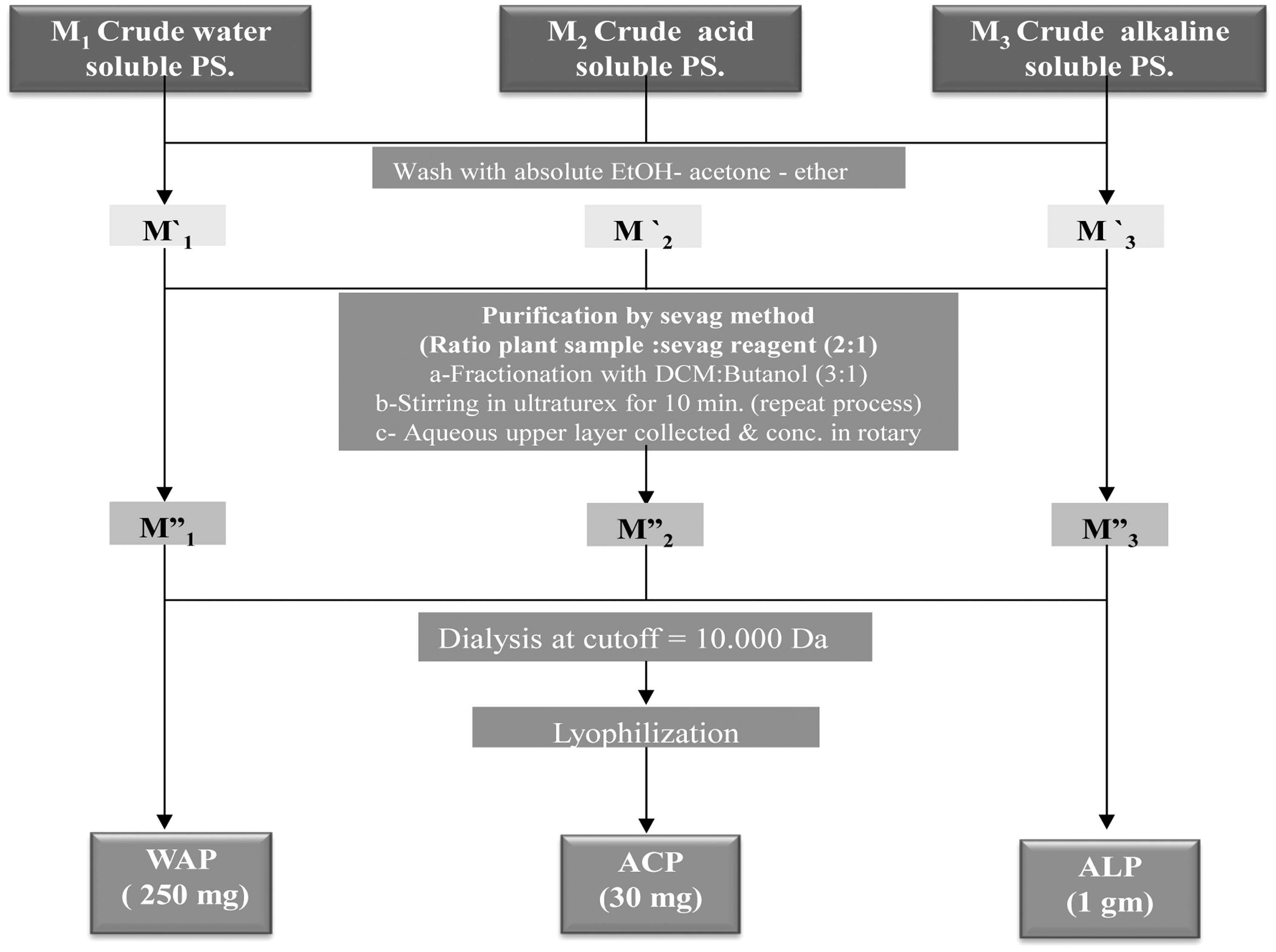
Purification steps of crude polysaccharides from A. arborescens.
Polysaccharide characterization by colorimetric phenol-sulfuric acid method
The content of polysaccharides was determined by the phenol-sulfuric acid method. Half a mL of standard grade sugar solution (glucose or mannose with serial dilutions ranging from 10 to 100 μg/mL) and 0.5 mL of purified polysaccharides at 100 μg/mL concentration were placed into a test tube, and 0.5 mL 5% phenol (in 0.1 M HCL) was added. After that 2.5 mL concentrated sulfuric acid was added rapidly. The tubes were immediately shaken and placed in a water bath at 30°C before readings were taken.
The colorimetric analysis of all solutions was carried out using a UV-Vis spectrophotometer. The absorbance of characteristic yellow orange color was measured at 490 nm for hexose monosaccharides. Blanks were prepared by substituting distilled water for the sugar solutions. The quantity of monosaccharides in different purified polysaccharide fractions was determined and expressed as hexose sugars using constructed standard curve with each standard sugar. 23 Results are shown in Table 1.
ACP, acid-soluble polysaccharide; ALP, alkaline-soluble polysaccharide; WAP, water-soluble polysaccharide.
In vitro proliferation and cytotoxicity assays
Cell culture
PC3 human prostate cancer cells, HepG2 human hepatocellular carcinoma cells, and MCF-7 human breast cancer cells were grown in RPMI-1640 medium, supplemented with 10% heat inactivated FBS, 50 U/mL of penicillin and 50 mg/mL of streptomycin, and maintained at 37°C in a humidified atmosphere containing 5% CO2. The cells were maintained as monolayer culture by serial subculturing.
Sulforhodamine-B cytotoxicity assay
Cytotoxicity was determined using the Sulforhodamine-B (SRB) method as previously described by Skehan et al. 24 Exponentially growing cells were collected using 0.25% Trypsin-EDTA and seeded in 96-well plates at 1000–2000 cells/well in RPMI-1640 supplemented medium. After 24 h, cells were incubated for 72 h with various concentrations of the tested polysaccharides. Following 72 h treatment, the cells were fixed with 10% trichloroacetic acid for 1 h at 4°C. Cells were stained for 10 min at room temperature with 0.4% SRB dissolved in 1% acetic acid. The plates were air-dried for 24 h, and the dye was solubilized with Tris-HCl for 5 min on a shaker at 1600 RPM. The optical density (OD) of each well was measured spectrophotometrically at 564 nm with ELISA microplate reader (ChroMate-4300). The IC50 values were calculated (Table 2) according to the equation for Boltzmann sigmoidal concentration–response curve using the nonlinear regression fitting models (GraphPad, Prism Version 5).
Data are presented as mean ± SD.
Statistically significant for the corresponding control group.
Assessment of immunomodulatory effects
Isolation of human peripheral blood mononuclear cell (PBMC)
Blood (20 mL) was taken from four different healthy donors throughout this research using the 5 mL syringe. The blood sample was diluted with the same volume of PBS-BSA-EDTA. Separation of blood cells was performed using density centrifugation. Briefly, the diluted blood sample was carefully layered on Ficoll-Paque Plus. The mixture was centrifuged at 400 g for 15 min at 20°C. The undisturbed lymphocyte layer was carefully transferred out. The cells were washed and pelted down with three volumes of PBS-BSA-EDTA twice and then resuspended in RPMI-1640 media with 100 IU/mL of penicillin, 100 μg/mL of streptomycin, and 10% v/v FBS. Cell counting was performed to determine the PBMC cell number with equal volume of trypan blue.
Phagocytosis assay
The reaction mixture consisted of 0.2 mL of peripheral monocyte (2 × 10⁷) in l mL of Hank's balanced salt solution containing 5% fetal calf scrum and 0.05 mL of polysaccharide samples. This mixture was preincubated in a water bath at 37°C for 7 min. A suspension of yeast particles was cultured with saline solution to 25 × 107 particles per mL and heated at 100°C for 30 min. To the preincubated reaction mixture, 0.1 mL of suspension of yeast particles was added and incubation continued at 37°C for 60 min. The number of yeast particles phagocytosed by neutrophilis was counted after staining with 5% Fuchsin in phenol solution.
Screening lymphocyte transformation test
Lymphocytes (1 × 10⁶cells/mL) were suspended in a solution of complete RPMI-1640 medium. Cell viability (tested by trypan blue dye exclusion) was always over 90%. The cells 100 μL were plated in 96-well plates and incubated with or without suboptimal dose (4 μg/mL) of PHA and the same concentration of the polysaccharides (200 μg/mL). Cells treated with PHA but not the extracts were used as a positive control. Control cells were all treated with DMSO in a final concentration equal to test wells. DMSO concentration never exceeded 0.1%. Cells were kept for 72 h at 37°C in a humidified incubator which maintained a constant atmosphere of 5% CO2. After 3 days of incubation, cells were collected by centrifugation and washed once in PBS. Then, they were fixed in 0.2% formaldehyde in PBS for 4 min (37°C), placed on slides, air-dried, and stained with Leishman dye, which distinguished between the nucleus and cytoplasm at a concentration of 0.033% with a staining period of 4 min. Shape and size of nucleus of lymphocytes observed under light microscope and comparative studies among negative control, positive control, and samples were observed (Table 3).
Control: The aloe sample was replaced with 0.05 mL of 0.15 M phosphate buffer, pH 7.2.
Data are represented as M ± SD n = 3. Statistical analysis was carried out using one-way ANOVA followed by Tukey post hoc test.
Statistically significant for the corresponding control group P < .05.
Statistically significant for the corresponding PHA group P < .05.
Modulation cytokine production test
Four cytokines were tested, including interleukin-2 (IL-2), interleukin-12 (IL-12), interferon-gamma (IFN-γ), and tumor necrosis factor-alpha (TNF-α). Most of them were upregulated significantly, compared with the negative (no treatment) and positive (treated with PHA) controls. Cytokine levels in culture medium of lymphocyte were treated with polysaccharide fractions (WAP, ALP) at 200 μg/mL concentration.
Chemical characterization of the bioactive polysaccharide fractions
Determination of molecular weight
The molecular weight of the polysaccharide was determined by gel permeation chromatography (GPC) on Agilent 1100 series, (Germany), Detector: Refractive Index FPl gel using dimethyl formamide (DMF) solvent. The sample 2 mg was dissolved in 4 mL of solvent, then it was filtrated by syringe filter 0.45, and then the sample was run on Styragel HRDMF column, 3 μm (7.8 × 300 mm), Water Company (Ireland). The polydispersity index was calculated from the Mw/Mn ratio. 25
Characterization monosaccharide composition of polysaccharides
WAP and ALP were hydrolyzed with 1 N HCL and converted to their alditol acetates. 26 The resulting alditol acetates were analyzed by gas chromatography (GC) using an Agilent 6890 N instrument fitted with flame ionization detector (FID) and equipped with a ZB-1701 column (30 m × 0.25 mm × 0.25 μm). The temperature of the column was kept at 150°C for 2 min and then increased to 200°C at the rate of 7°C/min. The rate of helium carrier gas was 1.2 mL/min. Monosaccharide composition was identified by comparison with authentic samples. The uronic acid content was determined by the carbazole-sulfuric acid method. 27
FT-IR analysis
FT-IR (Magna-IR 560) was analyzed using the KBr disc for detecting functional groups of WAP and ALP polysaccharides.
Linkage analysis (Periodate oxidation and Smith degradation)
The polysaccharides WAP and ALP (15.0 mg) were dissolved in 0.015 M sodium metaperiodate (30 mL), kept in the dark at 4°C, and then the absorption at 223 nm was being monitored every day. The reaction was completed after 120 h, and ethylene glycol (0.2 mL) was added to the solution with stirring for 30 min to decompose the excess of the reagent. Consumption of NaIO4 was measured by a spectrophotometric method by Montreuil et al. 28 Production of HCOOH was determined by titration with 0.01 M NaOH. The reaction mixture was dialyzed against distilled water, and the nondialysate was reduced with Sodium borohydride NaBH4 (25 mg, 12 h). The pH was adjusted to 5.0, the solution was dialyzed, and the nondialysate was lyophilized. Then hydrolyzed with 2 M trifluoroacetic acid TFA at 110°C for 2 h. The hydrolysate was analyzed by GLC.
Statistical analyses
Data are expressed as the mean ± standard deviation of the mean (SD). Statistical analyses and significance, as measured by the Mann–Whitney's test unpaired samples, were performed using GraphPad PRISM software version 5.0 (GraphPad Software). In all comparisons, P < .05 was considered statistically significant.
Results and Discussion
Extraction and purification polysaccharides
The conditions of polysaccharide preparation of A. arborescens were as described earlier in traditional scheme of stepwise extraction. 29 The current study dealt with some modifications used for polysaccharide purification, where low-molecular weight components were exhaustedly extracted with cold methanol followed by the separation process of different polysaccharides depending on pH gradient. This method usually allows highly reproducible and more purified separation of macromolecules according to their charge. During the separation of polysaccharides from the plant sample, protein has been removed by Sevag method for purification to avoid subsequent interference in analysis and characterization. Dialysis by distilled water was carried out to remove the salt from purified polysaccharides, which were lyophilized to dryness to yield WAP (95.8 mg), ACP (49.8 mg), and ALP (432.6 mg) as shown (Fig. 1). The total percentage of different polysaccharides after successive extraction and purification was about 0.4% of raw material dry weight.
Standardization of polysaccharides
There is no direct measurement of polysaccharides since they are mixed as complex and combination of a variety of monosaccharides. Phenol sulfuric acid method is a colorimetric method widely used to determine the total concentration of carbohydrates. The absorbance of the characteristic color was measured at 490 nm. Table 1 shows the amount of analytes derived from the calibration curve, where WAP extract possessed a higher content of mannose and glucose concentration than ACP and ALP.
In vitro antitumor activities of polysaccharides
Since various biological properties of antitumor activity of A. arborescens extract have been demonstrated in vivo, there were no reports concerning the effect of its polysaccharide fractions on tumor cells neither in vivo nor in vitro. The polysaccharides in combination with chemotherapy could exert a growth inhibition in case of metastasis. For example, polysaccharides from Monostroma and Patrinia scabra were found to possess anticancer activity or assist anticancer effects in the clinical trial. 30
The assay applied is based on the uptake of the negatively charged pink aminoxanthine dye (SRB) by basic amino acids in the cells. The greater number of cells, the greater amount of dye is taken up and, after fixing, when the cells are lysed, the released dye will give a more intense color and greater observance. 31 Chemopreventive effects of polysaccharides (WAP, ACP, and ALP) were tested on three cell lines (PC3, HepG2, and MCF-7) using different concentrations of 0.1, 1.0, 10.0, 100, and 1000 μg/mL. The IC50 values were calculated, and the results are shown in Table 2. The results showed that ALP and WAP fraction have more activity as tumor growth inhibitors against HepG2 cells tested, with IC50values 21.46 and 26.14 μg/mL, respectively. In contrast, all polysaccharide fractions exhibited weak cytotoxic effect against PC3 and MCF7.
Immunomodulatory effect of bioactive polysaccharides
Effects of polysaccharides on macrophage phagocytosis
Macrophages are the most important professional phagocytes, which play a critical role in the host defense against any type of invading cell, including tumor cells. Upon stimulation, they release chemoattractants that stimulate T cell accumulation as they also trigger its activation leading to the release of cytokines. 32 Results indicated that the cytotoxic active (WAP) and (ALP) polysaccharide fractions were capable of enhancing the in vitro phagocytosis of Candida spores by neutrophils (Table 3). The neutrophils were incubated with Candida spores in the presence of polysaccharides at concentrations 200 μg/mL. WAP caused a significant sevenfold increase in phagocytic activity, compared to vehicle control. Furthermore, WAP activity was comparable to PHA (positive control group). However, ALP showed only twofold increase of phagocytic activity compared to the control and was statistically different from PHA group.
Effect of polysaccharides on lymphocyte transformation
Biological response modifiers, which modulate the host biological responses against tumors, have been developed for cancer therapy. These are considered as a large biomolecule (immune polysaccharides) that interacts with the receptors on the surface of immune system cells. 33 The factors that determine whether a polysaccharide is likely to exhibit antitumor activity are unclear. 34 However, there is a direct association between the antitumor activity of polysaccharides and their interaction with the immune system.
Examination of the alterations in lymphocyte proliferation is considered as the most effective method for the study of polysaccharide activity and its mechanism of action in enhancing their immune functions of lymphocyte. Therefore, the transformed lymphoblast identification is based on staining technique and the morphology under light microscopy.
The current study revealed that the proliferation reaction of T lymphocyte occurs when it is stimulated by PHA. Notably, cultured T-lymphocyte proliferation was markedly increased in concentration (200 μg/mL) of WAP. The polysaccharide enhanced the in vitro transformation of lymphocytes when administered with a mitogen PHA. As shown in Fig. 3A, the negative control group showed nucleus and cytoplasm with faint bluish violet stain.
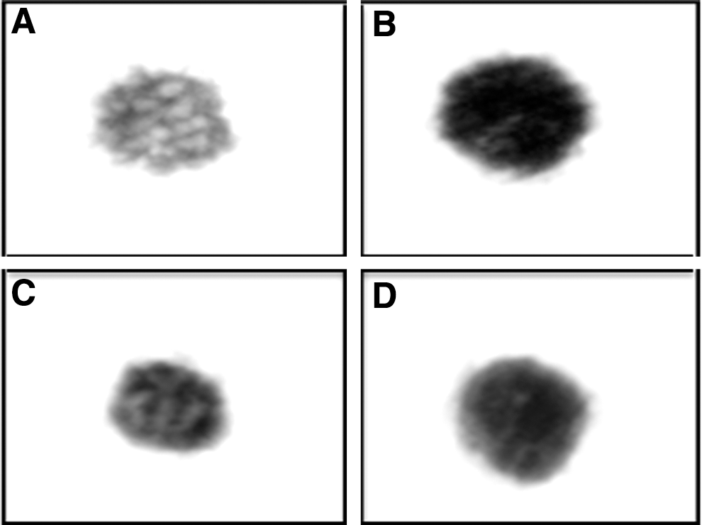
Lymphocyte transformation test
When T lymphocytes were incubated with PHA and polysaccharide samples in vitro, they were stimulated to increase the synthesis of nucleic acid and protein. In the positive control group (PHA), activated lymphocytes were detected with less distinct nuclei and more condensed chromatin. The nucleus was bound, at least on its concave side by a rim of nonglandular narrow cytoplasm (Fig. 3B); this was in accordance with a previous study. 35 Based on these morphological transformation changes, ALP exhibited higher growth and transformation (Fig. 3D) than WAP (Fig. 3C), indicating its potential as an immunomodulatory agent. In addition, cell diameters were measured microscopically, which was correlated with morphological transformation as shown in Table 3. Cells treated with PHA showed a significant twofold increase in cell diameter. Treatment with ALP significantly increased cell diameter comparable to PHA-treated cells. However, WAP did not cause any significant change from the control cells. These findings provided additional evidence that ALP has high potential as an immunomodulatory agent than WAP.
Elicitation of cytokine secretion
Cytokines are molecular messengers that allow the cells of the immune system to communicate with one another to generate a coordinated robust, but self-limited, response to a target antigen. They are produced by cells of innate and adaptive immunity in response to microbes and tumor antigens and directly stimulate immune effector cells and stromal cells at the tumor site and enhance tumor cell recognition by cytotoxic effector cells. 36 They are classified into several groups (e.g., interleukins, interferons, and chemokines) based on the structural homologies. 37,38 Modulation of cytokine secretion may offer new approaches in the treatment of cancer. Over the past two decades, harnessing the immune system to eradicate cancer has been accompanied by characterization of cytokines and exploiting their vast signaling networks to develop cancer treatments. 39,40 One of the strategies used in the modulation of cytokine expression is a herbal medicine; it has some direct effects on tumors and stimulation of cell growth in the immune system. 41 The following cytokines were chosen and investigated because of their vital role in immunotherapy especially in case of cancer disease.
Interleukin-2 (IL-2) stimulated lymphocytes which enhanced the growth of certain T cells (T cell growth factor) bearing the IL-2 receptor; IL-2 also supports the growth of natural killer (NK). It exerts potent immunoregulatory effects on effector and regulatory T and NK-T cells. 42 So it is considered to have effective immunomodulator action especially after investigation which described NK cells as the only effector cells which actively involved in killing of tumor cell target. 43,44 Interleukin-12 (IL-12) has emerged as a promising molecule for antitumor immunotherapeutic protocols; it has potent ability to activate T and natural killer (NK) cells. 45,46 Principally, IL-12 facilitates the presentation of tumor antigens through the upregulation of class I and II major histocompatibility complex, as well as the generation of T helper type I immune responses. These effects are tightly associated with the ability of IL-12 to induce expression of IFN-γ. 43 Interferon-γ (INF-γ) is produced by T-lymphocytes following stimulation of T cell mitogens, specific antigens, or IL-2. 47 It has been used both as an antiviral and antitumor agent, because of its potent ability to suppress tumor cell growth and angiogenesis. Moreover it has an important role to inhibit lymphomas and chemical carcinogenesis 48 and its modulation can affect the NK cell cytotoxicity. 49 TNF, also called cachectin or TNF-α, is a polypeptide hormone that affects many different cells of the body. It causes tumor cell necrosis and apoptosis. 50 From Table 4, the results showed that the polysaccharides WAP and ALP activated PBMC to release high quantity of IL-2 and IL-12. However, WAP possessed lower effect on IL-12 compared to PHA; these investigations suggest that these purified polysaccharides can be considered as biological response modifier. Therefore, the study provides a basis for polysaccharide use as an efficacious adjacent immunopotentiating therapy.
Data are presented as mean ± SD.
Control: The aloe sample was replaced by 0.05 mL of 0.15 M phosphate buffer, pH 7.2.
Statistically, analysis was carried out using one-way ANOVA followed by Tukey's post hoc test.
Statistically significant for the control (P < .05).
Statistically significant for the PHA group (P < .05).
Structural characterization of bioactive polysaccharides WAP and ALP
Average molecular weight (Mw) of 5.48 × 10⁶ g/mol and number average molecular weight (Mn) of 3.85 × 10⁶ g/mol (Fig. 4a). While WAP polydispersity index was 1.05 with overall weight average molecular weight (Mw) of 2.87 × 10⁶ g/mol and number average molecular weight (Mn) of 2.73 × 10⁶ g/mol (Fig. 4b).
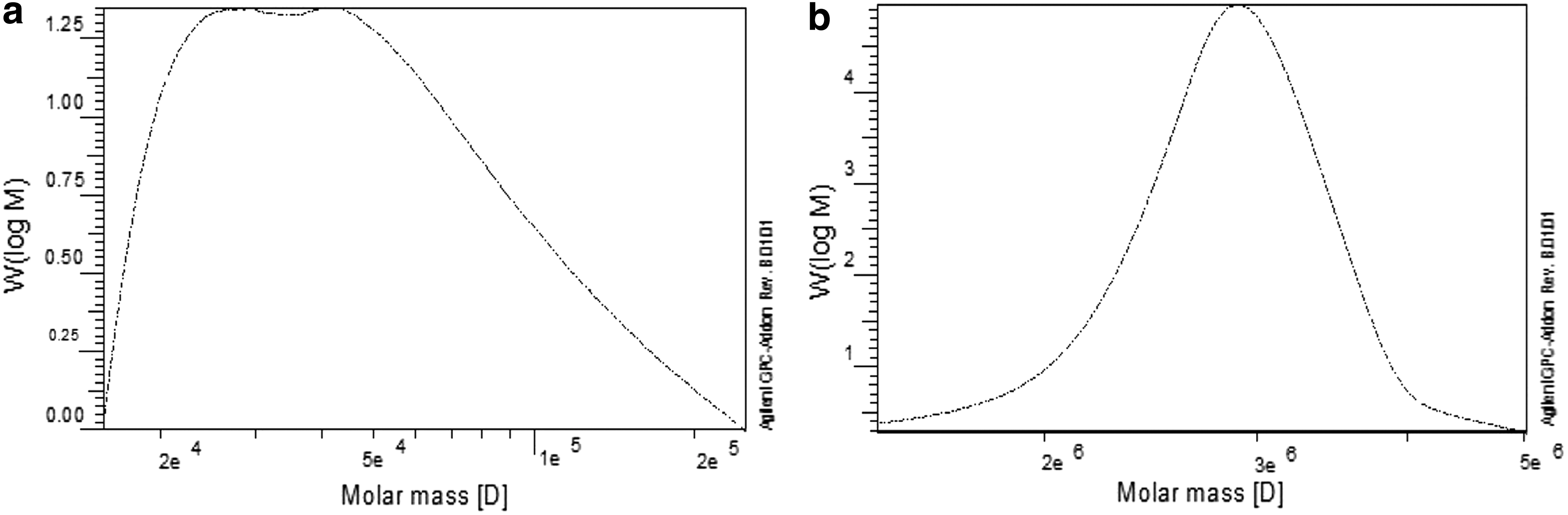
Molecular weight distributions of A. arborescens polysaccharide ALP
The GC analysis of the acetylated monosaccharides revealed that total monosaccharide contents were (34.8) in WAP and (70.8) in ALP as a molar ratio. WAP and ALP were composed of mannose, galactose, glucose, arabinose, rhamnose, and xylose with a molar ratio (3.1: 19.5: 3.9: 1.0: 3.7: 2.4) and (10.9: 2.7: 30.5: 4.4: 1.3: 14.8), respectively (Table 5). Glucose, galactose, and mannose were the major sugars in WAP and ALP, while Ribose was only found in ALP but not present in WAP. Moreover, fructose and sorbitol were found only in WAP. Carbazole-sulfuric acid spectrophotometric method revealed the presence of 27.53% uronic acid content in WAP, while in ALP only 15.46% was detected.
The current findings of monosaccharide composition are in accordance with previous studies referring to the anticancer and immunomodulatory activity of related polysaccharides. Whereas glucans, generally considered as biological response modifier candidate, are recognized as antitumor drugs, and also prevent cancer promotion and progression. 8 There are at least 12 glucan clinical trials in cancer therapy ranging from phase I to phase III. 51 Furthermore, mannans are considered as possible therapeutic tools for the therapy of cancer diseases and immune disorders. 52 In the present study, the WAP showed more phagocytic activity than ALP; this result could be explained by the previous report by Wang et al. stating that water soluble β-glucans seem to be stronger immunostimulators than insoluble ones. 53,54
FT-IR spectroscopy
The FT-IR spectra of WAP and ALP purified extracts are shown in Figure 5. Their infrared spectra were found to have different patterns suggesting that they possessed a bit dissimilar structures in addition to their diversity in the molecular weights. Infrared spectra showed the presence of many functional groups. The absorption at 1626 + 10 cm−1 revealed the presence of acetyl ester units in both extracts. It was suggested that the presence of acetyl groups is necessary for biological activity, possibly because they cover a number of hydrophilic hydroxyl groups and, thus, make the molecule more able to cross hydrophobic barriers in the cell. 55
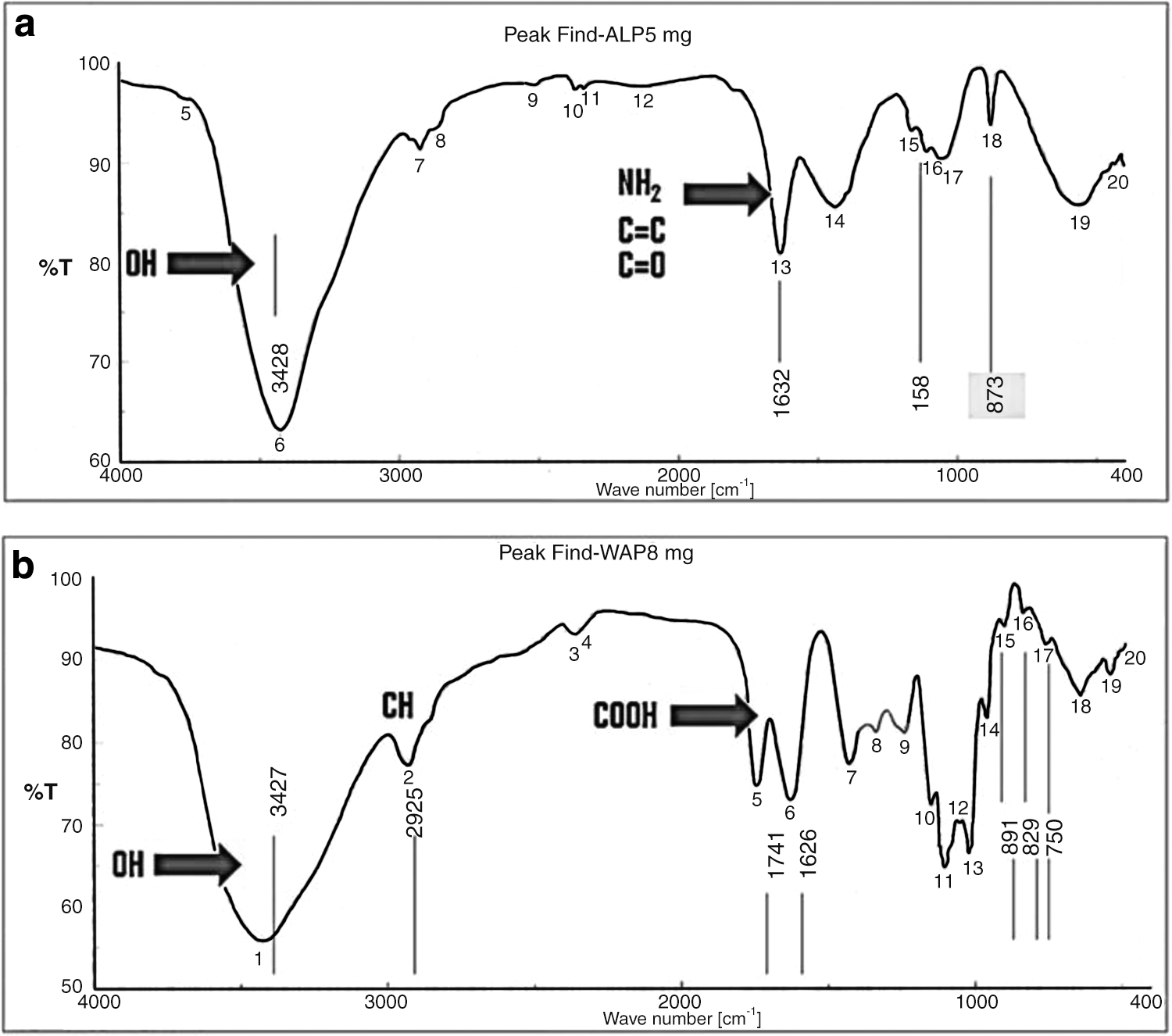
FT-IR spectrum of purified WAP
Furthermore, the absorption bands of carboxyl groups at around 1741 cm−1(asymmetrical stretching vibration) were most pronounced in the FT-IR spectrum of ALP polysaccharide, which is in accordance with the carboxyl group. Furthermore, the banding-like structure in the region of 2870–2930 cm−1 together with the v (C-H) vibrations, as well as a continuous absorption beginning at approximately the region of 3400 cm−1, is characteristic of a carbohydrate ring. The absorption around region ∼1200 cm−1 together with ∼1400 cm−1 is characteristic for v (C-O) and (C-O-H) of sugars. 56
In both polysaccharide types the weak bands at 873–891 cm−1 indicated the presence of β-D-glycoside linkages of glucose, galactose, and/or mannose. However, ALP was characterized by other different bands; the first at 829 cm−1 indicated the probability of presence of α-D-linkage in galactose and mannose sugar residues and β-linkage of arabinose. The second signal at 750 cm−1 was assigned to α-D-linkage of xylose. 57
Linkage analysis of bioactive polysaccharides
Periodate process was carried out under optimum conditions to permit titration of the released formic acid. The values of periodate consumption of the WAP and ALP polysaccharides are detected. HIO4 consumption and formic acid production of both polysaccharides were 0.97 mol/mol sugar residue and 0.008 mol/mol sugar residue, respectively; on periodate oxidation, the result indicated that WAP and ALP were polysaccharides with a (1–4)-glycosidic linkage. The lower value of formic acid in the analysis process supposed that both types of polysaccharides were linear not branched.
These results in addition to the results of both infrared spectra (IR) and Smith's degradation (small amount of glycerol and high quantities of liberated erythritol) partially proved the presence of the β-(1–4) linkages between the monosugars in the backbone. At the end of the periodate oxidation process, the resulting polyaldehydes were reduced to the corresponding polyalcohols, which were subjected to hydrolyses. The resulting polyalcohols hydrolyzed were then subjected to HPLC analysis. The sugar derivatives, such as glycerol, erythritol, and erythric acid, in addition to glucose were separated and quantitatively determined. The molar ratios of these sugars are shown in Table 6.
Erythritol was produced from C3, C4, C5, and C6 of the (1–4) glycosidic linkages of glucose and/or mannose after hydrolysis of the backbone, while erythric acid was liberated from C3, C4, C5, and C6 of the (1–4) glycosidic linkages in mannouronic acid. The presence of relatively small quantities of glycerol gave the information that glucose may be found at the nonreducing end. In addition, the disappearance of glyceric acid through the hydrolysis of the polyalcohol means that uronic acid did not exist in the nonreducing end, but probably was found inside the backbone. 58 The data showed that ALP mainly contained 1, 4-linked glucose residues. The backbone therefore was deduced to be composed of 1, 4-linked Glcp.
Summary
From A. arborescens leaves, three polysaccharides were separated using different pH gradients. Two of these, alkaline-soluble polysaccharide (ALP) and water-soluble polysaccharide (WAP), exhibited direct cytotoxic effects on hepatic tumor cells (HepG2). For WAP, it had ability to enhance phagocytosis, while ALP was able to increase cytokine release through stimulation of lymphocyte transformation. The study provides robust evidence for a new postulated mechanism of action for antitumor activity of A. arborescens polysaccharides, related to direct cytotoxicity and a potent biological modifier response. The chromatographic analysis and structure elucidation of these polysaccharides need further investigation.
Footnotes
Acknowledgment
The authors gratefully acknowledge botanist senior researcher A. Abdel-Mogali at the Flora and Taxonomy Research Department, Agricultural Museum, Giza, Egypt for the plant identification.
Author Disclosure Statement
The authors declare no conflicts of interest.