
Abstract
Osteoarthritis (OA) is a degenerative joint disease characterized by a progressive articular cartilage degradation manifested with significant functional impairment in consort with signs and symptoms of inflammation, stiffness, and loss of mobility. Current OA management is inadequate due to the lack of nominal therapies proven to be effective in hampering disease progression where symptomatic therapy focused intervention masks the primary etiology leading to irreversible structural damage. In this study, we describe the effect of UP1306, a composition containing a proprietary blend of two standardized extracts from the heartwood of Acacia catechu and the root bark of Morus alba, in maintaining joint structural integrity and alleviating OA associated symptoms in monosodium-iodoacetate (MIA)–induced rat OA disease model. Data from pain sensitivity, histopathology, and glycosaminoglycan (GAG) level were analyzed. Diclofenac at 10 mg/kg was used as a reference compound. Ex vivo proteoglycan protection model demonstrated 31.5%, 50.0%, and 54.8% inhibitions of proteoglycan degradations from UP1306 at concentrations of 50, 100, and 200 μg/mL, respectively. The merit of combining two bioflavonoid standardized extracts from A. catechu and M. alba was demonstrated in their Ex vivo synergistic proteoglycan protection activity. In the MIA in vivo OA model, administered orally at 500 mg/kg, UP1306 resulted in reductions of 17.5%, 29.0%, 34.4%, 33.5%, and 40.9% through week 1–5 in pain sensitivity, statistically significant improvements in articular cartilage matrix integrity, and minimal subchondral bone damage. Therefore, UP1306 could potentially be considered as an alternative remedy from natural sources for the management of OA and/or its associated symptoms.
Introduction
O
Despite recent advances in drug discovery, present-day OA management is inadequate due to the lack of primary therapies proven to be effective in modifying disease progression. The current approach focuses mainly on curtailing the sensitivity of disease-associated pain (like use of over-the-counter Nonsteroidal anti-inflammatory drugs, NSAIDs), which will only mask the actual etiology leading to irreversible damage to the articular structure. In addition, chronic usages of NSAIDs are also limited due to their gastrointestinal, renal, and cardiovascular side effects. 5 This calls for immediate attention for research and development by the pharmaceutical and nutraceutical industries to direct their resources and efforts in search of a cure or a slow down for this debilitating disease. In this regard, recently we reported a botanical composition designated as UP1306 (a proprietary blend of two bioflavonoid standardized extracts from the heartwood of Acacia catechu and root bark of Morus alba) with analgesic and anti-inflammatory effects, which was discovered through the in vivo screening of known traditional folk medicines suggesting its usage for OA associated symptom relief. 6 UP1306, administered orally at a dose of 300 mg/kg, resulted in 46.3–53.3% reductions in paw edema and 43.6–54.8% reductions in pain sensitivity in the carrageenan induced rat paw edema model, as well as a 34.4% reduction in visceral pain sensitivity in the Writhing's model in mice. 6 In vitro, UP1306 showed dose-dependent inhibition of the enzymatic activities of COX and LOX with IC50 values of 20.9, 49.2, and 11.1 μg/mL in COX-1, COX-2, and 5-LOX, respectively. 6 In a pilot human clinical trial, there was a statistically significant difference between the changes of urine CTX-II (C-telopeptide of type II collagen, a type II collagen biomarker frequently referenced for its close correlation with progression of articular cartilage degradation in OA patients) for UP1306 and placebo groups after 12 weeks of product use (P = .029) (Personal communication). Collectively, these findings suggest that UP1306 may have OA disease modifying activities which inspired the current study.
M. alba L (Family: Moraceae), the mulberry or white berry plant, is native to northern China and has been cultivated and naturalized elsewhere, from India through middle east to Southern Europe and recently to North America. The root bark of M. alba that is used in traditional medicine is known as Sang Bai Pi or Cortex Mori (Pharmacopoeia of the People's Republic of China, 2005). This herb is also known as Pong-na-moo in Korean and Sohakuhi in Japan. In contemporary pharmacological research, M. alba root bark has been reported to have antibacterial, 7 antioxidant and hypoglycemic, 8,9 hypolipidemic, neuroprotective, antiulcer, analgesic, 10,11 and anti-inflammatory activities. 12 Some of the prenylated flavonoids and stilbenoids such as morusin and mulberroside A are unique to Morus plants. 13 Extracts and prenylated flavonoids from Morus are known to inhibit Nitric oxide (NO) and IL-6 production, downregulate inducible nitric oxide synthase (iNOS), 14 inhibit activation of NF-κB, 15 and inhibit TNF-α 16 and IL-1β production, 17 suggesting its use in inflammation.
A. catechu (L.f.) Willd (also known as “black catechu” or “katha) is from the Leguminosae family native to India and Burma and also found in China and parts of Bangladesh. The heartwood hot water extract of A. catechu is mainly used in India as an ingredient of chewing gums. 18 Traditionally, A. catechu has been a component in Ayurveda for the purpose of relieving toothache, sore throat, gum ulceration, digestion aid, and hepatoprotection, 19,20 as well as healing bleeding gums and canker sores (Indus Herbal Extracts 2011). In the Chinese Pharmacopeia, the dry extract of stems and wood of A. catechu is used for healing ulcers, weeping skin disease, diarrhea, traumatic injuries, and hemorrhoids. Thai medicine also lists A. catechu for its antidiarrheic properties. 21 Catechin is a major flavan in Acacia plants. 22
Provided the above usages and activities of Acacia and Morus, there is a possibility for their combination to have a conceivable activity to relieve pain and to spare cartilage loss in OA. Therefore, the present study was designed to evaluate the potential analgesic and/or cartilage degradation protection activity of UP1306 using a monoiodoacetate (MIA)-induced OA disease model in rats. Ex vivo, sulfated GAG inhibition assays were also performed using rabbit articular cartilage explants.
Materials and Methods
The composition: The original dried root bark of M. alba L. var. multicaulis (Perr.) Loudon in the Moraceae family was collected from Chongqing, China and identified by professor Shou-Yuen Zhao from Si-Chuan Chinese Traditional Medicine Research Institutes. A voucher of specimen of M. alba (P00329) was deposited at the plant library of Unigen, Seattle, WA, USA. The recollected M. alba root barks were characterized and confirmed in comparison with the original voucher specimens.
The dried branch and trunk of A. catechu Willd. A. Suma (Senegalia catechu (L.f.) P.J.H Hunter & Mabb) in the Leguminosae family was initially collected from Yunnan, China and identified by professor Shou-Yuen Zhao from Si-Chuan Chinese Traditional Medicine Research Institutes. A voucher of specimen of A. catechu (P00530) was deposited at the plant library of Unigen, Seattle, WA, USA. The recollected heartwood of A. catechu from Distt Kangra Himachal Pradesh, India was characterized and confirmed in comparison with the original specimens.
Detailed procedure for the preparation of the composition has been described in the United States patent #20150072953. 23 Dried M. alba root bark was cut, crushed, and then extracted with approximately sevenfold volume of 70% ethyl alcohol in water at 100°C for 4 h thrice to give M. alba 70% EtOH extract powder after evaporation of solvent with a yield of 19.6% (w/w). The standardized Morus extract contains no less than 4% Mulberroside A and no less than 3% of total bioflavonoids, including Kuwanon G, Albanin G, and Morusin. Catechin enriched Acacia extract was obtained by repeated crystallization from the aqueous extract of A. catechu heartwood with a yield of 14.5% (w/w). (+)-Catechin was identified as the major active flavan in the A. catechu extract. The Acacia extract was standardized as no less than 65% catechin and a minor enantiomer epicatechin. Composition UP1306 was prepared by mixing the standardized extracts of A. catechu heartwood (no less than 65% catechins) and M. alba root bark (no less than 7% stilbenes and bioflavonoids) at a ratio of 1:2 by weight containing no less than 15% catechins and 2% stilbenes and bioflavonoids.
Ex vivo GAG releasing model
Articular cartilage from hock joints of rabbits (2.5 kg body weight) was removed immediately after each animal was sacrificed. The articular cartilage explants were obtained by following the method described by Cho et al. 24 Briefly, after the articular surfaces were exposed surgically under sterile conditions, approximately 200–220 mg articular surfaces per joint were dissected and submerged into complete medium (Dulbecco's modified Eagle's medium [DMEM], supplemented with heat-inactivated 5% Fetal Bovine Serum [FBS]; penicillin 100 U/mL; and streptomycin 100 μg/mL). They were then rinsed several times with the complete medium and incubated for 1–2 days at 37°C in a humidified 5% CO2/95% air incubator for stabilization. The complete medium was replaced with a basal medium (DMEM, supplemented with heat-inactivated 1% FBS, 10 mM hydroxyethyl piperazineethanesulfonic (HEPES), and penicillin 100 U/mL and streptomycin 100 μg/mL). Approximately 30 mg cartilage pieces (2 × 3 × 0.35 mm/piece) were placed in 24-well plates and treated with given concentrations of test agents. After pretreatment for 1 h, 5 ng/mL of recombinant human interleukin-1 alpha (rhIL-1α) was added to the culture medium and further incubated at 37°C in a humidified 5% CO2/95% air incubator. The culture medium was collected 24 h later and stored at −20°C until assay. The amount of sulfated GAGs in the medium at the end of reaction reflecting the amount of articular cartilage degradation was determined by 1,9-dimethyl-methylene blue method using commercially available kit (the Blyscan proteoglycan and GAG assay) according to the instructions of the manufacturer. Additional assay was also performed for individual extracts of A. catechu and M. alba at 33.3 and 66.7 μg/mL, respectively, to compare with the 100 μg/mL of UP1306 for synergy, if any. The merit of combining A. catechu and M. alba was then evaluated using the Colby's equation. 25 In this method, a formulation of two materials together will presume to have unexpected synergy, if the observed value of a certain end point measurement is greater or equal to the hypothetically calculated values. GAG releasing percent inhibition values of A. catechu and M. alba were used to determine the calculated efficacy values and compared to the observed GAG releasing percent inhibition values of UP1306.
In vivo MIA induced OA model
Animals
Sprague–Dawley rats were purchased from Charles River Laboratories at the age of 8 weeks and were acclimated upon arrival for a week before being assigned randomly to their respective groups. Rats (3/cage) were housed in a polypropylene cage and individually identified by numbers on their tails. Each cage was covered with a wire bar lid and filtered top. Each individual cage was identified with a cage card indicating project number, test article, dose level, group, and animal numbers. The Harlan T7087 soft cob beddings were used and changed at least twice a week. Animals were provided with fresh water and rodent chow diet # T2018 (Harlan Teklad; 370 W) ad libitum and were housed in a temperature-controlled room (22.2°C) on a 12-h light/12-h dark cycle. All animal experiments were conducted according to the institutional guidelines congruent with guide for the care and use of laboratory animals.
Model induction and treatment
Treatment started a week before MIA injection. Animals (body weight 215–229 g) were randomized into four groups of 10 rats per group as G1 = normal, G2 = vehicle (0.5% CMC-Na solution), G3 = Diclofenac (10 mg/kg; Lot # W08B043), and G4 = UP1306 (500 mg/kg) (Lot # AM14002) and were orally gavaged with respective treatment. UP1306 was tested in multiple dosages. In this study, only data from the 500 mg/kg have been depicted. On the induction day, isoflurane (Lot #B66H15A; Piramal Enterprise Ltd.) anesthetized rats were injected with 0.8 mg of MIA (Lot # A0352046; Acros Organics) in 50 μL saline solution into the intra-articular pocket of left femorotibial (knee) joint using 26 G needle an hour after treatment. 26 Normal control rats were injected with an equal volume of saline. Paw withdrawal thresholds as a result of constant pressure applied to the affected joint as a measure of pain sensitivity were taken once a week using Randall-Selitto Anesthesiometer (IITC) and treatment lasted for 6 weeks. Body weights were measured once a week to calculate the respective weekly dosage of each group.
Histopathology
At necropsy, animals were asphyxiated with CO2, and the femorotibial joint was carefully dissected out, fixed in 10% buffered formalin, and sent to Nationwide Histology for further histopathology analysis. The fixed specimens were then decalcified with Calci-Clear Rapid for 1 and a half days and embedded in paraffin. Standardized 5 μm serial sections were obtained at the medial and lateral midcondylar level in the sagittal plane and were stained with hematoxylin and eosin (HE) and Safranin O-fast green to enable evaluation of proteoglycan content. A modified Mankin system 27 was used to score structural and cellular alterations of articular components as indications of disease progression and/or treatment efficacy. The histological analysis was conducted by a certified Pathologist at Nationwide Histology.
Statistical analysis
Data were analyzed using SigmaPlot (Version 11.0; Systat Software, Inc.). The results are represented as mean ± standard deviation. Statistical significance among groups was calculated by means of single factor analysis of variance (ANOVA) and by t-test. P ≤ .05 was considered statistically significant. When normality test failed, for nonparametric analysis, data were subjected to Mann–Whitney sum ranks for t-test and Kruskal–Wallis one-way ANOVA on ranks for ANOVA.
Results
Glycosaminoglycan
Ex vivo, rabbit cartilage explants were cultured for 24 h with IL-1α (5 ng/mL) in the absence or presence of test agents to examine the protective effects on proteoglycan (PG) degradation. Results were expressed as μg GAG released into the medium. As depicted in Figure 1, significant cartilage protection activity was observed by the composition UP1306. When the rabbit cartilage explants were treated with rhIL-1α, the amount of GAG released into the culture medium increased significantly (i.e., 125%) compared to the untreated normal control. UP1306 composition reduced rhIL-1α -mediated degradation of proteoglycan in a concentration-dependent manner (P < .05). A total of 31.5%, 50.0%, and 54.8% inhibitions of proteoglycan degradation were observed for UP1306 at concentrations of 50, 100, and 200 μg/mL, respectively, compared to rhIL-1α exposed untreated explants. The positive control, diclofenac, resulted in 67.4% reductions in release of GAG compared to the rhIL-1α challenged explants. Based on the level of proteoglycans in culture medium at the highest concentration (200 μg/mL), it could be judged that UP1306 almost totally inhibited the rhIL-1α-induced proteoglycan degradation to the level of the normal control without rhIL-1α. Further analysis of data from the components of UP1306 against the composition using Colby's equation demonstrated greater inhibition of GAG release to the cultured media for explants treated with the composition UP1306 than A. catechu or M. alba alone (Table 1).
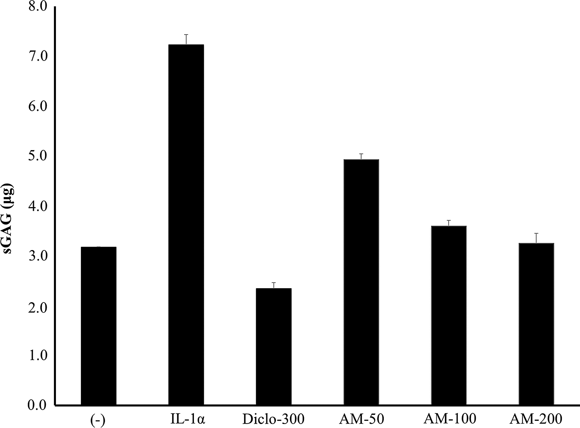
Effect of UP1306 composition on GAG releasing. Cartilage explants were incubated for 24 h in DMEM with 1% heat-inactivated FBS. Each set of data represents the mean ± SD (−): control cultured medium without IL-1α; IL-1α: cultured medium treated with IL-1α (5 ng/mL); Diclofenac-300: cultured medium treated with IL-1α plus 300 μg/mL of Diclofenac; UP1306–50/100/200 μg/mL: cultured medium treated with IL-1α plus UP1306 at doses of 50/100 or 200 μg/mL. GAG, glycosaminoglycan.
Rabbit cartilage explants were cultured for 24 h with rhIL-1α (5 ng/mL) in the absence or presence of a gambir extract, Morus extract, or UP1306 at the disclosed doses to examine the potential protective effect on proteoglycan degradation. Whether a synergistic effect was present was calculated using Colby's formula. Expected value: theoretically calculated inhibition value based on Colby's formula for synergy; experimental value: inhibition observed at 100 μg/mL of UP1306; synergy = experimental value ≥ expected value.
Pain sensitivity
Pain, one of the main cardinal symptoms of OA, was evidenced a week following model induction. As seen in Figure 2, rats with an intra-articular injection of MIA without treatment showed progressive increase in pain sensitivity as exhibited by the mean pain sensitivity values. Compared to the vehicle treated normal control animals, rats with intra-articular 0.8 mg/joint MIA showed 43.4%, 46.5%, 47.1%, 43.4%, and 46.5% increase in pain sensitivity from week-1 to week-5, respectively. In contrast, rats treated orally with a daily dose of 500 mg/kg of UP1306 showed statistically significant reductions in pain sensitivity as of week-1 of oral treatment. Reductions of 17.5%, 29.0%, 34.4%, 33.5%, and 40.9% in pain sensitivity were observed for rats treated with UP1306 from week-1 to week-5, respectively, compared to vehicle treated MIA injected rats. These reductions were statistically significant at each data point examined. Reductions of 36.8%, 35.9%, 30.2%, 30.5%, and 36.9% in pain sensitivity were observed for rats treated with diclofenac from week-1 to week-5, respectively, compared to vehicle treated MIA injected rats. These reductions were also statistically significant at each data point examined.
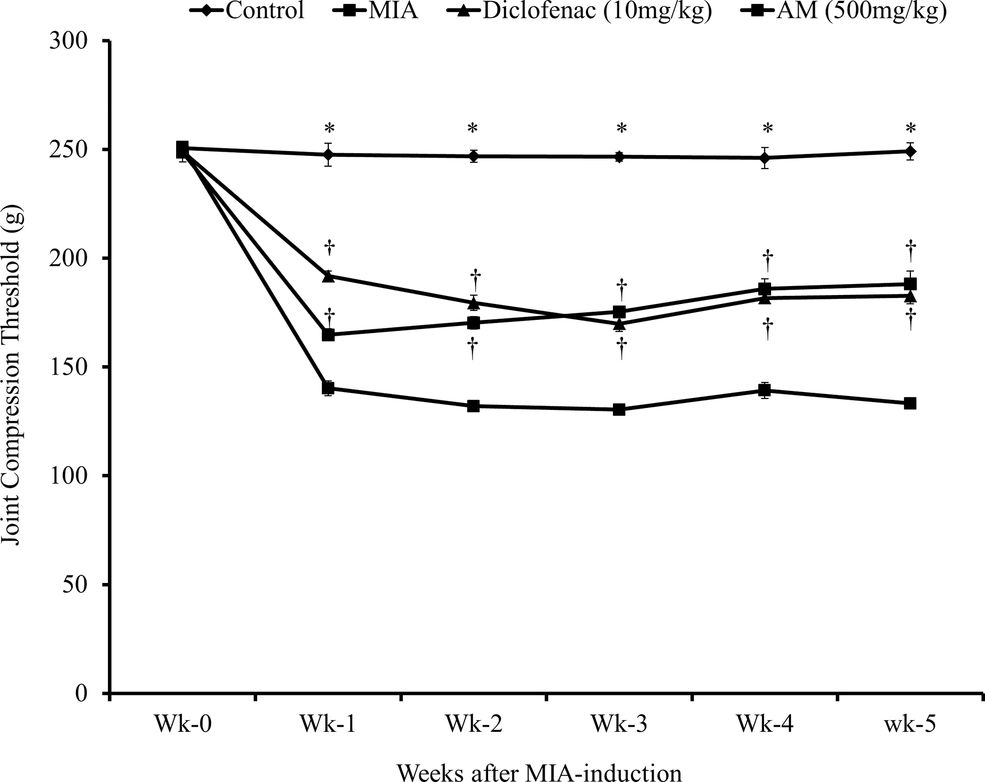
Compression threshold for MIA-injected rats treated with AM. Osteoarthritis disease model was induced by intraarticular injection of 0.8 mg/joint MIA to the left femorotibial joint of SD rats (N = 9). Rats were treated with diclofenac and UP1306 at oral doses of 10 mg/kg and 500 mg/kg, respectively, for 6 weeks. Compression threshold was assessed every week using Randall–Selitto. “AM” is an acronym for the constituents of the composition UP1306: Acacia and Morus. Data are expressed as Mean ± SD. † P ≤ .00001; *P ≤ .000001.
Histopathology findings
Complementing the pain sensitivity reduction data, statistically significant improvements in articular cartilage matrix integrity were shown as reflected by the modified total Mankin score for animals treated with UP1306 at a daily oral dose of 500 mg/kg for 6 weeks (Table 2 and Fig. 3). Structural abnormalities and fibrovascular proliferation were also significantly reduced in this group. The extent of osteoclast activities and subchondral bone damages was minimal. However, unlike the pain sensitivity data, a positive trend without statistical significance was observed only in fibrovascular proliferation from the positive control, Diclofenac, compared to vehicle control (Table 2 and Fig. 3). In contrast, various degrees of histopathological changes, including cellular degeneration and disorganization of the articular cartilage chondrocytes, depletion and collapse of the intracellular matrix, articular surface irregularities, osteophyte remodeling, and fibrillation of the subchondral bone were observed for MIA-injected rats treated with vehicle. These changes are similar to the most common findings in human OA biology. 28 In Safranin O staining, articular cartilage of UP1306 revealed minimum loss of staining intensity indicating its ability to spare cartilage degradation (Table 2 and Fig. 3). Rats in the normal control groups treated with vehicle showed negligible changes in all the parameters examined (Table 2 and Fig. 3). Normal structure of the articular cartilage, subchondral bone of both tibia plateaus and femoral bone, and the surrounding joint structure appeared intact in this group of rats.
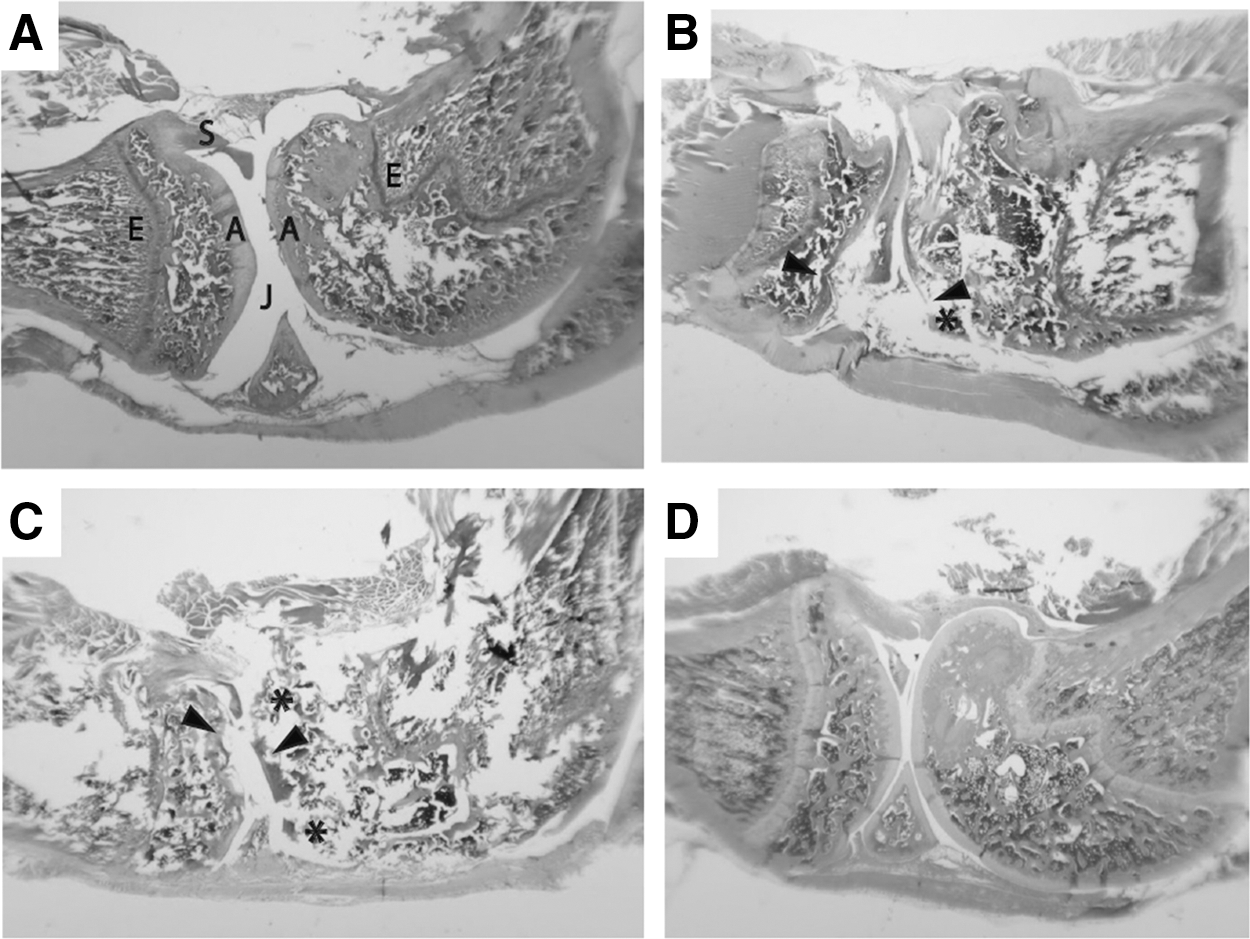
Histopathology: The femorotibial joint was carefully dissected out, fixed in 10% buffered formalin, then decalcified with Calci-Clear Rapid for 1 and a half days, and embedded in paraffin. Standardized 5 μm serial sections were obtained at the medial and lateral midcondylar level in the sagittal plane and were stained with hematoxylin and eosin.
Structural abnormality (0–6): Cartilage thickness/thinning, irregular surface frayed/fissure loss, degeneration, ulcerative necrosis/fragmentation, severe disorganization/chaotic; Bone at the articular surfaces (0–6): Subchondral bone thickness/volume & density, osteoclastic activity, subchondral bone damage; Inflammation/Cellular infiltration (0–6): Cellular Infiltration/Inflammation & Proliferation, hypercellular, cluster/hypocellular; Fibrovascular proliferation (0–6): Fibrovascular Proliferation replacing periarticular/capsule/bone (Pannus), condyle &/or tibial plateau, menicus reduction, fusion, adhesion; Matrix GAGs (0–6): Matrix GAG reduction: radial, interterritorial to pericellular loss of staining, femoral condyle/tibial plateau integrity, & thickness of articular Cartilage. †P ≤ .001; * P ≤ .05; ‡P ≤ .07
GAG, glycosaminoglycan.
Discussions
The MIA-induced OA disease model in rats is a standardized model most frequently used to mimic the human OA. 14 The model involves inoculation of MIA into a femorotibial joint pocket that induces pain responses in the ipsilateral limb accompanied by progressive cartilage degradation. Intra-articular injection of MIA disrupts chondrocyte glycolysis by inhibiting glyceraldehyde-3-phosphatase dehydrogenase and results in chondrocyte death, neovascularization, subchondral bone necrosis and collapse, as well as inflammation. 29 These characteristics make the model very attractive to evaluate compounds for their anti-inflammatory, analgesic, and/or potential disease modifying activities as it shares similar disease pathology to the human OA. As a result we selected this mature model to investigate the effect of UP1306 in mitigating pain sensitivity and maintaining articular structural integrity administered orally at 500 mg/kg for 6 weeks. To supplement the in vivo study, ex vivo GAG releasing assay was also carried out using rabbit articular cartilage.
Mounting evidence has been documented describing the beneficial use of A. catechu and M. alba in OA. For instance, the major flavan in Acacia, catechin, has been shown to inhibit 5-lipoxygenase (5-LOX) and cyclooxygenase-2 (COX-2) gene expression, phospholipase A2 (PLA2), pro-inflammatory cytokines such as TNF-α, and multiple interleukins, such as IL-1, 2, 6, 8, and 12. 30 Similarly, a variety of bioactive compounds from M. alba root bark have shown in vivo or in vitro anti-inflammatory activity. For example, inhibition of NO production, reduction of iNOS expression, inhibition of Prostaglandin E2 (PGE2) production and inhibition of nuclear factor κB (NFκB) by Oxyresveratrol, 31 and inhibition of both NO production and iNOS, as well as reduction of pro-inflammatory mediators such as COX-2, IL-1β, and IL-6 by total flavonoids from the root bark 32 and by prenylated flavonoids 33 from M. alba, were reported. Using human chondrocytes, recently Joeng et al. have also showed inhibition of IL-1β induced expression of matrix metalloproteinases (MMP-1 and −13) and inflammatory mediators (NO, PGE2, iNOS, and COX-2 expression) by suppressing the activation of NF-κB and mitogen-activated protein kinase (p38 MAPK). 34 In vivo, oxyresveratrol and mulberroside A have been reported to have an anti-inflammatory effect on a carrageenan-induced paw edema model in rats at dosages of 7.5 and 50 mg/kg, respectively. 31 In a collagen induced arthritis model in mice, Morus extract administered orally for 2 weeks at a dose of 300 mg/kg caused significant suppression of immune cell infiltration, synovial hyperplasia, cartilage destruction, and bone erosion. In the same study, inflammatory cytokines (such as TNF-α, IL-1β, and IL-6) were significantly suppressed by the extract in a dose-dependent manner. The number of osteoclasts in the hind tibia was also significantly decreased. 15 Similarly, catechin administered orally at doses of 60 and 120 mg/kg for 2 weeks significantly suppressed secondary inflammatory paw swelling, pain response, and polyarthritis index in adjuvant induced arthritis in rats. It also inhibited production of IL-1, TNF-α, and PGE2. 35 Hence, the hypothesis of formulating these historically well-known plant based bioflavonoids into a well-defined standardized composition for indication of OA associated symptoms and/or disease modification was tested in the present study, and promising results were documented.
Articular cartilage degradation occurs as a result of an imbalance in the homeostasis of fundamental matrix components such as GAGs and type II collagen. 36 This pathogenesis is triggered, in part, by the action of inflammatory cytokines, primarily IL-1 37,38 that also mediates the production of pro-inflammatory mediators (including NO and PGE2) and matrix degrading enzymes, aggrecanase and MMP. While the catabolic enzymes and MMPs disrupt collagen fibers, 39 members of a disintegrin and metalloprotease with thrombospondin (ADAMTS) family degrade aggrecan and both cases result in the release of GAGs. 40 GAGs, besides serving as a building block for cartilage, also exert specific pharmacologic effects such as decreasing IL1-induced gene expressions by inhibiting the cytokine intracellular signaling cascade and NF-κB activation. 41 GAG loss from articular cartilage into the synovial fluid in human OA patients has been reported 42 with a direct and strong correlation (r = 0.65) of disease progression. 43 This clearly implies that the reduction in the sGAG ex vivo assay observed as a result of UP1306 treatment could have a potential therapeutic advantage in maintaining structural integrity of articular cartilage beyond curtailing associated pain. From the synergy data, it can also be inferred that combining the two extracts from Acacia and Morus could provide a greater protection against proteoglycan degradation than either of the components given alone. In agreement with these findings, in a pilot human clinical trial, statistically significant reduction in urine CTX-II (a biomarker for cartage degradation) was observed for OA subjects who received 400 mg oral treatment of UP1306 for 12-weeks compared to the placebo group. In support of our study, previously prenylated flavonoids extracted from M. alba have shown inhibition of the catabolic enzymes ADAMTS1 44 and matrix metalloproteinase (MMP-1 and MMP-3). 15,34 Catechin, the major flavan of Acacia, also inhibited the degradation of human and bovine cartilage proteoglycan and type II collagen 45 and hindered IL-β induced cartilage proteoglycan degradation and expression of matrix metalloproteinases (MMP1 and MP-13) in human chondrocytes. 46 Therefore, the current study could be considered as a bridge for the transition from the preclinical ex vivo and in vivo studies to the human clinical trial by providing an indication to the cartilage protection activity of UP1306.
The composition also played a major role in suppressing OA disease associated pain. This analgesic effect of the composition was believed to be attained as a result of its individual components which are historically known for their strong anti-inflammatory activity contributing to alleviate pain sensitivity. This mitigation of disease associated symptoms was also shared by the common NSAID, Diclofenac, as expected. Higher inhibition of pain sensitivity was observed for rats treated with Diclofenac for the first 2 weeks of the study compared to either UP1306 or vehicle treated MIA challenged rats. These observations were reversed as of week 3 of the study. There was slightly higher inhibition of pain sensitivity as a result of oral treatment of UP1306 compared to the Diclofenac treated rats.
Furthermore, significant improvements in maintenance of the articular structural integrity of rats treated with the composition UP1306 were observed in vivo. These effects were demonstrated in the histopathology data as exhibited by limited loss, degeneration, or necrosis of chondrocytes, smoother articular cartilage surface, deeper and uniform stain of intracellular matrix, and close to normal contour of the subchondral bone. However, diclofenac failed to show noteworthy significance in the Mankin score. This lack of significance in cartilage protection observed for Diclofenac indicates the limitation of NSAIDs for prolonged usage in OA patients restricting their usage only for symptomatic relief without the benefits of disease modifying activity. Substantiating our findings, previously, orally administered Diclofenac caused significant analgesic activity, 47,48 but inadequate cartilage and proteoglycan degradation protection. 49
Through the years, augmented reports have been documented describing the role of inflammation in OA disease initiation and progression through the network of articular components that includes cartilage, subchondral bone, and synovial membrane. Researches have shown that stimulation of chondrocyte surface receptors by mechanical stress, aging factors, and pro-inflammatory mediators, such as TNF-α or IL-1β, will lead to induction of the NF-κB signaling pathway that triggers the secretion of several matrix degrading proteinases, 50 which in turn causes cartilage degradation. The NF-κB pathway could also be activated by synoviocytes upon stimulation by similar factors such as mechanical stress or cytokines (TNF-α and IL-1β), which subsequently produce additional MMPs, ADAMPTS, apoptotic molecules (COX2, NO, and PGE2), chemokines (IL-8), and cytokines (TNF-α, IL-1β, and IL-6) ensuring cartilage destruction and synovium membrane inflammation. 37 As a matter of fact, almost all of the listed biomarkers have been modulated by either of the constituents of UP1306. Hence, the OA related beneficial effects of UP1306 could be, in part, by protection of cartilage degradation (as shown by its greater inhibition of sGAG release) possibly by inhibiting multiple matrix degrading proteinases and/or aggrecanases. Additional cartilage protection activity of UP1306 could also be through interventions at the inflammatory process (possibly by inhibiting primary pro-inflammatory mediators) and, hence, disrupting the perpetual cycle that links cartilage degradation to the synovitis. These moderations may perhaps hamper the recurrent attack of the joint either by the degradants from the cartilage or pro-inflammatory cytokines from the synovial membrane, which ultimately provides pain relief and improved joint mobility and stability. Nevertheless, additional preclinical studies are warranted to understand the specific mechanisms of action of the composition.
Considering the multifactorial nature of OA, it has previously been suggested that the ability to slow the progression of articular cartilage degeneration is greater with a combination therapy compared with any single component alone. 51 The composition of bioflavonoid standardized extracts from A. catechu and M. alba may suit very well in this category as synergistic effects in alleviating pain sensitivity and reducing paw edema, which were observed when they were administered together in a standardized formulation (UP1306) in carrageenan induced rat paw edema model. 6 In this study again, as demonstrated by (a) the inhibition of GAG release, (b) the suppression of pain sensitivity, and (c) the maintenance of closer to normal joint structures, these medicinal plants may have complementary effects on each other in preventing articular cartilage degradation and mitigating associated pain, which could be translated to improved joint mobility and function.
In conclusion, we have evaluated the efficacy of orally administered UP1306, a composition containing a proprietary blend of two bioflavonoid standardized extracts from the heartwood of A. catechu and the root bark of M. alba, in MIA-induced OA disease model in rats. Ex vivo, treatment of rabbit cartilage explants with rhIL-1α resulted in significant degradation of proteoglycan, which was inhibited by UP1306 in a dose-dependent manner. And, perhaps most importantly, in vivo, significant pain alleviation and maintenance of articular structural integrity were observed. These findings, in conjunction with the promising uCTX-II data from a pilot human clinical trial, suggest that UP1306 could potentially be considered as an alternative therapy from natural sources for the treatment of OA and/or its associated symptoms. However, additional human clinical studies with a much larger patient population tested for a longer duration are needed to further validate these findings in human.
Footnotes
Acknowledgments
The authors express their best gratitude to Dr. Wenwen Ma, Dr. Min Chu, and Unigen team for their incalculable support for the completion of this research. The authors also thank Nationwide Histology (Veradale, WA, USA) for the histopathology analysis.
Author Disclosure Statement
All authors are currently associated with Unigen Inc. and, therefore, have financial interests.