
Abstract
The effect and mechanisms of Myristica fragrans on blood clotting were evaluated by evaluating blood coagulation time and the fibrinolytic system. The compounds
Introduction
T
Coagulation of blood after vascular spasm and platelet aggregation involves formation of prothrombinase and the conversion of prothrombin into thrombin, which helps the conversion of fibrinogen into fibrin (clot). 15 Prothrombin time (PT), activated partial thromboplastin time (APTT), thrombin time (TT), and fibrinogen (FIB) tests are the most commonly clinically used coagulation tests.
PT is used in the screening test for the extrinsic coagulation system. 16 APTT is a commonly used screening test for the endogenous coagulation system. 17 TT reflects the time required for fibrinogen to be converted to fibrin in the common pathway of the coagulation process. 18 FIB mainly reflects the content of fibrinogen, which is an acute-phase protein produced during the process of coagulation. 19
Plasminogen and fibrinolytic enzyme are the main components of the fibrinolytic system, and fibrinolytic enzyme is an important protease involved in many physiological and pathological processes. 20 Urokinase-type plasminogen activator is a multifunctional multidomain protein that is not only a regulator of fibrinolysis but also associated with several acute and chronic pathological conditions. 21
Abnormal changes in the blood coagulation and fibrinolysis system are closely related to the damage from diabetes mellitus. 22 AMPK (protein kinase), 23 PPARγ receptor, 9 and protein tyrosine phosphatase PTP1B 24 might be potential mechanistic targets for antidiabetic agents for the treatment of type 2 diabetes. By slowing down the digestion and absorption of dietary carbohydrates either by means of dietary manipulations or intestinal, 25 α-glucosidase inhibitory drugs such as acarbose, miglitol, and voglibose have shown the ability to reduce blood glucose levels in patients with type 2 diabetes. 26 So, α-glucosidase inhibitors have been used as a new class of antidiabetic drugs, and many are found in medicinal plants. 27
Our study was undertaken to investigate the blood clotting mechanism of compounds from M. fragrans by observing the effects on blood coagulation time and the fibrinolytic system. In addition, the α-glucosidase inhibitory activities of M. fragrans were screened.
Materials and Methods
Collection and identification of plant samples
The pericarp parts of M. fragrans were collected in December 2012 from the Wanning Region, Hainan Province, China, and a voucher specimen was identified by Prof. Chang-qin Li of Henan University and deposited in the Herbarium of the Institute of Natural Products, Henan University.
General
Column chromatography was performed with different types of D-101 macroporous resin (Tianjin Haiguang Chemical, Inc., China), Sephadex LH-20 (Pharmacia, Sweden), 200–300, 40–80 mesh silica gel (Ding Kang Silica Gel Co., Ltd., Qingdao, China), silica gel H (Qingdao Marine Chemical Co., Qingdao, China), and 40–63 μm silica gel (Merck, Germany).
Deuterated chloroform (Beijing SeaSky Bio Technology Co., Ltd.) and analytical grade solvents were purchased from Kemiou Chemical Reagent Co., Ltd. (Tianjin, China). α-Glucosidase (22003401), 4-N-trophenyl-α-D-glucopyranoside (PNPG, D00129651), and acarbose (100434) were from Sigma (USA). Saline (Qi Pharmaceutical Co., Ltd., Shandong, 2012053107); vitamin K1 injection (Tianjin Pharmaceutical Group Co., Ltd., Xinzheng, 1109051); injection breviscapine (Hang Sheng Pharmaceutical Co., Ltd., Hunan, 20110202); PT (Lot: 105227), APTT (Lot: 112163), TT (Lot: 121116), FIB (Lot: 132058) assay kits (Shanghai Sun Biotech Co., Ltd., China); bovine fibrinogen (Sigma, F8630); thrombin (Sigma, T4648); urokinase (Biotechnology Co., Ltd., Hangzhou, Macao and Asia, Lot: 1201166); agarose (GENE Company, 122015); and complex tablet (Hutchison Whampoa Guangzhou Baiyunshan Chinese Medicine Co., Ltd., China, 20130326).
Animals
The animal experimental protocol was approved by the Ethics Committee on Animal Experiments in Henan University. The rabbit license of the laboratory (No. 15-2-6) was issued by the Experimental Animal Center of Henan Province (Zhengzhou, China). All the procedures were carried out according to the guidelines of the National Institutes of Health for Care and Use of Laboratory Animals and were approved by the Bioethics Committee of Henan University.
There was one male Rex rabbit (Institute of Henan University of Traditional Chinese Medicine, Henan University) weighing from 2.0 to 2.5 kg. The rabbit, given free access to food and water, was raised in a controlled environment at 25°C, 45–65% humidity, and 12 h of light/dark.
Extraction and isolation
The extraction was executed according to published methods with some modifications. The dried pericarp parts of M. fragrans (1.2 kg) were extracted twice (1 h each time) with ethanol (70%) at 60°C. 28 After evaporation of ethanol, the residue was dispersed in water and adsorbed in D-101 (Tianjin Haiguang Chemical) macroporous resin column chromatography with water, and then eluted using a stepwise gradient of 20%, 40%, and 60% methanol. The solution was concentrated under reduced pressure to yield the methanol fraction (10.7 g). The methanol fraction was subjected to medium pressure liquid chromatography (Beijing ChuangXinTongHeng Science and Technology, China) over silica gel H and developed with petroleum ether/acetone to yield five fractions.
Fraction 1 was chromatographed on Sephadex-LH-20 (petroleum ether-CHCl3-MeOH 9:9:2, v/v/v) and further separated on silica gel H with petroleum-EtOAc (8:2, v/v) to yield compound
Fraction 4 was separated on silica gel H with petroleum ether/CHCl3/acetone (8:1:1, v/v/v) and further chromatographed on Sephadex-LH-20 (petroleum ether-CHCl3-MeOH 9:9:2, v/v/v) and separated on silica gel H with petroleum ether/acetone (8:2, v/v) to yield compound
Biological study
Coagulation time test
All tested compounds and the positive control were dissolved by a mixed solution (propanediol:ethanol:saline = 1:1:3). Blood samples were taken from the rabbit's auricular vein, and we disinfected the site of collection with physiological saline and then covered with multilayer sterile gauze bandage tightly wound. The blood was then decalcified by sodium citrate (38 g·L−1) to prevent blood clotting and serum was separated from the plasma by centrifugation (894.4 g) in TGL-16 high-speed centrifuge (Zhongda Instrument Factory, Jintan, China) for 15 min at 25°C. 29 APTT and PT were determined according to the method as described previously. 30 In brief, serum (50 μL) was mixed with 25 μL of samples, after APTT assay reagent (50 μL) was added, and incubated in LRH-150 incubator (Shanghai Yiheng Technology Co., Ltd., China) for 5 min at 37°C, and then 25 mM CaCl2 (2.775 g·L−1, 100 μL) was added. Clotting times were recorded. In PT assays, serum (50 μL) was mixed with 25 μL of samples and incubated for 3 min at 37°C. PT assay reagent (50 μL), which has been preincubated for 10 min at 37°C, then was added and clotting time was recorded.
TT and FIB were determined according to the manufacturer's recommendations (Shanghai Sun Biotech). In the above tests, blank solvent was used as the blank control group; breviscapine and vitamin K1 were used as positive controls. PT, APTT, TT, and FIB tests were conducted using a HF6000 Semi-Automated Coagulation Analyzer (Chinese Prescription Medical Instrument Co., Ltd., Jinan, China).
Data are expressed as mean ± standard deviation of three independent experiments and subjected to ANOVA to assess significant differences using SPSS (Version 19.0; SPSS, Inc., Chicago, IL, USA).
Effect on fibrinolytic and urokinase fibrinolytic activity
Activity was measured by the fibrin plate method 31 as described previously. 32 It was performed as follows: 30 BP/mL thrombin (500 μL) was used to cover the entire surface of a sterile Petri dish (9 cm in diameter). Fibrinogen solution (8 mL) and agarose solution (7 mL), which were preincubated at 37°C, were added. After the sample was solidified into the fibrin plate by rapid mixing, it was allowed to sit for 30 min, incubated for 1 h at 37°C, and holes were punched in the surface.
Fibrinolytic activity
A twenty-microliter sample was added to each hole in the fibrin plates at different positions, and then, 20 μL of blank solvent and 20 μL of urokinase solution were added as blank group and positive group, respectively. After being cultured for 16 h at 37°C, a Vernier caliper was used to measure the vertical circle diameter for the average.
Urokinase fibrinolysis activity
A ten-microliter sample and 20 μL of urokinase solution were added to each hole in the fibrin plates' different positions, and then, 10 μL of blank solvent and 20 μL of urokinase solution were added again in the blank group. After being cultured for 16 h at 37°C, a Vernier caliper was used to measure the vertical circle diameter for the average.
According to the above method, the standard urokinase was diluted into five concentrations with gradient dilution, then diameter square as the abscissa and circle soluble vitality as the ordinate were to make a standard curve and calculate the regression equation. Taking the sample's circle soluble diameter into the equation to draw the circle soluble vitality (A) value, the inhibitory rates were calculated according to the formula: inhibition rate = 1-A (sample)/A (blank).
α-Glucosidase inhibitory screening assay
The bioassay was executed according to a method described previously. 33 Briefly, α-glucosidase inhibitory activity was measured spectrophotometrically in a 96-well plate based on PNPG as substrate. The assay mixture (160 μL) containing 8 μL of a sample in dimethyl sulfoxide (DMSO) (or DMSO itself as blank control), 112 μL phosphate buffer (pH 6.8), and 20 μL enzyme solution (0.2 U·mL−1 α-glucosidase in phosphate buffer) was mixed and incubated at 37°C for 15 min, and then, 20 μL substrate solution (2.5 mmol·L−1 PNPG prepared in the same buffer) was added. The reaction was processed at 37°C for 15 min and stopped by adding 80 μL of 0.2 mol·L−1 Na2CO3 solution. The amount of p-nitrophenol released from PNP-glycoside was quantified on a 96 microplate reader at 405 nm by 1510-Multiskan Spectrum (Thermo Fisher Scientific, Inc., USA).
The inhibitory rates (%) were calculated according to the formula: I% = [1-(ODtest–ODtest blank)/(ODcontrol–ODcontrol blank)] × 100%. Acarbose was used as positive control. All the field experiments were carried out with three replications and then the data were expressed as mean ± standard deviation.
Results
Isolation
Six compounds were isolated from episperms of M. fragrans. Compounds
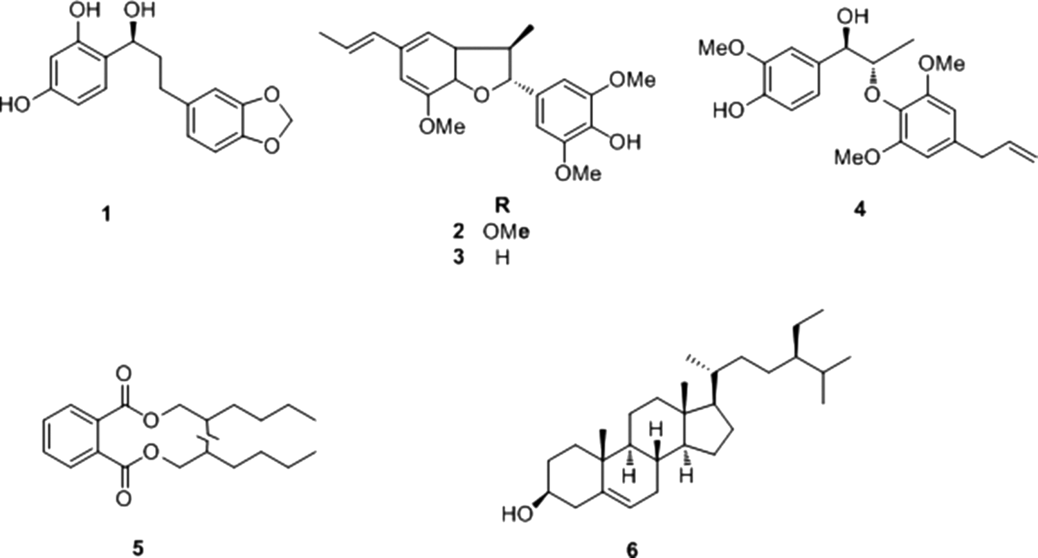
Chemical structures of compounds
(benzo[3′,4′]dioxol-1′-yl)-7-hydroxypropyl)benzene-2,4-diol (1 )
C16H16O5; Colorless oil; EI-MS m/z: 288[M]+; 1H-NMR (400 MHz, CDCl3, δ, ppm, J/Hz): 6.38 (1H, d, J = 6.8 Hz, H-5), 6.80 (1H, d, J = 8.0 Hz, H-5′), 6.86 (1H, d, J = 8.0 Hz, H-6), 6.92 (1H, d, J = 8.0 Hz, H-6′), 4.93 (1H, d, J = 9.6 Hz, H-7), 2.12 (1H, m, H-8), 1.98 (1H, m, H-8), 2.85 (1H, m, H-8′), 2.70 (1H, d, J = 1.6 Hz, H-8′), 5.96 (2H, s, O-CH2-O). 13C-NMR (100 MHz, CDCl3, δ, ppm): 135.7 (C-1), 156.0 (C-2), 103.7 (C-3), 156.0 (C-4), 108.3 (C-5), 130.3 (C-6), 77.9 (C-7), 30.2 (C-8), 114.3 (C-1′), 106.9 (C-2′), 148.0 (C-3′), 147.4 (C-4′), 108.3 (C-5′), 119.7 (C-6′), 25.4 (C-8′), OCH2O (101.2).
Odoratisol A (2 )
C21H24O5; Yellow oil; EI-MS m/z: 356 [M]+; 1H-NMR (400 MHz, CDCl3, δ, ppm, J/Hz): 6.67 (2H, s, H-2,6), 5.06 (1H, d, J = 9.6 Hz, H-7), 6.34 (1H, d, J = 16.0 Hz, H-7′), 3.45 (1H, m, H-8), 6.08 (1H, m, H-8′), 1.38 (3H, d, J = 6.8 Hz, H-9), 1.86 (3H, d, J = 6.0 Hz, H-9′), 3.89 (6H, s, MeO-3, and MeO-5), 3.88 (3H, s, MeO-3′). 13C-NMR (100 MHz, CDCl3, δ, ppm): 133.2 (C-1), 103.5 (C-2), 147.1 (C-3), 134.9 (C-4), 147.1 (C-5), 103.5 (C-6), 93.9 (C-7), 45.6 (C-8), 17.5 (C-9), 132.2 (C-1′), 109.4 (C-2′), 144.1 (C-3′), 146.5 (C-4′), 134.9 (C-5′), 113.3 (C-6′), 131.2 (C-7′), 123.4 (C-8′), 18.3 (C-9′), 56.3 (MeO-3), 56.3 (MeO-5), 53.4 (MeO-3′).
Dehydrodiisoeugenol (3 )
C20H22O4; Yellow crystalline; EI-MS m/z: 326 [M]+; 1H-NMR (400 MHz, CDCl3, δ, ppm, J/Hz): 1.39 (3H, d, J = 6.4 Hz, 3-Me), 1.87 (3H, d, J = 6.0 Hz, H3-10), 3.45 (1H, t, H-3), 3.88 (3H, s, 3′-OMe), 3.89 (3H, s, 7-OMe), 5.08 (1H, d, J = 9.2 Hz, H-2), 5.58 (1H, s, 4′-OH), 6.11 (1H, m, H-9), 6.37 (1H, d, J = 15.6 Hz, H-8), 6.76 (1H, s, H-6), 6.78 (1H, s, H-4). 13C-NMR (100 MHz, CDCl3, δ, ppm): 94.1 (C-2), 45.2 (C-3), 131.4 (C-3a), 112.1 (C-4), 130.4 (C-5), 109.4 (C-6), 144.3 (C-7), 146.7 (C-7a), 130.9 (C-8), 123.6 (C-9), 17.8 (C-10), 132.4 (C-1′), 103.5 (C-2′), 147.2 (C-3′), 130.4 (C-4′), 146.7 (C-5′), 103.5 (C-6′), 17.7 (C3-Me), 56.1 (3′-OMe), 56.3 (7-OMe).
Erythro-2-(4-allyl-2,6-dimethoxyphenoxy)-1-(4-hydroxy-3-rnethoxyphenyl)propan-1-ol (4 )
C21H26O6; Yellow oil; EI-MS m/z: 374 [M]+; 1H-NMR (400 MHz, CDCl3, δ, ppm, J/Hz): 1.12 (3H, d, J = 6.4 Hz, γ-H × 3), 3.36 (2H, d, J = 6.8 Hz, α′-H × 2), 3.87 (6H, s, MeO × 2), 3.89 (3H, s, MeO), 4.33 (1H, q, β-H), 4.80 (1H, brs, OH), 5.10–5.15 (2H, t, γ′-Hb, and γ′-Ha), 5.84–6.03 (1H, m, β′-H), 6.46 (2H, s, 3′-H, and 5′-H), 6.67–6.97 (3H, ABX, 2-H, 5-H, and 6-H). 13C-NMR (100 MHz, CDCl3, δ, ppm): 133.2 (C-1), 108.8 (C-2), 146.6 (C-3), 146.6 (C-4), 114.0 (C-5), 119.0 (C-6), 82.5 (C-α), 73.0 (C-β), 12.9 (C-γ), 136.3 (C-1′), 153.6 (C-2′), 105.7 (C-3′), 132.2 (C-4′), 105.7 (C-5′), 153.6 (C-6′), 40.7 (C-α′), 137.2 (C-β′), 116.3 (C-γ′), 56.1 (MeO × 3).
Bis(2-ethylhexyl)phthalate (5 )
C24H38O4; Yellow oil; EI-MS m/z: 390 [M]+; 1H-NMR (400 MHz, CDCl3, δ, ppm): 7.69 (2H, q, H-3,6), 7.54 (2H, q, H-4,5), 4.23 (4H, m, H-1′), 1.70 (2H, m, H-2′), 1.44 (4H, m, H-1″), 1.35 (4H, m, H-3′), 1.32 (4H, m, H-4′), 0.94 (6H, m, H-2″), 0.88 (6H, m, H-6′). 13C-NMR (100 MHz, CDCl3, δ, ppm): 167.9 (COO-), 132.7 (C-1,2), 131.0 (C-4,5), 129.0 (C-3,6), 68.3 (C-1′), 38.9 (C-2′), 30.5 (C-3′), 29.1 (C-4′), 23.9 (C-1″), 23.1 (C-5′), 14.2 (C-6′), 11.1 (C-2″).
β-Sitosterol (6 )
C29H50O; White needle crystals; mp 138–141°C; EI-MS m/z: 414.7 [M]+; 1H-NMR (CDCl3, 400 MHz) δ:5.36 (1H, brs, H-6), 3.52 (1H, m, H-3α), 0.68 (3H, s, CH3-18), 0.80 (3H, s, CH3-27), 0.82 (3H, d, J = 8 Hz, CH3-26), 0.86 (3H, s, CH3-29), 0.93 (3H, d, J = 8 Hz, CH3-21), 1.01 (3H, s, CH3-19). 13C-NMR (CDCl3, 100 MHz) δ:37.4 (C-1), 32.1 (C-2), 72.0 (C-3), 42.5 (C-4), 140.9 (C-5), 121.9 (C-6), 31.8 (C-7), 32.1 (C-8), 50.3 (C-9), 36.7 (C-10), 21.2 (C-11), 39.9 (C-12), 42.5 (C-13), 56.9 (C-14), 24.5 (C-15), 28.4 (C-16), 56.2 (C-17), 12.0 (C-18), 20.0 (C-19), 36.3 (C-20), 19.5 (C-21), 34.1 (C-22), 26.3 (C-23), 46.0 (C-24), 29.3 (C-25), 18.9 (C-26), 19.2 (C-27), 23.2 (C-28), 12.1 (C-29).
The effects on blood coagulation time
Compared with the blank group, compounds
The value of TT showed that compounds
Mean value ± standard error of mean. Compared with Blank (0.01 < a P < .05, 0.001 < b P < .01, c P < .001); Compared with Breviscapine (0.01 < d P < .05, 0.001 < e P < .01, f P < .001); Compared with vitamin K1 (0.01 < g P < .05, 0.001 < h P < .01, i P < .001).
: significant difference.
: very significant difference.
: highly significant difference.
Urokinase fibrinolytic activity assay
The fibrin plate assay was used to assay the urokinase fibrinolytic activity (Fig. 2). There was a good linear relationship between the urokinase activity and dissolved ring indicating the mass concentration (3.125–50 IU·mL−1). The thrombolysis properties are also revealed in Figure 2.
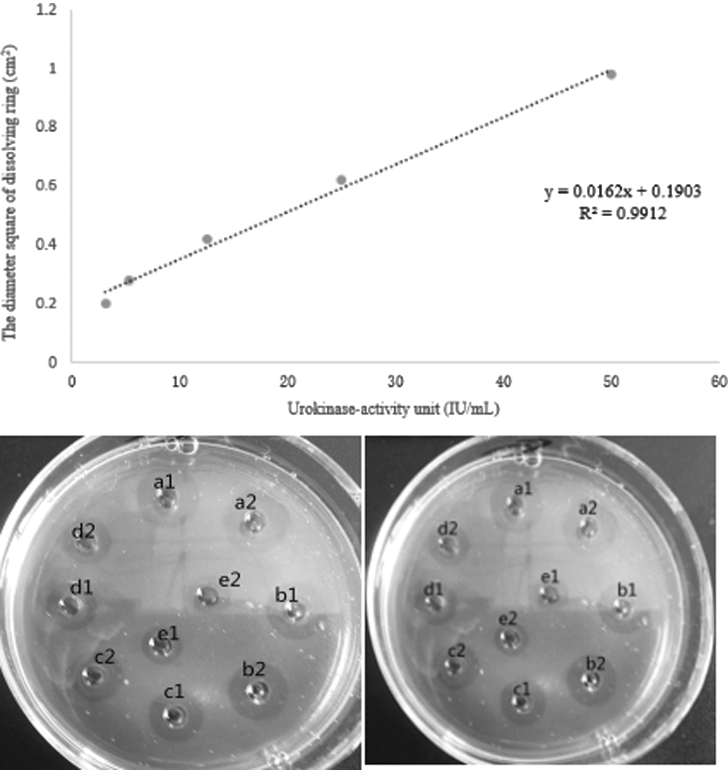
The urokinase activity standard curve and the fibrin plate images a1, a2: 50 IU•mL-1; b1, b2: 25 IU•mL-1; c1, c2: 12.5 IU•mL-1; d1, d2: 6.25 IU•mL-1; e1, e2: 3.125 IU•mL-1.
The series of Compound Danshen tablet concentrations and its fibrinolytic inhibitions are presented in Table 2. Origin 8.0 software was used to make a scatter diagram and mark the half-maximal inhibitory concentration (IC50) value of 6.577 mg/mL. The fibrinolytic inhibition activity was described in the fibrin plate (Fig. 3).

Fibrinolytic activity inhibition by complex tablets in the fibrin plate a1, a2: 1.875 mg/mL; b1, b2: 3.75 mg/mL; c1, c2: 7.5 mg/mL; d1, d2-15 mg/mL; e1, e2: blank control.
NT, unable to provide data or calculation.
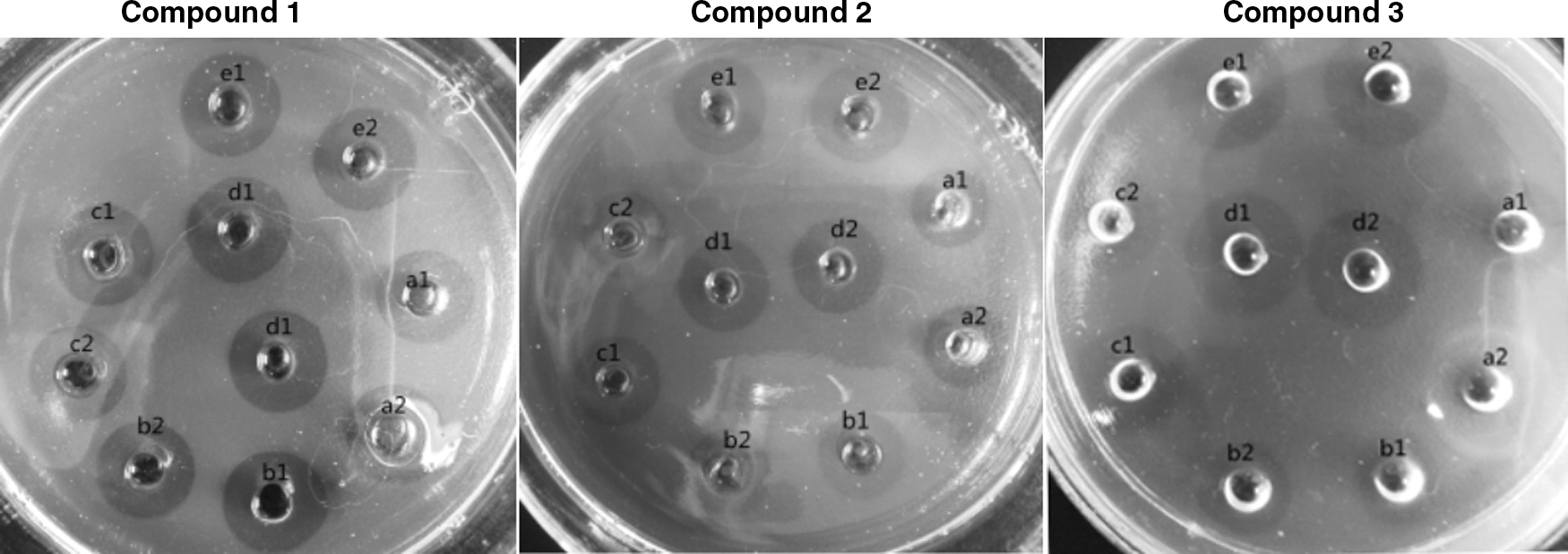
The fibrinolytic activity from M. fragrans
Three compounds did not show any fibrinolytic activity (Fig. 4).

Fibrinolytic activity from Myristica fragrans in the fibrin plate a1, a2: urokinase; b1, b2: blank control; c1, c2: compound 1; d1, d2: compound 2; and e1, e2: compound 4.
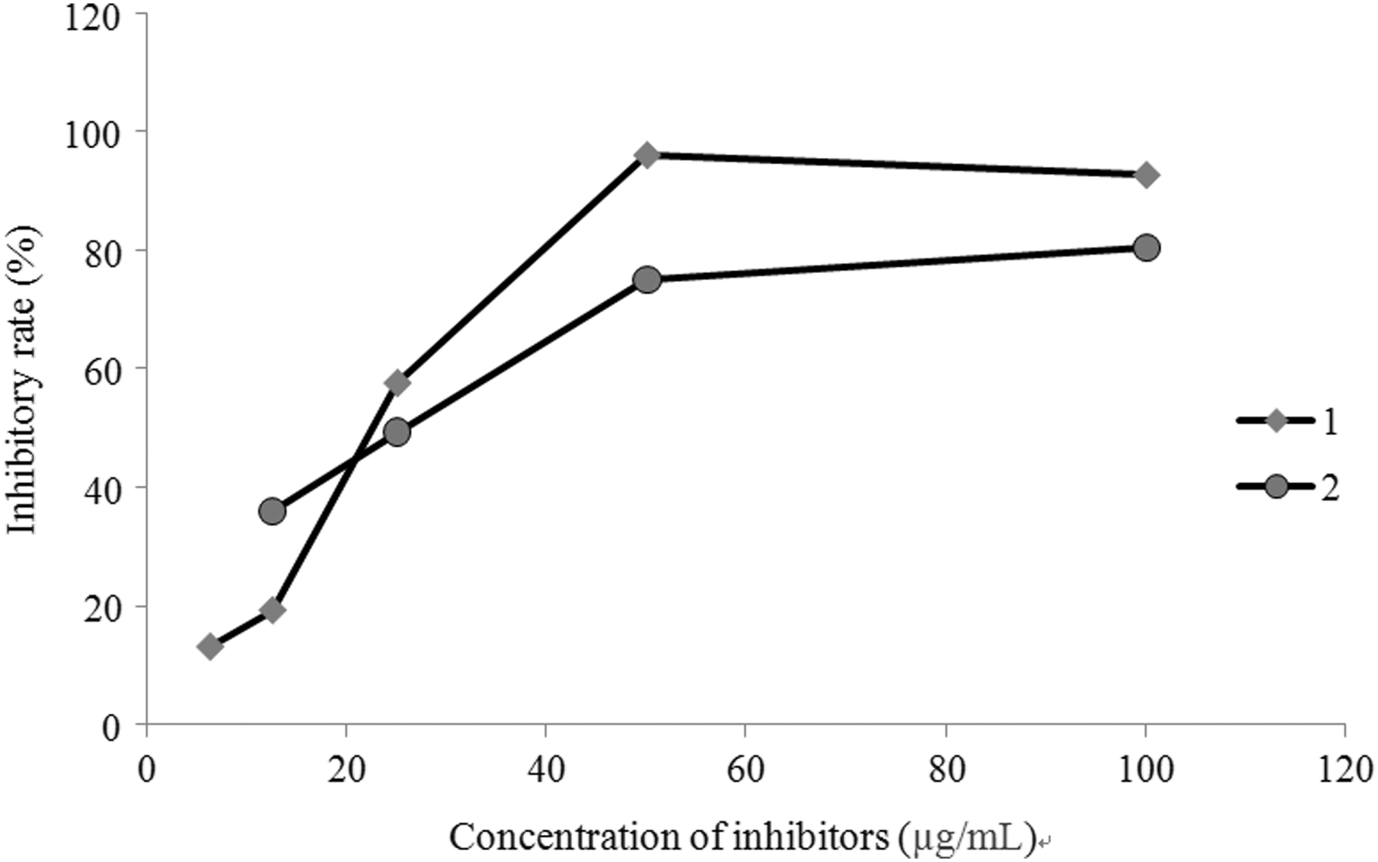
The fibrinolytic inhibition activity from M. fragrans
According to the method, prescreening fibrinolytic inhibition rates of the three compounds exceeded 50% and entered into secondary screening (Fig. 5; Table 3).

Prescreening fibrinolytic inhibition activity from Myristica fragrans in the fibrin plate a1, a2: blank control; b1, b2: compound 1; c1, c2: compound 2; d1, d2: compound 4; and e1, e2: complex tablets.
All three compounds had high inhibitory effects on the fibrinolytic pathway activated by urokinase. Compound
Secondary screening of fibrinolytic inhibition activity from Myristica fragrans in the fibrin plate a1, a2: 1/2 initial concentration; b1, b2: 1/4 initial concentration; c1, c2: 1/8 initial concentration; d1, d2: 1/16 initial concentration; and e1, e2: blank control.
α-Glucosidase inhibitory activity
Compounds
Effect at different compound concentrations of Myristica fragrans on α-glucosidase inhibitory activity.
Prescreening concentration 100 μg·mL−1.
NT, not available because of low activity (I% < 50%).
Discussion
The body's blood clotting response is normally precisely controlled by three systems: (1) serine protease inhibitors, such as the plasminogen activator inhibitor; (2) the protein C/S anticoagulant system; and (3) surface binding and coagulation inhibitors. 39 Urokinase as one plasminogen activator launches the fibrinolytic function by activating plasminogen into plasmin. 32 Figure 8 shows the schematic diagram of blood coagulation.
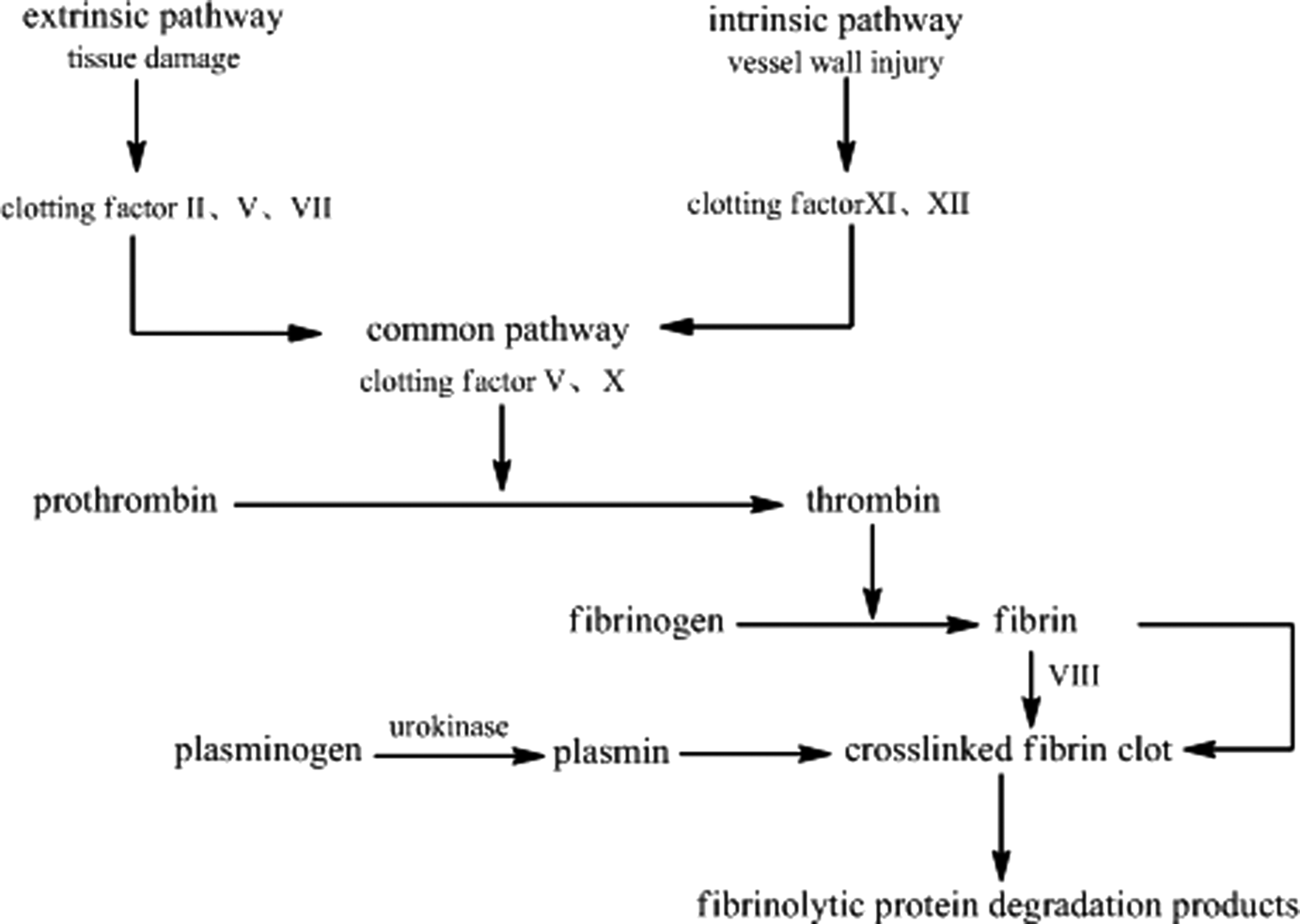
Schematic diagram of blood coagulation.
The three active compounds identified in the study belong to phenol and lignan, however, no studies have been conducted to investigate their procoagulant effects. On the contrary, several studies have reported on the anticoagulant activity of lignans. It has been reported that the inhibitory mechanism of erythro-(7S,8R)-7-acetoxy-3,4,3′,5′-tetramethoxy-8-O-4′-neolignan (EATN) on platelet aggregation might increase cyclic adenosine monophosphate levels and subsequently inhibit intracellular Ca2+ mobilization by interfering with a common signaling pathway rather than by directly inhibiting the binding of thrombin or platelet activating factor (PAF) to their receptors. 40 Specifically, bicycle (3,2,1) octane neolignans, 8,3′-neolignans, and 8-O-4′-neolignans were demonstrated to possess potent PAF antagonistic properties. However, it also revealed that 8-O-4′-neolignan activities were not obvious, especially when C-7 of the compounds was replaced by a hydroxyl group. 41
The reason for the coagulation activity difference may be that they belong to a different type of lignans. In a previous study, three phenolic acids, vanillic acid, p-coumaric acid, and protocatechuic acid, possessed procoagulant activity. 42 Lignans are the most common constituents of M. fragrans. Our research objectives of the procoagulant ingredients in pericarps of M. fragrans and the structure–activity relationship are further discussed.
Clinically used procoagulant medicines can be divided into four categories according to their mechanisms, including promoting clotting factor activity, inhibition of fibrinolytic system, effect on blood vessels, platelets, and topical hemostatic or others, such as vitamin K, aminomethylbenzoic acid, adenosine, and thrombin, respectively. 43
Currently, various types of hemostatic agents have been explored; unfortunately, each of them has its own shortcomings. For example, the fibrinogen or thrombin is mainly obtained from animal or human blood; they are expensive and include the potential risk of viral infection. The adhesive properties of collagen and gelatin sponges to tissues are poor. Water-soluble collagen is a toxicoid, and the use of this material in human body may cause inflammatory responses. 44,45
Therefore, it is particularly important to develop safe and new hemostatic agents for clinical use with better hemostatic properties and reduced tissue response.
This is the first study to report the procoagulant mechanism of (benzo[3′,4′]dioxol-1′-yl)-7-hydroxypropyl)benzene-2,4-diol (
Pericarps peeled off after maturity, while the isolated pericarp compounds are still active, could promote coagulation, and inhibit α-glucosidase activity, This research suggests that M. fragrans pericarp has the potential to be developed into a new hemostatic therapeutic intervention that is effective, inexpensive, and derived from easily obtainable sources.
Footnotes
Acknowledgments
This work was supported by the Henan Province University Science and Technology Innovation Team (16IRTSTHN019), Industry & University Research Project in Henan Province (162107000038 and 152107000051), Kaifeng City Science and Technology Innovation Talent (1509010), National cooperation project of Henan Province (2015GH12), Key project in Science and Technology Agency of Zhengzhou City (20150341), and Science Research Foundation of Henan University (ZZJJ20140047).
Author Disclosure Statement
No competing financial interests exist.