
Abstract
Malus toringoides (Rehd.) Hughes (MT) leaves are traditionally used as a medicine for treating or preventing cardiovascular disease in Tibet. In addition to the effect of this medicinal plant on thrombosis, we tested its effect on dyslipidemia in a hypolipidemic rat model. A total of 60 healthy Sprague-Dawley rats were randomly divided into six groups, as follows: normal control, model control, simvastatin groups, and MT low-, medium-, and high-dose groups. The normal controls were fed with a normal diet, whereas all other groups were fed with a high-fat diet. After 6 weeks, the high-fat diet had induced hyperlipidemia in the rats, which were then orally administered with different doses of MT leaf extract (50, 100, and 200 mg/kg) for an additional 6 weeks. Serum levels of total cholesterol (TC), triglycerides (TG), low- and high-density lipoprotein cholesterol (LDL-c and HDL-c, respectively), as well as the antioxidant capacity of glutathione peroxidase (GSHP-x), superoxide dismutase (SOD), and malondialdehyde (MDA) were measured at the end of the study. MT significantly reduced serum TC, TG, and LDL-c and increased the HDL-c content in MT-treated rats compared with the model group. These changes were dose dependent. MT treatment also significantly elevated the activity of SOD and GSHP-x, and decreased the serum levels of MDA compared with untreated hyperlipidemic rats, thereby increasing serum antioxidant capacity. In addition, MT reduced liver steatosis in hyperlipidemic rats. Overall, MT exerts considerable hypolipidemic and antioxidant properties.
Introduction
W
The level of circulating cholesterol is associated with many pathways of cholesterol metabolism, including absorption, synthesis, clearance, and excretion; when one of these pathways is altered, the balanced cholesterol level in vivo may be disrupted. 5 Reducing the level of cholesterol in plasma by reducing cholesterol input can effectively prevent or treat atherosclerosis and CVD. Although antihyperlipidemia drugs exhibit rapid lipid-lowering effects and good efficacy, 6 the use of these drugs is restricted because of their potential side effects. 7 –9
Malus toringoides (Rehd.) Hughes (MT) is a rosaceous plant that is used as a traditional folk medicine in Tibet, China. This plant originally grows on snowy mountains at an attitude of ∼3000–3700 m and is primarily distributed in the western part of Sichuan, Gansu, Qinghai, and Tibet in China. In ancient Tibet, a concoction of fresh MT leaves was consumed as a herbal drink or medicine to treat diseases, such as hypertension, indigestion, liver injury, hyperlipidemia, and hyperglycemia. 10,11 The medicinal effects of MT are associated with the complex components of MT. Phytochemical studies have shown that MT contains flavonoids, polysaccharides, fatty acids, and amino acids, which are generally considered components that contribute biological and therapeutic activities to MT. 12 MT was recently reported to contain a special medicinally relevant flavonoid 13 that exerts antidiabetic and antioxidant effects in streptozotocin-induced diabetic rats. 14 However, to date, no studies have described the protective effects of MT in hyperlipidemic rats. This study found that MT exerts hypolipidemic and antioxidant effects. These results indicate that MT may be used to prevent hyperlipidemia and thus is possibly useful as a therapeutic agent against CVD.
Materials and Methods
Reagents and chemicals
Standards of phlorizin and phloetin were purchased from National Institute for the Control of Pharmaceutical and Biological Products (Beijing, China). Kits used to determine TC, TG, LDL-c, HDL-c, superoxide dismutase (SOD), malondialdehyde (MDA), glutathione peroxidase (GSHP-x), total antioxidant capacity (T-AOC), apolipoprotein A (ApoA), and apolipoprotein B (ApoB) were purchased from Jiancheng Biotechnology Science, Inc. (Nanjing, China). Other reagents and chemicals used were of analytical grade and obtained from Sinopharm Chemical Reagent Co., Ltd. (Shanghai, China).
Plant material and preparation of MT extract
Leaves of MT were purchased from Tibet Hongxiang Pharmaceutical Technology Co., Ltd. (Tibet, China) in March 2015, and a voucher specimen (No. Qust-1510) was deposited at the Herbarium of the Department of Pharmacy, Qingdao University of Science & Technology, China. Pulverized leaves were extracted twice with 70% EtOH under reflux for 1.5 h each round. The extract was filtered with filter paper and then extracted with petroleum ether 10 times. The petroleum ether extracts were discarded; the other solution was purified on a polyamide column and initially eluted with 2000 mL of water and then evaporated in vacuo at 60°C. The yield obtained was 1.51%.
HPLC analysis of MT
High performance liquid chromatography (HPLC) was performed on an Ultimate 3000 equipped with a sample injector, diode-assay UV/Vis detector, and Hypersil BDS C18 column (250 × 4.6 mm, 5 μm) that was used as the stationary phase. The mobile phase consisting of methanol-2% phosphoric acid (20:80) was used for elution for 36 min, and the flow rate was maintained at 1.0 mL/min. The column temperature was maintained at 30°C, and the injection volume was 20 μL. The detection wavelength was 280 nm.
Animal and experimental design
All animal experiments were performed according to the rules and regulations of the Animal Ethics Committee at Qingdao University of Science & Technology. Male Sprague-Dawley rats aged 6–8 weeks (160 ± 20 g) were purchased from Qingdao Institute for Food and Drug Control (Qingdao, China). All rats were housed individually in stainless steel cages and maintained at 22°C ± 2°C under a relative humidity of 40–60% and under a 12-h light–12-h dark cycle, with free access to food and water.
The rats were fed with commercial diet for 5 days to allow them to adapt to the experimental environment. A total of 60 rats were randomly divided into six groups (10 rats for each), namely, normal control group (NC), model control group (HFC), positive control group (PC), MT low-dosage group (LMT), MT medium-dosage group (MMT), and MT high-dosage group (HMT). The rats in NC were provided with standard chow, whereas the other groups were fed with a high-fat diet (Table 1). After 6 weeks, the rats in PC were orally administered with simvastatin at a dose of 2.5 mg/kg body weight; the three MT-treated groups were orally administered MT extract dissolved in physiological saline for 6 weeks at doses of 50, 100, and 200 mg/kg body weight. The rats in NC and HFC received the same volume of normal saline solution at the same period. At the end of the experimental period, overnight fasted rats were anesthetized with 10% chloral hydrate (0.3 mL/100 g) intra-abdominally and then sacrificed. Blood samples were collected by cardiac puncture and separated by centrifugation at 3000 g for 15 min to obtain the serum. The livers were immediately excised, weighed, and stored in a buffer solution of 10% formalin until further analysis.
Hepatic morphology
The tissue slices of liver were fixed in 10% buffered neutral formalin for 24 h and then embedded in paraffin. Thin sections (4 μm) were stained with hematoxylin and eosin, and the stained areas were viewed under an optical microscope at × 100.
Biochemical analysis
The serum concentrations of TC, TG, LDL-c, HDL-c, SOD, MDA, GSHP-x, T-AOC, ApoA, and ApoB were determined using commercially available kits (Jiancheng Biotechnology Science, Inc.) according to the manufacturer's instructions.
Statistical analysis
All data were expressed as mean ± SD and tested for normality using SPSS version 17.0 for Windows. Data were analyzed by one-way analysis of variance followed by testing differences between pairs by Tukey's range test. A P-value of <0.05 was considered statistically significant.
Results
HPLC analysis of MT
The peaks of each marker substance in the dried MT extract were identified by comparing the retention time and UV spectra of the standards of phlorizin and phloetin, respectively (Fig. 1). The extract contains two major components, phloridzin and phloretin, at 10.3% and 1.5%, respectively. Although MT was used as a traditional herbal medicine in Tibet, the pharmacological effect of MT extract has been rarely reported. We intend to extensively investigate the chemical components of MT in our future studies to identify its biological activities.
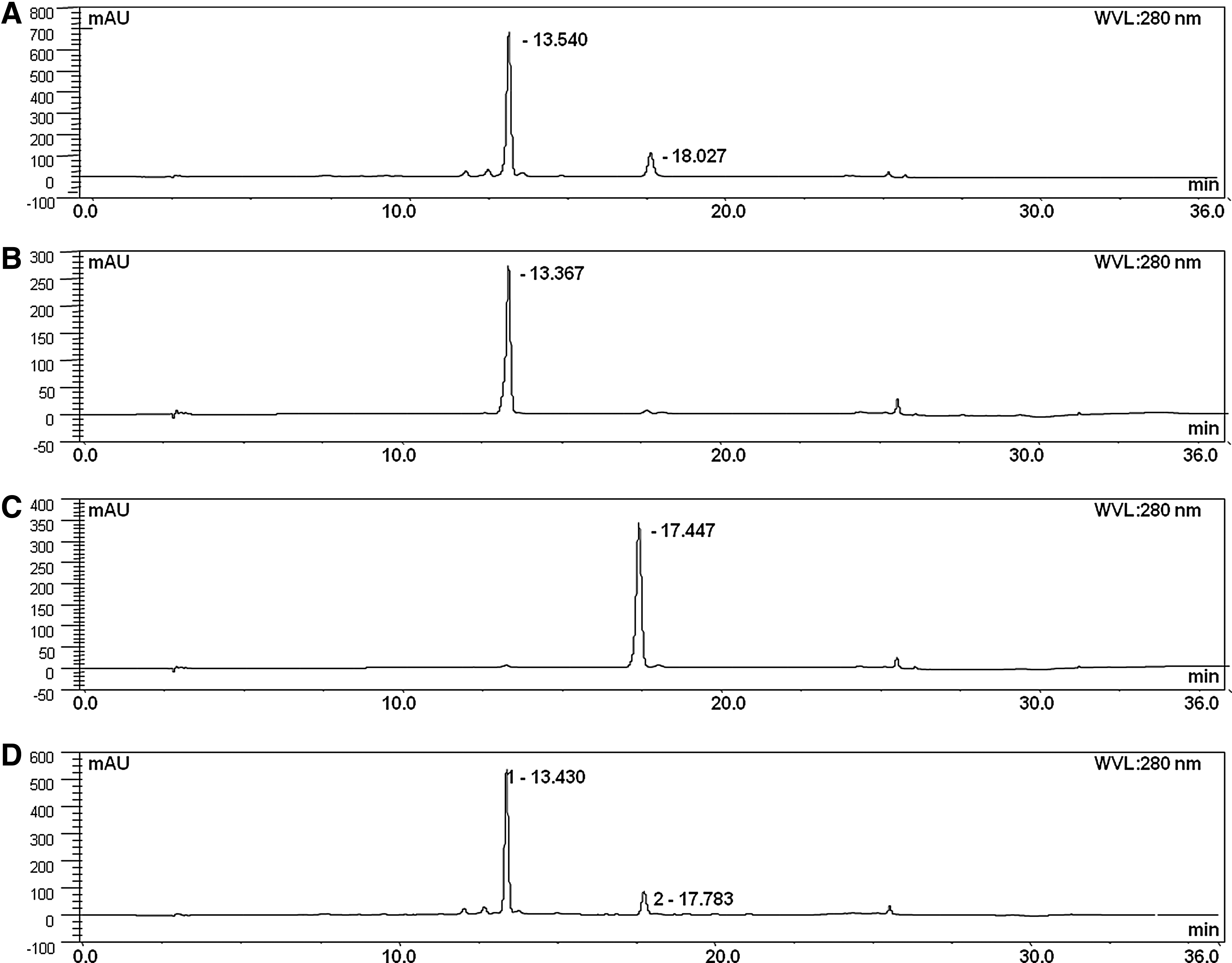
Results of high performance liquid chromatography (HPLC) analysis. Absorption peaks: Total MT extract
Effect of MT on the serum levels of lipids and apoproteins
The hypolipidemic effect of MT extract was evaluated in high-fat-diet-induced hyperlipidemic rats. The blood lipid levels among the rats across groups did not obviously vary before feeding the rats with cholesterol-rich diets. After induction with a high-fat diet, the hyperlipidemic diet apparently altered the serum lipid concentrations and thus markedly causing hyperlipidemia, resulting in significantly elevated blood levels of TC, TG, and LDL-c, and in decreased serum HDL-c levels (Table 2).
Values in the same column with different superscript letters indicate significant difference between groups (P < 0.05) by the Tukey's test.
HDL-c, high-density lipoprotein cholesterol; HFC, model control group; HMT, MT high-dosage group; LDL-c, low-density lipoprotein cholesterol; LMT, MT low-dosage group; MMT, MT medium-dosage group; MT, Malus toringoides (Rehd.) Hughes; NC, normal control group; PC, positive control group; TC, total cholesterol; TG, triglycerides.
After the three MT-treated rat groups were orally administered with different MT doses for 6 consecutive weeks, the TC, TG, and LDL-c levels in the blood were lower (P < .05) than those in the HFC, whereas their HDL-c level was higher (P < .05) than that in the hyperlipidemic rats not treated with MT extract. Although the three MT-treated groups exerted a significant decrease in the level of serum TG compared with HFC, no significant difference was found among MT-treated groups. The TG-lowering effect was similar between MT-treated groups and PC. The serum levels of TC in LMT were similar to PC, but higher (P < .05) than those levels in the MMT and HMT. HMT had the lowest (P < .05) LDL-c levels, and the levels were also lower (P < .05) than PC, but the LDL-c levels in PC were similar to LMT and MMT. The HMT had higher (P < .05) HDL-c levels compared with LMT and MMT, but were similar to PC.
Table 3 shows that the serum levels of ApoA in the HFC group were obviously lower than in the NC group, whereas the ApoB levels were higher in the NC group (P < .05). However, after treatment of the rats with different doses of MT extract, the ApoA and ApoB levels significantly changed compared with those in the HFC group. The serum ApoA levels were higher for all groups orally administered with MT compared with HFC. HMT had higher (P < .05) ApoA levels compared with LMT, and those levels were not different from MMT and PC. The three MT-treated groups had similar serum levels of ApoB compared with PC, but HMT and MMT had lower P < .05) ApoB levels than HFC.
Values in the same column with different superscript letters indicate significant difference between groups (P < 0.05) by the Tukey's test.
MT showed significantly decreased serum in the levels of TG, TC, LDL-c, and ApoB and significantly increased in the serum levels of HDL-c and ApoA compared with HFC (P < .05); the above-mentioned changes were dose dependent, and only the TC was significantly different. Therefore, the high-dose MT (200 mg/kg) showed considerable hypolipidemic effect.
Effect of MT on liver function
Figure 2 shows the result of histological examination of rat liver. The slices obtained from the control rats fed with standard diet exhibited grossly pink staining and their cell structures (Fig. 2A) were normal compared with those in the HFC group. By contrast, the liver of those rats in HFC showed significant changes in morphology and fat content, as well as grossly white coloration (Fig. 2B). Lipid dispositions were extensively distributed in nearly all hepatocytes, and greater number of lipid vacuoles was observed in HFC than in NC. When the hyperlipidemic rats received MT treatment for 6 consecutive weeks, the lipid disposition in the hepatocytes of LMT (Fig. 2D) and MMT (Fig. 2E) was obviously less. The lipid vacuoles were replaced by cytoplasm. The nucleus gradually showed a central location. No significant differences were found between LMT and MMT. When the dose of MT extract was increased to 200 mg/kg body weight, the deposited lipid in the cells nearly completely disappeared (Fig. 2F) and the liver sections exhibited a normal hepatic histological appearance similar to that in the control groups and positive control groups (Fig. 2C). Therefore, the high-dose MT exhibited a considerable hypolipidemic effect in MT-treated groups.
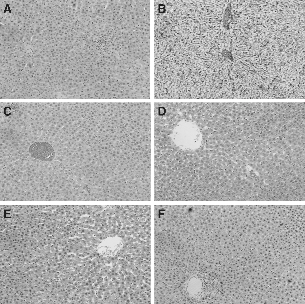
Morphological change in hepatic tissue in high-fat-diet-induced hyperlipidemic rats. Few lipid vacuoles were observed in the liver of NC
Effect of MT on antioxidant activity in serum
Table 4 shows that the antioxidant capacity of the rats in HFC declined and is characterized by lower levels of SOD, T-AOC, and GSHP-x, as well as higher MDA levels than in NC. After treatment with MT extract for 6 consecutive weeks in HFC, the activity of SOD and GSHP-x was significantly elevated, the MDA level was decreased (P < .05), and the T-AOC increased (P < .05) compared with HFC untreated with MT. Although the antioxidant capacity of MT was dose dependent, it was not significantly different among MT-treated groups. Both MMT and HMT had similar serum levels of SOD to PC, but these levels were higher (P < .05) than LMT. In addition, all MT-treated groups had similar serum levels of T-AOC, GSHP-x, and MDA compared with PC, but high-dose MT (200 mg/kg) exerted greater antioxidant capacity in MT-treated groups, which demonstrated the considerable antioxidant capacity of MT.
Values in the same column with different superscript letters indicate significant difference between groups (P < 0.05) by the Tukey's test.
GSHP-x, glutathione peroxidase; MDA, malondialdehyde; SOD, superoxide dismutase; T-AOC, total antioxidant capacity.
Discussion
This study established a hyperlipidemic rat model by feeding the rats with foods rich in cholesterol and saturated fats to evaluate the hypolipidemic potential and antioxidant status of the ethanol extract of MT. The MT extract exhibited considerable antihyperlipidemic and antioxidant effects. Table 2 shows that the rats fed with a high-fat diet displayed a higher plasma concentration of TC, TG, and LDL-c, as well as lower concentration of serum HDL-c, than the control group maintained on a standard chow. This result indicates that a high-fat diet can increase the incidence of hyperlipidemia, consistent with previous findings. 15 High concentrations of blood lipid contributes to the increase in the incidence of hyperlipidemia and in the subsequent onset of CVD. 16 Moreover, LDL-c is associated with the cholesterol deposit in arteries and aorta that culminates in coronary heart diseases. 17 HDL-c concentrations are speculated to reflect the removal rate of excess peripheral cholesterol, and increased serum HDL-c is therefore associated with reduced risk of atherosclerosis. 18 Increasing HDL-c concentration potentially contributes to antiatherogenesis possibly through the ability of HDL-c to inhibit LDL-oxidation and through its capacity to protect endothelial cells from the cytotoxic effects of oxidized LDL-c. 19 When the rats received a diet rich in cholesterol and saturated fats supplemented with MT extract, the serum concentrations of TC, TG, and LDL-c significantly decreased, whereas the plasma level of HDL-c increased. In addition, the MT extract could reduce the accumulation of hepatic lipid droplets, indicating that the MT extract can lower the serum lipid profiles and accumulation of lipid droplets in hepatic tissue cells, and can enhance the HDL-c level, demonstrating the potential of MT extract as therapeutic agent against hyperlipidemia and as preventive agent against CVD. These effects of MT extracts can be associated to its components, namely, phlorizin and phloetin, which possess an antioxidant effect. 20,21
Oxidative stress is a causative factor that links hyperlipidemia to the pathogenesis of atherosclerosis. 22,23 Oxidative stress occurs when free radical production exceeds the capacity of the natural antioxidant system. 24 Feeding rats with a high-fat diet can promote production of free radicals, increasing the amount of lipid peroxides and the formation of reactive oxygen species (ROS), which contains superoxide anion radical, hydroxyl radical, and lipid peroxyl radicals that can cause irreversible damage to cellular macromolecules, including membrane lipids. 25 In the human body, the antioxidant enzyme system mainly includes two factors, SOD and GSHP-x, which serve as the primary defense systems against ROS generated in vivo during oxidative stress. SOD exists at high amounts in all cells and can disrupt superoxide anion radical, whereas the GSHP-x activity contributes to the easy detoxification of H2O2 in the plasma; these antioxidant enzymes act cooperatively at different sites in the metabolic pathway of free radicals to prevent oxidative damage. 26 MDA is the main product of lipid peroxidation and an indicator of lipid peroxidation. Low MDA level suggests that less lipid peroxidation occurs and weak oxidant stress exists. 27 This research found that the MT extract could significantly increase SOD and GSHP-x levels and reduce MDA content in hyperlipidemic rats, indicating that the MT extract could reduce oxidative stress, resulting in low lipid peroxidation in serum. In addition, the MT extract exhibits a considerable hypolipidemic effect that is associated with its antioxidant potential.
This study indicates that the MT extract can lower lipid concentrations in serum and in organs of high-fat-diet-induced hyperlipidemic rats. In addition, MT increased the activity of antioxidant enzymes and decreased lipid peroxidation, possibly preventing the progress of hyperlipidemia; this effect is related to high antioxidant capacity of MT extract. Our results suggest that MT is a potential food additive or pharmaceutical agent to treat or prevent hyperlipidemia.
Footnotes
Acknowledgment
The project was supported by the National Natural Science Foundation of China (Grant Nos. 81360686 and 31500288).
Author Disclosure Statement
No competing financial interests exist.