
Abstract
Inflammatory bowel disease, including Crohn's disease and ulcerative colitis (UC), is a group of inflammatory conditions of the colon and small intestine. UC is a chronic inflammatory disorder of the colon and rectum that includes intervals of acute exacerbation. Although recent studies have suggested that proinflammatory cytokines might have initiated the inflammatory responses in UC, its etiology remains unclear. Aronia berries are rich in dietary polyphenols such as phenolic acids, anthocyanins, flavonoids, and proanthocyanidins with various health benefits, including antioxidant, anti-inflammatory, and antiaging activities. The objective of this study was to determine whether Aronia berry can be an effective intervention for the treatment of UC. BALB/c mice were administered 5% dextran sulfate sodium (DSS) to induce UC. They were then given Aronia berry extracts at concentrations of 10 or 100 mg/kg. During the induction of UC, the expression levels of nuclear factor-kappa B were increased in colonic epithelial cells and immune cells, leading to increased proinflammatory cytokine levels. Aronia berry extract significantly improved the clinical signs of DSS-induced UC, including body weight loss, colon length shortening, and disease activity index increase, with histological markers of colon injury. Furthermore, oral administration of Aronia berry extract inhibited prostaglandin E2 production in DSS-induced colitis and decreased the levels of nitric oxide, interleukin-6, and tumor necrosis factor-α in lipopolysaccharide-stimulated macrophages. These results suggest that Aronia berry extract could efficiently ameliorate clinical signs and inflammatory mediators of UC. Therefore, Aronia berry might be a promising natural treatment for UC.
Introduction
I
Aronia berries (chokeberries) in the Rosaceae family are perennial herbs native to North America. 5 Recently, Aronia extracts have gained a lot of attention in Korea and farmers have increased production of Aronia species. Chokeberries have three species: red chokeberry (Aronia arbutifolia), black chokeberry (A. melanocarpa), and purple chokeberry (A. prunifolia). Black chokeberries have been used as food and medicine. 6 Aronia berries have several chemical components, including phenolic acids, anthocyanins, flavonoids, and proanthocyanidins. In particular, Aronia berries are known to have a higher content of anthocyanin than other fruits. 7 Aronia berries also contain many tannins. Therefore, it is highly astringent when eaten raw. Aronia berries are usually taken as a powder or extract. Anthocyanins in Aronia berries have remarkable antioxidant and anticancer effects. 8 Some studies have reported that anthocyanins can be used to treat cardiovascular diseases such as stroke and heart disease. 9
Berries have shown promise in the treatment of chronic inflammatory diseases. For example, black chokeberry can decrease the expression levels of nuclear factor-kappa B (NF-κB) p65 in dextran sulfate sodium (DSS)-administered mice as an anti-inflammatory agent. 10 Aronia berry extract can also inhibit the secretion of interleukin-6 (IL-6) in lipopolysaccharide (LPS)-induced murine splenocyte, but induce the secretion of IL-10. 11
Although the etiology of UC remains unknown, recent studies have suggested that an inflammatory response initiated by the interaction between the immune system (including macrophages and dendritic cells) and antigens is involved. 12 The inflammatory response in UC is induced by the activation of macrophages by bacterial products such as LPS. Activated macrophages will release proinflammatory cytokines such as IL-1β, tumor necrosis factor-α (TNF-α), and IL-6. Patients with UC have been reported to express high levels of inflammatory cytokines such as IL-6 and TNF-α. 13
Although Aronia berries are known to have various health benefits, whether they have beneficial effect on UC remains unclear. Therefore, the objective of this study was to determine whether Aronia berry could inhibit clinical signs and inflammatory mediators in DSS-induced UC.
Materials and Methods
Reagents
DSS (mol wt: 36,000–50,000) was purchased from MP Biochemicals (Solon, OH, USA). Anti-phospho-JNK, anti-p38, and anti-NF-κB antibodies were purchased from Cell Signaling Technology (Woodland, CA, USA). Anti-mouse IL-6 and TNF-α antibodies were purchased from BD Pharmingen (San Diego, CA, USA). Anti-JNK and anti-NF-κB antibodies were purchased from Santa Cruz Biotechnology (Santa Cruz, CA, USA). Anti-GAPDH antibody was purchased from Thermo Scientific (Pittsburgh, PA, USA). Hematoxylin was purchased from Muto Pure Chemicals Co. (Bunkyo-ku, Tokyo, Japan). Eosin was purchased from Sigma-Aldrich (St. Louis, MO, USA). Prostaglandin E2 (PGE2) assay kits were purchased from Enzo Bioscience (Farmingdale, NY, USA).
Experimental animals
All experimental protocols (CBNU2016-0016) were approved by the Committee on the Care of Laboratory Animal Resources, Chonbuk National University. All experiments were conducted in accordance with the Guide for the Care and Use of Laboratory Animals. Male BALB/c mice (8 weeks old, 21–25 g) and C57BL/6 mice (5 weeks old, 19–20 g) were purchased from Samtako (Osan, Korea). These mice were group-housed under a controlled temperature (25–27°C) with a 12 h light–dark cycle. They were provided free access to a standard diet and tap water (or specified drinking solution). All mice were acclimated under these conditions for at least 7 days before inclusion in experiments. Mice were sacrificed on day 10, and their colons were removed. After length measurement, the colons were cut into small pieces (about 2–3 cm in length) and kept in 15 mL tubes. The tips of colons were fixed in formalin for histological analysis.
Extraction of Aronia
Aronia berries were purchased from cooperative farmer's market in Sunchang village (Jeonbuk, Korea). They were washed twice with distilled water and dried. Aronia berry extract was prepared by decocting with 70% ethanol for 1 h 30 min at 80°C. The solution in ethanol was then filtered and allowed to evaporate using a rotary evaporator at a temperature of 40–45°C. The extract was diluted in 0.9% saline and filtered through a 0.22 μm syringe filter (HYUNDAI Micro, Seoul, Korea).
Induction of colitis with DSS
Mice were separated into five groups: CON (normal), DSS (5% w/v DSS), AR10 (5% DSS with oral administration of Aronia 10 mg/kg), AR100 (5% DSS with oral administration of Aronia 100 mg/kg), and ASA (5% DSS with oral administration of 5-ASA). The DSS groups were given 5% DSS (MP Biochemicals) in drinking water for 7 days. Body weight was monitored daily.
Disease activity index
Diarrhea with blood and mucus are used to clinically identify UC.
14
Disease activity index (DAI) scores were used to calculate intestinal disease activity in mice as described in Table 1.
15
Enzyme-linked immunosorbent assay
At the end of experiment, serum samples were prepared and subjected to enzyme-linked immunosorbent assay (ELISA) to determine serum levels of IL-6 and TNF-α using ELISA kits from BD Pharmingen. In brief, microwells were coated with 100 μL per well of capture antibodies and incubated at 4°C overnight. After aspiration of the media, 100 μL of each standard or sample was added into each well and the plates were incubated at room temperature (RT) for 3 h. Plates were then washed and 100 μL of Working Detector (Detection Ab + SAv-HRP) was added to each well. Plates were incubated at RT for 1 h. Then, 100 μL of substrate solution was added to each well, and plates were incubated at RT for 30 min in the dark. Absorbance at 450 nm was measured using a VersaMax microplate reader (Molecular Devices, Sunnyvale, CA, USA).
Western blot analysis
Colons were homogenized in lysis buffer (iNtRON Biotech, Seongnam-Si, Republic of Korea) and centrifuged by high speed centrifuge 1248R (Labogene, Daejeon, Republic of Korea) at 16,582 g for 10 min at 4°C. The supernatants were collected and transferred to fresh tubes. Protein concentrations were quantified using the BCA protein assay reagent (Sigma-Aldrich). Lysates (50 μg of protein) were separated by 10% sodium dodecyl sulfate-polyacrylamide gel electrophoresis and transferred to polyvinylidene difluoride membranes (Roche Diagnostics, Indianapolis, IL, USA). After blocking with 5% skim milk in phosphate-buffered saline-Tween-20 (PBST) for 1 h at RT, membranes were incubated with primary antibodies against mitogen-activated protein kinases (MAPK), NF-κB, and phospho-IκB at 4°C overnight. After washing three times with PBST, membranes were then incubated with secondary antibodies at RT for 1 h. After washing again with PBST three times, the protein-antibody complexes were then developed with ECL western blotting luminol reagent (Santa Cruz Biotechnology) and recorded with an LAS-4000 Image Reader (Fujifilm Life Sciences, Tokyo, Japan).
Histological analysis
Cross sections of the distal colons (2 cm) were fixed in 10% paraformaldehyde solution. They were then cut into small fragments, dehydrated in an ethanol series (70–100%), washed with xylene, and embedded in paraffin. The fragments were sliced into 5 μM-thick sections and stained with hematoxylin-eosin (H&E) stain. These sections were visualized with an optical microscope equipped with a camera to capture images at 100 × magnification.
PGE2 assay
Whole mouse colons were washed with cold PBS and immediately stored at −80°C until use. Colon tissues were homogenized in 1 mL ice-cold lysis buffer (10 mM Tris-HCI [pH 7.4], 1 mM ethylenediaminetetraacetic acid, 10 μM indomethacin, and 1% protease and phosphatase inhibitor cocktail [Sigma-Aldrich]) using a homogenizer. Tissue homogenates were transferred to microcentrifuge tubes, vortexed, and centrifuged at 4°C for 10 min at 16,582 g. The supernatant (5 μL) from each sample was used to determine protein concentration using a DC Protein Assay Kit (Bio-Rad, Hercules, CA, USA) according to the manufacturer's protocol. Determination of PGE2 levels was carried out using a PGE2 Monoclonal Enzyme Immunoassay Kit (Enzo Bioscience). All samples were diluted by Assay Buffer in 1:100. Experiments were run in triplicates and a standard curve was created at the same time. Absorbance at 405 nm was measured using the VersaMax microplate reader.
Isolation and culture of peritoneal macrophages
Mice were given an intraperitoneal injection of 4% thioglycolate solution (3 mL) and sacrificed on the third day after injection. Peritoneal cavities were rinsed with Dulbecco's Modified Eagle Medium (DMEM) (5 mL). The peritoneal lavage fluid was centrifuged at 4000 rpm for 5 min. Cells were then resuspended in DMEM and seeded into 24-well microplates. These cells were cultured in 24-well plates at 37°C for 24 or 48 h in DMEM plus 10% fetal bovine serum.
Cell viability assay
To determine cell viability, thiazolyl blue tetrazolium bromide (MTT) was used. Mice peritoneal macrophages were pretreated with Aronia berry extract (0.001, 0.01, 0.1, or 1 mg/mL) for 24 h. MTT solution at final concentrations of 0.5 mg/mL was then added to each well and incubated for 4 h. After the incubation, the supernatant was removed, and the formazan crystals were dissolved in 500 μL of dimethyl sulfoxide. The absorbance of each well at wavelength of 540 nm was then measured using the VersaMax microplate reader.
Measurement of nitric oxide level
Mice peritoneal macrophages were pretreated with Aronia berry extract (0.001, 0.01, 0.1, or 1 mg/mL) for 1 h. After that, they were treated with 1 μg/mL of LPS and incubated for 48 h. After the incubation, the supernatants were collected for nitric oxide (NO) determination. To measure nitrite, an equal volume of Griess reagent (1% sulfanilamide/0.1% naphtylethyenediamine dihydrochloride in 2.5% H3PO4) was mixed with cell culture supernatant at RT for 10 min. Nitrite concentration was determined by measuring the absorbance at wavelength of 540 nm using the VersaMax microplate reader. NaNO2 was used to obtain the standard curve.
Statistical analysis
Results are shown as a summary of data from at least three experiments. Data are presented as means ± standard errors of the mean. Statistical evaluation of results was performed by independent t-test. For all experiments, statistical significance was defined when P-value was <.05.
Results
Effect of Aronia berry extract on clinical signs of UC induced by DSS
To measure the effect of Aronia berry extract on UC, Aronia berry extract was given to an animal model of UC induced by DSS, and changes in body weight were measured. Mice were administered DSS followed by Aronia berry extract at a concentration of 10 or 100 mg/kg for 10 days. 5-ASA was used as a positive control. As shown in Figure 1, the greatest weight loss was observed in the group administered with DSS alone. In the AR10 and AR100 groups, body weight loss was significantly (P < .05) suppressed compared with that in the group administered with DSS alone. Mice given Aronia berry extract experienced less weight loss than those given 5-ASA, the positive control (Fig. 1A). DAI scores were also significantly lower in the AR100 group compared with those of the DSS group. The effect of Aronia berry extract on DAI in DSS-induced UC model was better than 5-ASA administered group. (Fig. 1B).
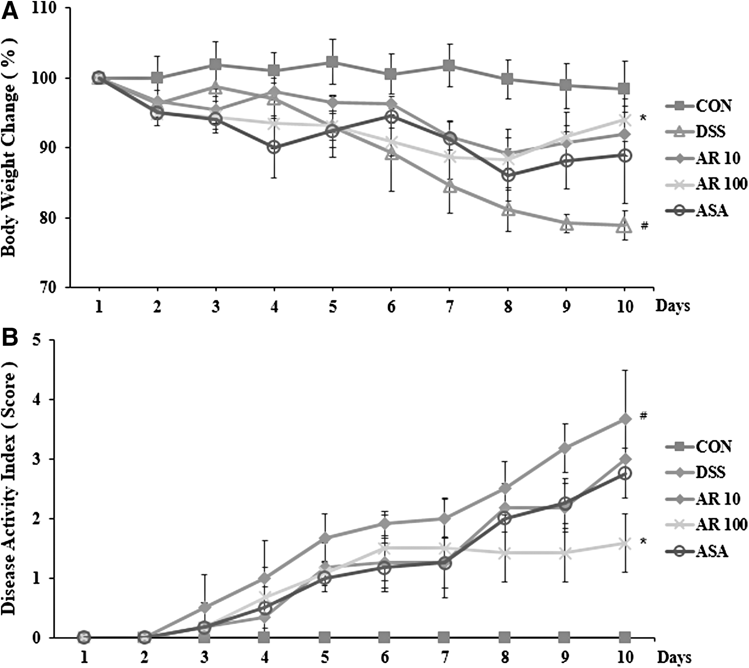
Effects of Aronia berry extract on body weight and DAI in DSS-induced colitis mice. Experimental colitis in mice was induced by treating animals with 5% DSS for 10 days. 5-ASA (50 mg/kg/day) was administered orally as a reference drug. Aronia berry extract was administered orally at doses of 10, 100 mg/kg once a day for 10 days before 5% DSS supplement.
Effect of Aronia berry extract on colon length shortening in UC induced by DSS
In DSS-induced animal models, various physical symptoms will appear, including changes in the length of the large intestine. The length of the large intestine can be used to indirectly assess the degree of inflammation. 14 In this study, the average colon length of the control group colon was 10.2 ± 0.52 cm. The length of the colon in the DSS-induced group was significantly (P < .05) decreased to 6.21 ± 0.53 cm compared with that in the control. Colons from the AR10 and AR100 groups were significantly (P < .05) increased to 7.38 ± 0.45 cm and 8.35 ± 0.46 cm, respectively (Fig. 2A, B), compared with that in the DSS alone group.
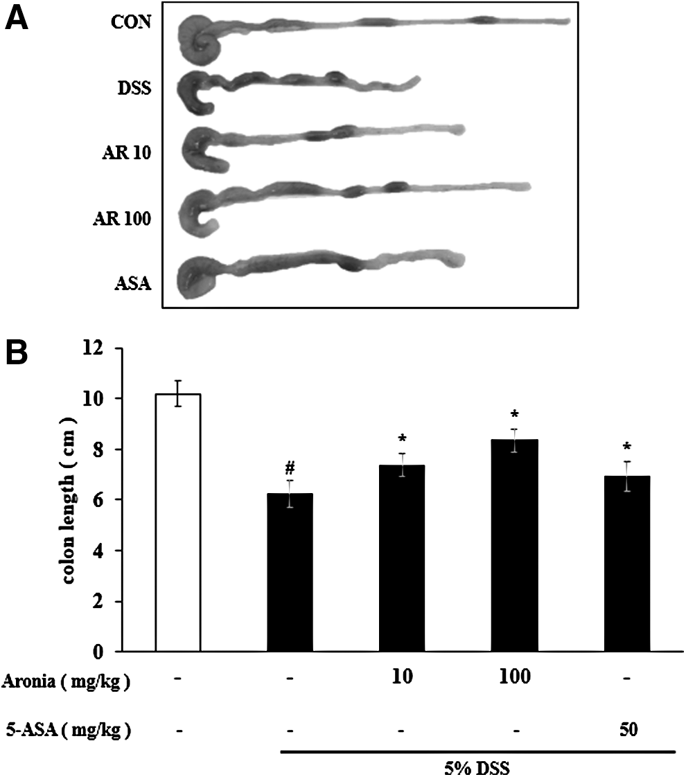
Effect of Aronia berry extract on colon length in DSS-induced colitis mice. Experimental colitis in mice was induced by treating animals with 5% DSS for 10 days. Aronia berry extract was administered orally at 10 and 100 mg/kg per day during the experiment.
Effect of Aronia berry extract on serum levels of inflammatory cytokines
Inflammatory cytokines appear early in the inflammatory response of UC. 16 To determine the effect of Aronia berry extract on serum levels of TNF-α and IL-6, ELISA was conducted. Serum IL-6 levels were found to be significantly (P < .05) lower in the AR100 group (49.41 ± 7.94 pg/mL) than that in the DSS group (85.40 ± 10.71 pg/mL) (Fig. 3A). Serum TNF-α levels were also significantly (P < .05) decreased in the AR100 group (601.52 ± 54.98 pg/mL) compared with those in the DSS group (828.62 ± 110.38 pg/mL) (Fig. 3B).
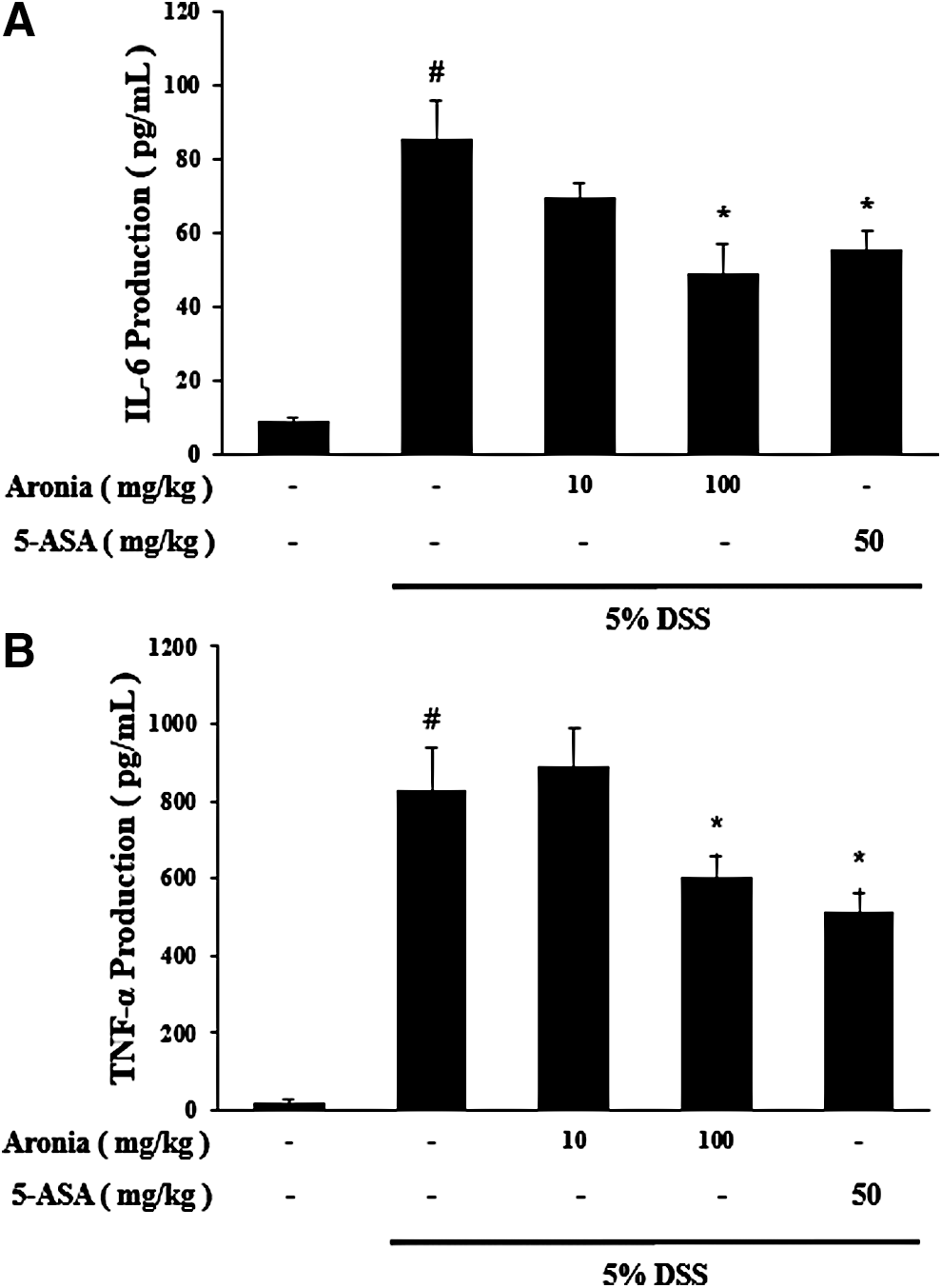
The effect of Aronia berry extract on production of proinflammatory cytokines in serum of mice with DSS-induced colitis. Ulcerative colitis was promoted in male BALB/c mice by administering 5% DSS for 10 days. Cytokine production was determined by ELISA.
Effect of Aronia berry extract on PGE2 production in colon tissue
The levels of PGE2 have been reported to be elevated in the intestines of UC patients. 17 We used ELISA to determine the effect of Aronia berry extract on inflammatory mediator PGE2 in DSS-induced UC mouse colon tissue. PGE2 levels in the DSS group (20615.5 ± 2403.7 pg/mL) were found to be significantly (P < .05) higher compared with those of the CON group (10806.1 ± 1131.6 pg/mL). However, PGE2 levels in the AR100 group (11082.4 ± 2686.7 pg/mL) were significantly (P < .05) lower than those in the DSS group (Fig. 4).
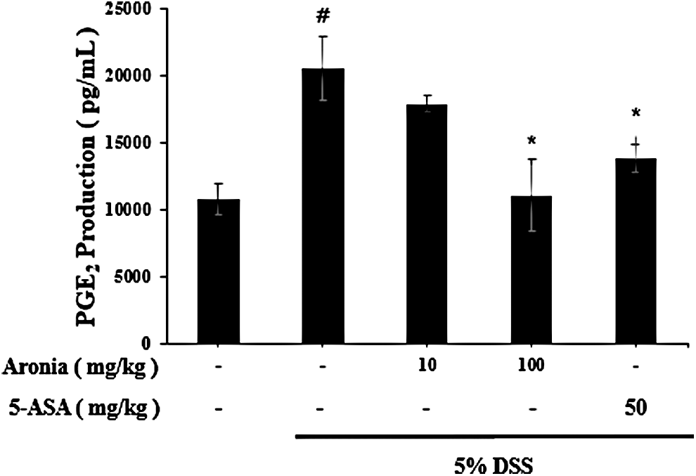
Effect of Aronia berry extract on PGE2 production in colon tissues. PGE2 levels were measured using PGE2 assay kits. PGE2 production in colon tissues. Data were representatives of three independent experiments. Values are mean ± SEM. Data were analyzed by Student's t-test. (# P < .05 vs. control group, *P < .05 vs. DSS group). PGE2, prostaglandin E2.
Effect of Aronia berry extract on epithelial injury in DSS-induced colitis
For histological examination of the colon, tissues were stained with H&E and observed at ×40 and ×100 magnification using an optical microscope. The epithelium is composed of an epithelial cell layer and lamina propria. DSS treatment caused epithelial injury. Results of H&E staining of colon tissue revealed that the epithelial cells of the CON group were normal. No inflammation was observed. However, the epithelial cell and crypt structures were destroyed in the DSS group. On the contrary, 100 mg/kg Aronia berry extract or 5-ASA treatment attenuated these injuries (Fig. 5A, B).
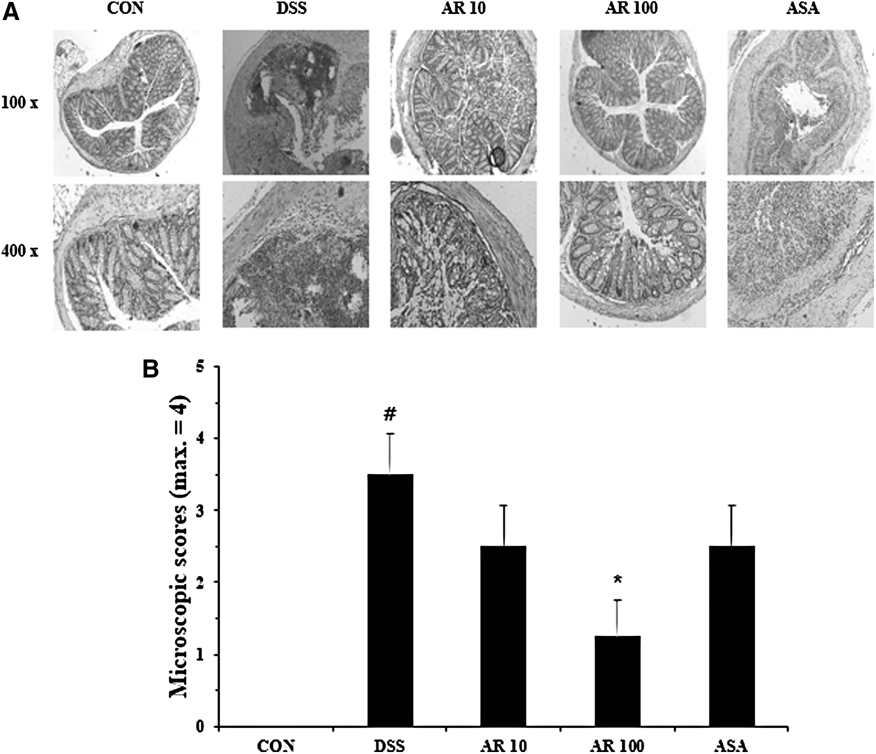
Effect of Aronia berry extract on epithelial injury in colon tissues of DSS-induced colitis mice. Over the same period, Aronia (10, 100 mg/kg) and the positive control 5-ASA (50 mg/kg) were orally administered once daily.
Effects of Aronia berry extract on inflammatory cytokine production in LPS-challenged peritoneal macrophages
MTT assay was used to examine the effect of Aronia berry extract on inflammatory cytokine production in LPS-challenged peritoneal macrophages. Cells were treated with Aronia berry extract (0.001, 0.01, 0.1, or 1 mg/mL) for 24 h, and cell viability was measured. Although cell viability was decreased slightly, to 96% compared with that of the untreated group (100 ± 0.49%), there was no significant difference in cell viability (Fig. 6A). Macrophages challenged with LPS after pretreatment with Aronia berry extract did not show significant decreases in cell viability either (Fig. 6B). Inflammatory cytokine is an important indicator of inflammation. Therefore, we investigated the effect of Aronia berry extract on the production of inflammatory cytokines in LPS-treated macrophages. IL-6 level was significantly reduced by Aronia berry extract treatment at a concentration of 1 mg/mL (1.43 ± 0.39 ng/mL) (Fig. 6C). In addition, TNF-α level in the group treated with Aronia berry extract 1 mg/mL (1.53 ± 0.14 ng/mL) was significantly (P < .05) lower compared with those in groups treated with other concentrations (0.001, 0.01, or 0.1 mg/mL) of Aronia berry extract (Fig. 6D). These results showed that Aronia extract significantly suppressed the generation of LPS-induced inflammatory cytokines in a concentration-dependent manner.
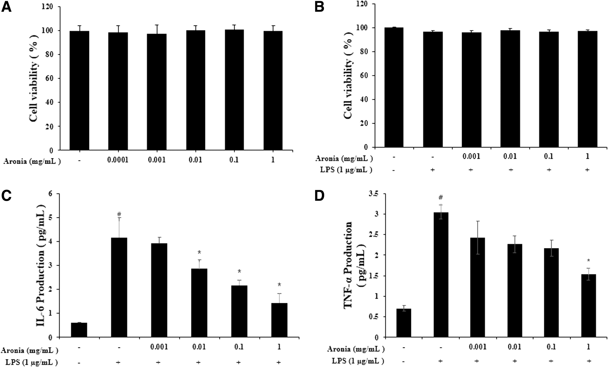
Effects of Aronia berry extract on inflammatory cytokines production on LPS-stimulated peritoneal macrophages. Macrophage were treated with various concentrations (0, 100 μg/mL, 1, 10, 100 mg/mL) of Aronia berry extracts for 4 h before being incubated with LPS (1 μg/mL) for 24 h. Cell viability was measured by MTT assay.
Effect of Aronia berry extract on NO production in LPS-stimulated peritoneal macrophages
NO has been considered as an important proinflammatory mediator that plays a key role in the pathogenesis of IBD. 18 After macrophages were pretreated with Aronia berry extract (0.01, 0.1, or 1 mg/mL) for 4 h, the production of LPS-induced NO was measured. LPS at a concentration of 1 μg/mL was used to stimulate microphage for 48 h. Using Griess reagent, the NO production was dose-dependently decreased by Aronia berry extract treatment (Fig. 7).
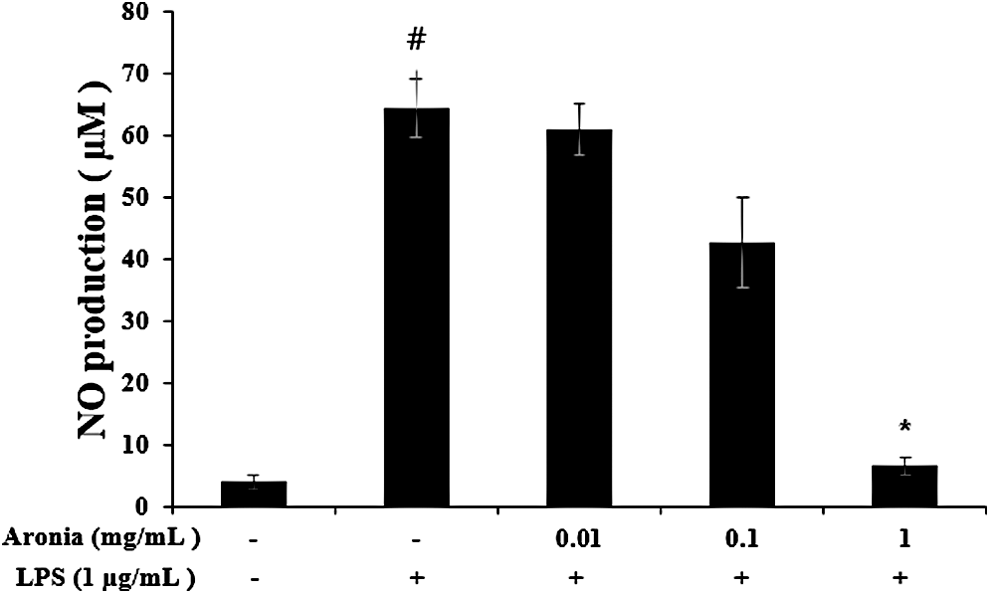
Inhibition of LPS-induced NO production by Aronia berry extract. Macrophage were preincubated with 0.01, 0.1, 1 mg/mL of Aronia berry extracts for 4 h and treated with 1 μg/mL of LPS for 48 h. The NO production was measured by Griess reagent system. Values are mean ± SEM. (# P < .05 vs. blank group, *P < .05 vs. LPS-induced group). NO, nitric oxide.
Effect of Aronia berry extract on phosphorylation of MAPK in peritoneal macrophages
We hypothesized that MAPKs could mediate the anti-inflammatory effects in the colon. To determinate the potential implication of MAPKs in the anti-inflammatory effects observed in macrophages after Aronia berry extract treatment, phosphorylation levels of ERK, JNK, and p38 were examined by western blot analysis using ERK, JNK, and p38 phosphor-specific MAPKs antibodies. Results are shown in Figure 8A. Treatment with LPS significantly activated ERK, JNK, and p38 proteins, indicating that MAPK proteins are activated at acute stage of colonic lesion. However, after treatment with AR at 1 mg/mL, phosphor-ERK and JNK activations were significantly lower (Fig. 8B).
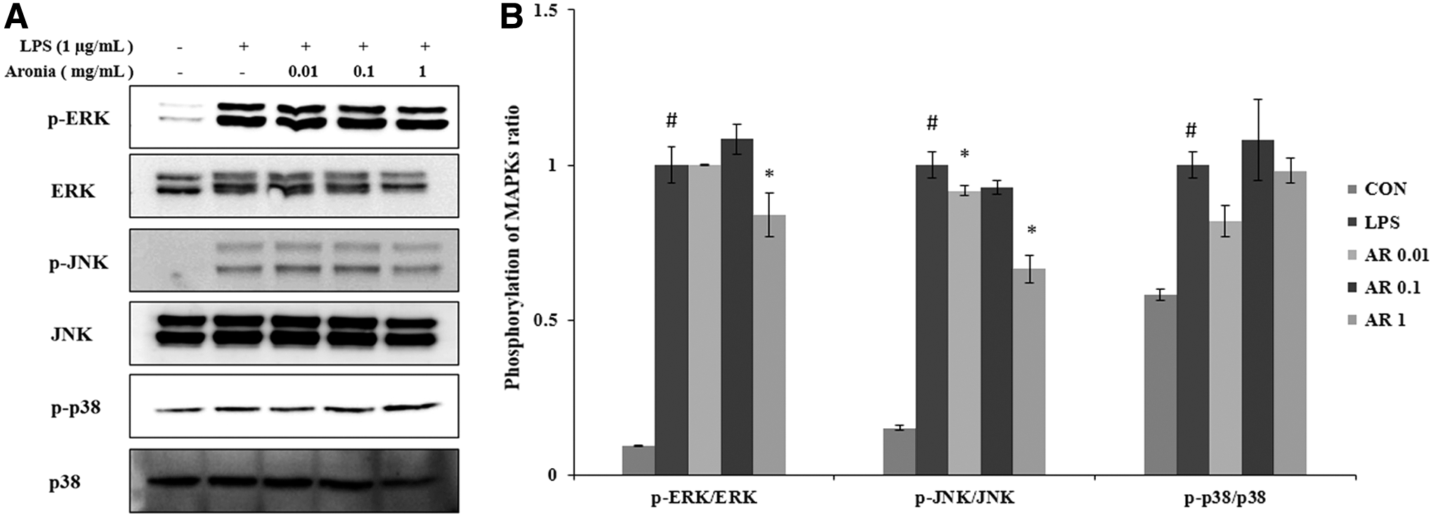
Effect of Aronia berry extract on activation of MAPK in macrophage.
Discussion
UC presents with symptoms such as weight loss, abdominal pain, and bloody stools. It is a form of IBD that causes inflammation and ulceration of the colon. 19,20 Treatment of UC entails the use of glucocorticoids, sulfasalazine, and immunosuppressive drugs. 21 However, these drugs are associated with severe side effects. There is a need for treatment options that do not have serious side effects. Recently, there is an increasing interest in the use of traditional herbal medicines to inhibit inflammation. Herbal medicines used to treat UC include Scutellaria baicalensis and Ixeris dentata. However, these herbs have failed to show clear benefits in clinical trials. 22,23 We hypothesized that anthocyanin-rich Aronia could decrease the symptoms associated with UC. We therefore administered Aronia berry extract orally to BALB/C mice with DSS-induced colitis and observed histological and immunohistological changes as well as macroscopic morphological changes that might indicate benefits. A previous study on the effect of an Aronia species (A. melanocarpa) on inflammation has reported that Aronia extract can inhibit the inflammatory response in macrophages by directly blocking the expression of iNOS and COX-2, thereby suppressing uveitis induced in the RAW264.7 macrophage cell line. 8 Aronia extract can also ameliorate inflammation caused by histamine and serotonin treatment. 24 It has also been shown that orally administered Aronia extract protects the liver following acute liver damage caused by carbon tetrachloride in mice. 25 In this study, we established an animal model of UC to evaluate the effect of Aronia berry extract.
Inflammatory cytokines such as TNF-α and IL-6 are known factors that can mediate the inflammatory response in models of UC. 26 Excessive secretion of these inflammatory cytokines causes sepsis and a variety of IBDs. 12,27 In particular, TNF-α levels have been reported to be significantly increased in patients with UC. 28 Thus, modulators of these inflammatory cytokines may provide important clues to the treatment of UC. TNF-α plays an essential part in suppressing tumors and viruses. However, when TNF-α is overexpressed, a continuous inflammatory reaction occurs. 16 Therefore, suppressing the level of TNF-α might ameliorate chronic inflammatory diseases. In this study, we determined the effect of Aronia berry extract on IL-6 and TNF-α expression in the serum and colon tissue in a DSS-induced UC mouse model. Aronia berry extract inhibited IL-6 and TNF-α production more than 5-ASA administration, a drug currently used to modulate secretion of IL-6 and TNF-α in patients with UC. Based on these results, we conclude that Aronia berry extract can ameliorate DSS-induced UC by modulating the production of inflammatory cytokines.
Various inflammatory mediators are increased in colon tissue from patients with UC. PGE2 produced by COX-2 is an important inflammatory factor that causes inflammatory reactions and vasodilation. 29 When overexpressed, PGE2 may cause tumors. 30 Inhibition of PGE2 can reduce the symptoms of colitis in vivo and in vitro in a DSS-induced mouse model. 31 –33 We showed that PGE2 was reduced in DSS-induced UC following Aronia extract administration. We also confirmed that Aronia berry extract exerted a modulating effect on the MAPK pathway and the secretion of inflammatory cytokines in murine macrophages. Our results indicated that Aronia berry extract might be useful herbal medicine in IBD.
In summary, we found that Aronia berry extract suppressed the symptoms of UC in a DSS-induced mouse model. We also examined inflammation-related activities of Aronia berry extract using mouse peritoneal macrophages. Aronia berry extract was found to be more effective than the anti-inflammatory drug 5-ASA, which is currently used to treat IBD. In addition, Aronia extract reduced the expression of COX-2 in peritoneal macrophages. Given these results, Aronia extract might be a safe and effective treatment to suppress inflammation by adjusting the levels of inflammatory mediators. Therefore, Aronia may have potential as a natural treatment for colitis that can replace current drug therapies. Further studies are needed to identify the active ingredients in Aronia berry extract using component analysis and to confirm the results in human clinical trials.
Footnotes
Acknowledgments
This research was supported by Basic Science Research Program through the National Research Foundation of Korea (NRF) funded by the Ministry of Science, ICT and Future Planning (2015R1C1A1A01054675) and the Industrial Technology Research Infrastructure Program (N0000004) funded by the Ministry of Trade, Industry and Energy (Sejong, Korea) and Sunchang Research Institute of Health and Longevity.
Author Disclosure Statement
No competing financial interests exist.