
Abstract
Coenzyme Q10 (CoQ10) is a powerful antioxidant substance synthesized in the body. The current study aimed to determine whether CoQ10 suppresses inflammation and inhibits p-STAT3 expression in an experimental colitis mouse model. The mice were orally fed with CoQ10 once a day for 13 days. Histological analysis of the colons was performed by immunohistochemistry. Expression of IL-17, FOXP3, p53, AMPK, and mTOR and activation of p-STAT3 and p-STAT5 in lymph node and spleen tissues were detected by confocal microscopy of stained tissue sections. The relative mRNA expression was measured with real-time PCR, and protein levels were examined by western blot. CoQ10 reduced the disease activity index score and the colon histological score. It also reduced inflammatory mediators in the colon and increased the colon length. The expression of IL-17 and p-STAT3 was decreased with CoQ10 treatment. In contrast, CoQ10 treatment increased the expression of p-AMPK and FOXP3. Expression of anti-inflammatory cytokines was shown to increase in colitis mice treated with CoQ10. These results suggested that CoQ10 may reduce the severity of colitis and suppress inflammation through the inhibition of p-STAT3 and IL-17. These results support the use of CoQ10 as a potential targeted therapy for the treatment of colitis.
Introduction
I
IL-17 is a pro-inflammatory cytokine that causes an immune response and inflammation and activates signal transducer and activator of transcription (STAT) 3. Previous reports have shown that the promotion of IL-17 expression increases p-STAT3 levels and its downstream signals such as IL-6. 5,6 The suppression of STAT3 activation has been suggested as a promising strategy for the control of various autoimmune diseases, due to the regulation of numerous pro-inflammatory cytokines by STAT3, a transcription factor. 7 It is well documented that STAT3 inhibitors attenuate experimental autoimmune diseases, including IBD, through the suppression of p-STAT3 expression. Previous investigations have demonstrated that the inhibition of p-STAT3 induces an anti-inflammatory response and decreased IL-17 expression. 8,9
Oxidative stress is an imbalance between reactive oxygen species production and antioxidant defenses to detoxify the steady state of oxidative stress. It has been demonstrated that oxidative stress plays an important role in the pathogenesis of various diseases. 10 In addition, oxidative stress participates in various inflammatory diseases. For instance, oxidative stress is related with the pathogenesis of chronic inflammation and autoimmune diseases, including IBD. 11 –14
Antioxidants play a critical role in the anti-inflammatory mechanism that decreases inflammation and prevents the generation of toxic oxidants. 15 As antioxidants are used to reduce inflammation, 16,17 there is a relationship between antioxidants and IBD. It has been demonstrated that antioxidant treatments for IBD show the possibility of attenuating symptoms of the disease. 18,19 Coenzyme Q10 (CoQ10), also known as ubiquinone, is a lipid-soluble antioxidant. It has been reported that CoQ10 reduces inflammatory mediators and attenuates oxidative stress. 20,21 Furthermore, CoQ10 induces the activation of AMP-activated protein kinase (AMPK), reducing inflammation in various inflammatory diseases. 22 –24 Recently, CoQ10 also revealed therapeutic effects downregulating inflammation in experimental graft versus host disease. 25 CoQ10 can be synthesized in most human tissue. Indeed, biosynthesis of CoQ10 occurs in multiple sites of animal cells because CoQ10 synthesis steps are generated in mitochondria, endoplasmic reticulum, and peroxisomes. 26 The absorption of exogenous CoQ10 in the human body occurs in the intestine. It has been demonstrated that secretion of pancreatic enzymes and bile into the intestines increases the absorption of lipophilic substances such as CoQ10. 27 Although reports on the metabolism of CoQ10 in the body are limited, CoQ10 metabolism occurs in several tissues. Previous report demonstrates that CoQ10 supplementation increased its concentration, but the levels return to normal state within a few days. 28
We hypothesized that CoQ10 has an anti-inflammatory effect in colitis. The purpose of the present study was to determine whether CoQ10 reduces inflammation and has an anti-inflammatory effect and therapeutic activity in DSS induced colitis mice. Thus, we aimed to elucidate the therapeutic function of CoQ10 in colitis and its underlying effect on inhibiting IL-17 and p-STAT3-mediated inflammation in a mouse model of colitis.
Materials and Methods
Animals
Eight-week-old male C57BL/6 mice (SLC, Inc., Shizuoka, Japan) were maintained in groups of five animals in polycarbonate cages within a pathogen-free environment and were fed standard mouse chow (Ralston Purina, St. Louis, MO, USA) and water ad libitum. All experimental procedures were reviewed and approved by the Animal Research Ethics Committee at the Catholic University of Korea. Mice were euthanized at the end of a study for the purpose of sample collection and histologic examination by CO2 chamber.
Induction of colitis and CoQ10 treatment
Colitis was induced exclusively in C57BL/6 mice by oral ingestion of 2.5% DSS (dextran sulfate sodium, 36–50 kDa; MP Biomedicals) for 4 days. Mice were orally fed with CoQ10 (Sigma; 303-98-0, 0.4 mg/mouse) each day for 13 days after colitis induction. CoQ10 was dissolved in cotton seed oil. During the DSS and drug treatments, the body weights of the mice were recorded daily.
Assessment of colitis
During the experimental period, the severity of colitis was assessed daily by measuring the percentage of body weight change and disease activity index (DAI). The DAI was calculated as previously described by summarizing the score for body weight loss (0 points, <5% weight loss; 1 point, 5–10% weight loss; 2 points, 10–15% weight loss; 3 points, 15–20% weight loss; and 4 points, >20% weight loss), stool consistency (0 points, formed pellets; 2 points, pasty/semiformed stool; and 4 points, liquid stool), and rectal bleeding (0 points, no rectal bleeding; 2 points, hemoccult positive; and 4 points, visible gross bleeding). 29 –31
Histopathological analysis
Colon tissue was harvested and fixed in 10% formalin solution. Formalin-fixed colon sections were paraffin embedded, and 7-μm sections were stained with hematoxylin and eosin (H&E). Inflammation severity was scored on a 0–3 scale (0: none; 1: slight; 2: moderate; and 3: severe), as was the extent of injury (0: none; 1: mucosal; 2: mucosal and submucosal; and 3: transmural); crypt damage was scored on a 0–4 scale (0: none; 1: basal one-third damaged; 2: basal two-thirds damaged; 3: only surface epithelium intact; and 4: loss of entire crypt and epithelium). 32 Each value was multiplied by an extent index ranging from 1 to 4, which reflected the amount of involvement for each section. At least three sections from each colon were analyzed to produce each score value.
Confocal microscope
Spleen tissue was obtained surgically from sacrifice and snap-frozen in liquid nitrogen and stored at −80°C. For immunostaining, 7-μm tissue sections of the spleens or mesenteric lymph nodes were stained with FITC-conjugated anti-IL-17, FITC-conjugated anti-p-STAT3 Y705, FITC-conjugated anti-p-STAT5, FITC-conjugated anti-Foxp3, FITC-conjugated anti-p53, APC-conjugated anti-CD25, PE-conjugated anti-CD4 (all from eBioscience, San Diego, CA, USA), rabbit-anti-mTOR, rabbit-anti-AMPK, and rabbit-anti-P53 (all from Cell Signaling) overnight at 4°C for 24 h. The tissues were blocked with goat or rabbit serum. These sections were washed with PBS and incubated with fluorescence-conjugated secondary Ab, a goat anti-rabbit IgG. The stained sections were analyzed using a Zeiss microscope (LSM 510 Meta; Carl Zeiss, Oberkochen, Germany) at 3400 × magnification.
Immunohistochemistry
The endogenous peroxidase activity was quenched with methanol and 3% H2O2. Immunohistochemistry was performed using the VECTASTAIN ABC Kit (Vector Laboratories, Burlingame, CA, USA). The tissues were first incubated with the primary anti-TNF-α, anti-IL-17, anti-IL-6, anti-IL-10, or anti-TGF-β (all from Santa Cruz Biotechnology, Santa Cruz, CA, USA) overnight at 4°C and a biotinylated secondary linking Ab and a streptavidin–peroxidase complex for 1 h. The final color product was developed using 3,3-diaminobenzidine chromogen (DAKO, Carpinteria, CA, USA). All histological assessments were determined by two independent and blinded observers. Images were captured using a DP71 digital camera (Olympus, Center Valley, PA, USA) attached to an Olympus BX41 microscope at 3400 × magnification.
Western blotting
Freshly isolated total colonic cells from the mice treated with CoQ10 or left untreated were washed with cold saline, and the total proteins were extracted with lysis buffer (Applied Science, Mannheim, Germany). The harvested lysates were centrifuged for 15 min at 4°C to pelletize cellular debris. The protein lysate was loaded onto a gel for 10% SDS-PAGE, followed by transfer to nitrocellulose membranes (Invitrogen Life Technologies). The blots were then blocked with 5% nonfat dry milk in tris-buffered saline and Tween 20 (TBST) for 1 h at room temperature. Then, the blots were incubated overnight at 4°C with Abs specific for p-STAT3 Y705, p-STAT3 S727, STAT3 p-AMPK, AMPK, and β-actin, followed by incubation with goat anti-mouse or anti-rabbit HRP-conjugated secondary Abs. The resulting bands were visualized with ECL reagents (Amersham Biosciences, Piscataway, NJ, USA).
Statistical analysis
All data are expressed as the mean ± standard deviation. The statistical analysis was performed using SPSS 10.0 for Windows (IBM Corp., Armonk, NY, USA). The comparison of numerical data between groups was performed with a nonparametric Mann–Whitney test. Statistical analysis was performed using SPSS 10.0 for Windows (SPSS, Chicago, IL, USA). P values < .05 were considered significant.
Results
CoQ10 decreased the development of DSS induced colitis
To investigate the anti-inflammatory effects of CoQ10, the mice with DSS induced colitis were fed orally with either CoQ10 (0.4 mg/mouse) once a day or with carrier. CoQ10 treatment prevented DSS induced colitis onset, as shown by the significant maintenance of weight (Fig. 1A) and colon length (Fig. 1C). CoQ10 treatment was also associated with a significant decrease in the DAI score (Fig. 1B) and in colon tissue destruction evident with H&E staining (Fig. 1D).
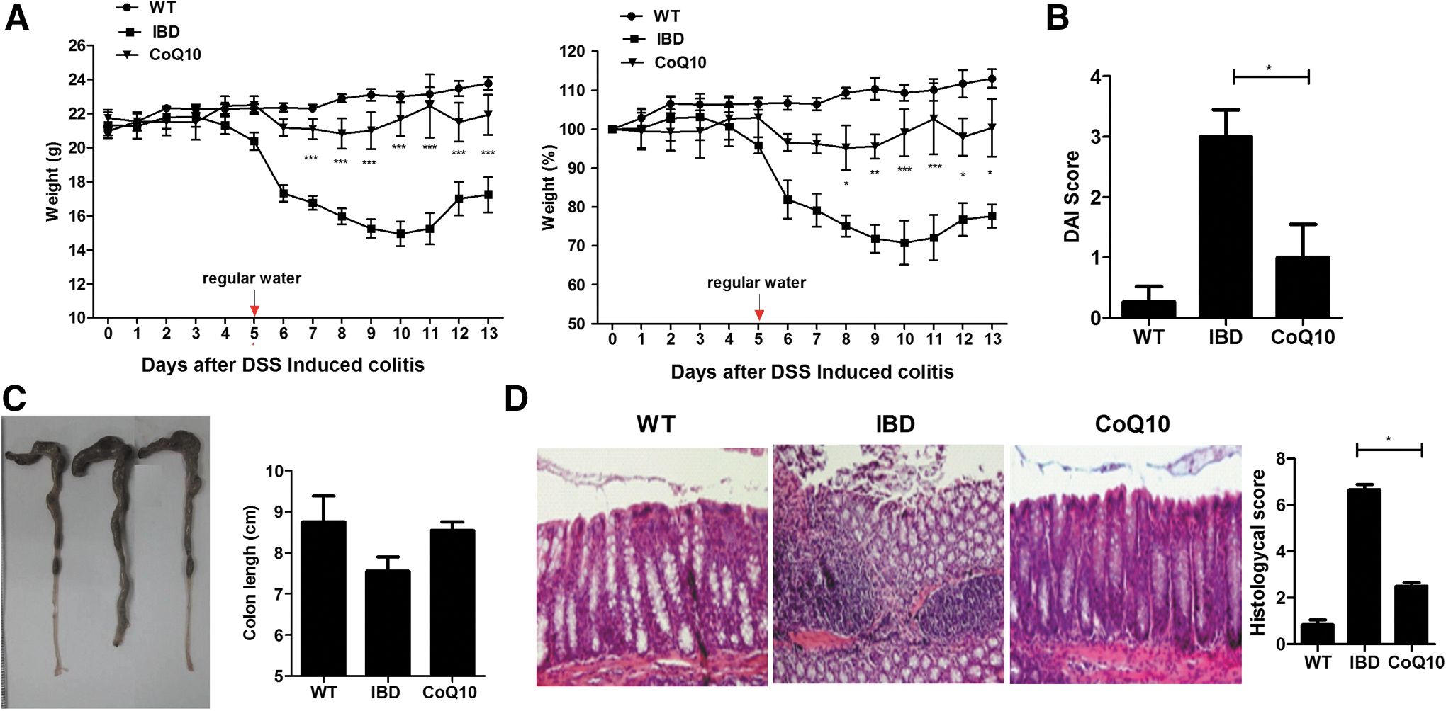
CoQ10 prevented the induction of colitis. CoQ10 (0.4 mg/mouse) was injected orally once daily into mice with colitis. The mice were sacrificed on day 14 after the induction of colitis (n = 5).
CoQ10 suppressed colitis through the inhibition of inflammatory cytokines
To investigate the effect of CoQ10 on inflammation of colon tissue, we performed immunochemical staining for pro- and anti-inflammatory cytokines. The expression of TNF-α, IL-6, and IL-17 was decreased in the CoQ10-treated mice (Fig. 2A). In contrast, CoQ10 slightly increased the expression of IL-10 and TGF-β in mucosa (Fig. 2B).
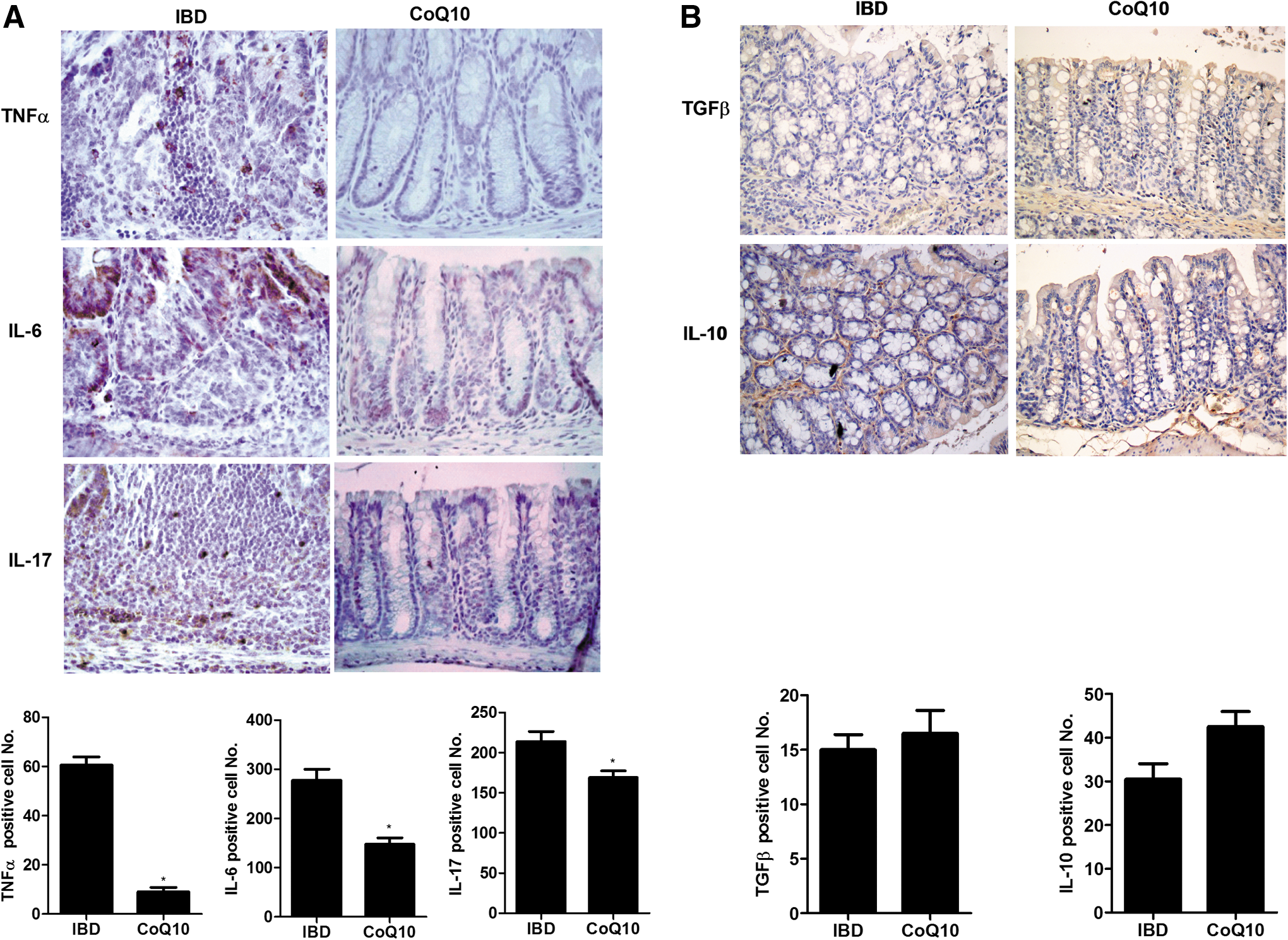
CoQ10 treatment reduces intestinal inflammation in mice with colitis.
CoQ10 reduced the expression of p-STAT3 in colon tissue
For evaluation of the protein level of p-STAT3, we conducted a western blot analysis. CoQ10 treatment suppressed significantly p-STAT3 expression in the colon tissue (Fig. 3A). On the contrary, the expression of AMPK was upregulated significantly in the colon tissue of CoQ10-treated colitis mice (Fig. 3B).
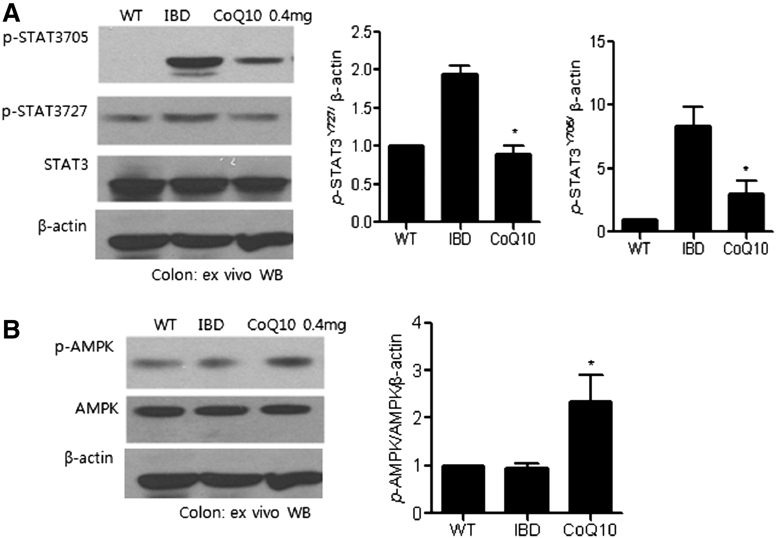
CoQ10 inhibits the expression of p-STAT3 tyr705 and ser727, but enhances the activation of AMPK.
CoQ10 regulated the differentiation of Th17 (CD4+IL-17+) and Treg (CD4+CD25+Foxp3+) cells through STAT3 in the lymph nodes and spleen
Total RNA was isolated from lymph nodes and spleen of either untreated or CoQ10-treated mice with DSS induced colitis. The expression of pro-inflammatory and anti-inflammatory cytokine mRNA was measured by real-time PCR. In the lymph node, the mRNA level of pro-inflammatory cytokines such as IL-17 significantly decreased with CoQ10 treatment. Gene expression of IFN-γ was also reduced, but not significantly. However, the mRNA expression of FOXP3 and anti-inflammatory cytokines IL-10 and TGF-β increased significantly with CoQ10 treatment (Fig. 4A). In the spleen, the mRNA level of pro-inflammatory cytokines such as TNF-α, IL-8, IL-17, and IFN-γ was significantly reduced with CoQ10 treatment (Fig. 5A). In confocal scanning, the expression of IL-17 and p-STAT3 tyr705 decreased significantly; however, the production of FOXP3, p-STAT5, and p53 was upregulated significantly in the lymph node and spleen (Figs. 4B and 5B).
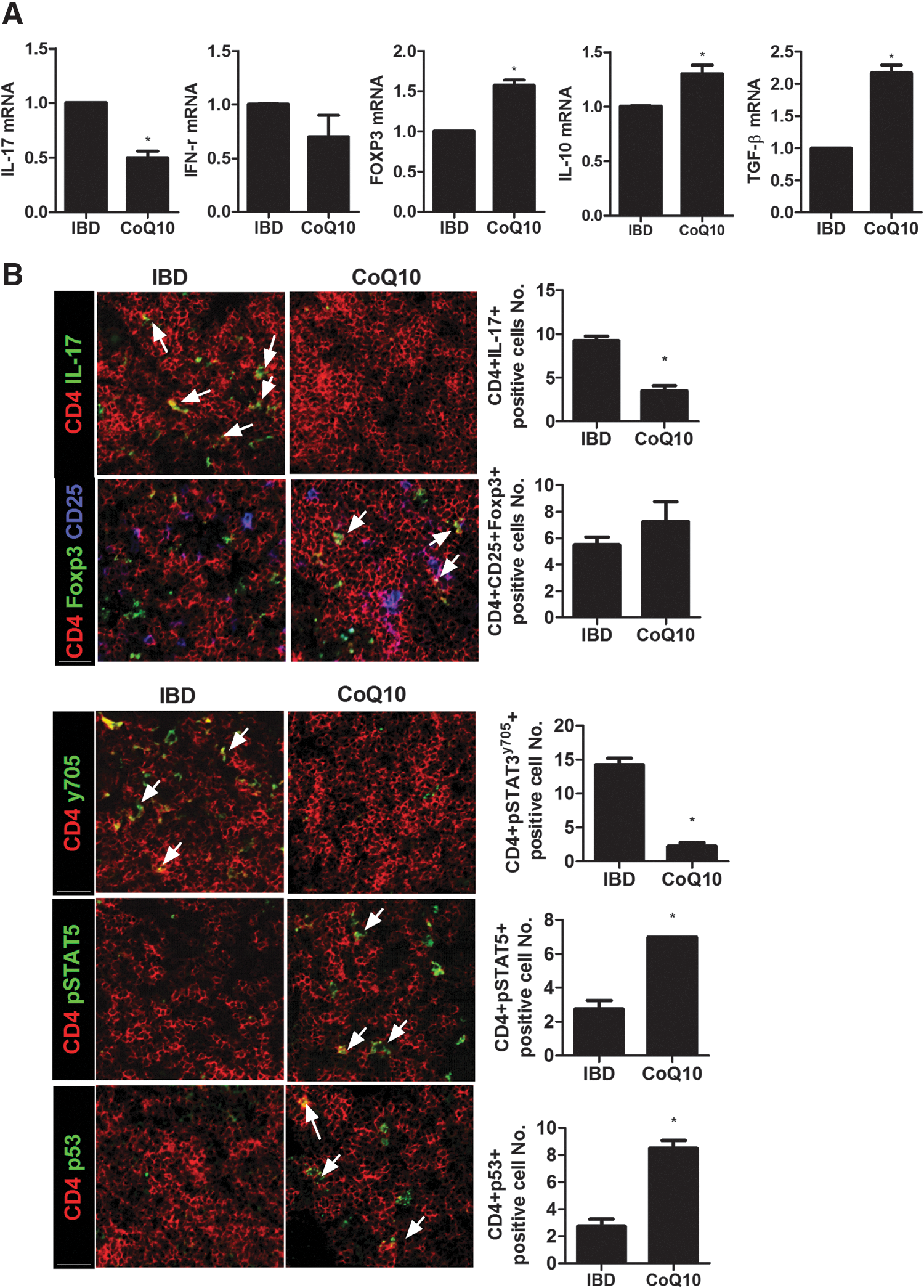
CoQ10 regulates the mRNA expression of inflammatory cytokines and differentiation of Th17 and Treg cells through the regulation of p-STAT3 and p-STAT5 in lymph nodes.

CoQ10 regulates the mRNA expression of inflammatory cytokines and differentiation of Th17 and Treg cells through the regulation of p-STAT3 and p-STAT5 in the spleen.
Discussion
Although CoQ10 has been recognized as an antioxidant, there is minimal evidence regarding CoQ10 activity in the inflammation of colitis. In the present investigation, the anti-inflammatory function of CoQ10 on a colitis mouse model was demonstrated.
The most meaningful observation in this study was that the anti-inflammatory activity of CoQ10 inhibited p-STAT3 production. It is well known that p-STAT3 causes the upregulation of several pro-inflammatory cytokines 33 and that the expression of IL-1β, IL-6, and TNF-α also enhances colitis. 34 –36 A significant decrease in inflammatory mediators, including IL-6 and TNF-α, was detected in the colon tissue of the colitis mice treated with CoQ10. But the expression of anti-inflammatory cytokines such as IL-10 and TGF-β was promoted slightly in the colon tissue of the DSS induced colitis mice treated with CoQ10. The inhibition of pro-inflammatory cytokine expression and p-STAT3 expression and the induction of anti-inflammatory cytokine production are notable features in reduced inflammation. Therefore, our findings have demonstrated that CoQ10 can perform an anti-inflammatory function in colitis.
AMPK performs a key role in protein synthesis and transcription and reduces the activation of p-STAT3. 37,38 Conversely, AMPK activation increases the expression of p53 and reduces mTOR activity. 39 In addition, p53 inhibits the expression of mTOR. 40 In this study, there was an increase in AMPK and p53 expression in DSS induced colitis mice treated with CoQ10. However, p-STAT3 expression decreased with CoQ10 treatment. Thus, CoQ10 exerts a regulatory effect on the balance between AMPK and p-STAT3 in the colitis mouse model.
It is well documented that Th17 cells induce inflammation; however, Treg cells also perform an anti-inflammatory function. 41,42 The activation of STAT3 and STAT5 is involved in regulating the Treg population. For instance, p-STAT3 suppresses the proliferation of Treg cells. 43 In contrast, the activation of STAT5 protracts FOXP3 expression in Treg cells. 44 CoQ10 treatment also has a direct impact on T cells. There is evidence that CoQ10 downregulates Th17 cell differentiation in CD4+ T cells isolated from mice and human. 45 Our results reveal that CoQ10 treatment reduced the expression of p-STAT3 and IL-17 and increased the production of p-STAT5 and FOXP3. These results suggest that CoQ10 is involved in anti-inflammatory activity through the regulation of the expression of p-STAT3 and IL-17 and the production of p-STAT5 and FOXP3.
CoQ10 is administered orally and reveals antioxidant effect. It is well documented that oral CoQ10 dosing is effective for clinical application. 46 In this study, the oral administration of CoQ10 showed therapeutic effect in a colitis mouse model. We fed with CoQ10 (0.4 mg/mouse) once a day, and the dose is approximately equivalent to 1200 mg in humans, which is appropriate since CoQ10 supplementation at doses as high as 1800 mg/day has been safely used in previous clinical trials. 47 Therefore, the dose in this study can be applied safely to colitis patients. The results of this study suggest that the oral administration of CoQ10 can be used in patients with colitis.
Although CoQ10 is an antioxidant, no adverse effects occurred with its use in this study. CoQ10 may dampen tissue damage by decreasing ROS in colon cells. Thus, in vivo animal studies conducted over a long time period are required to confirm the safety of the clinical application of CoQ10 in colitis.
In this study, CoQ10 simultaneously reduced the Th17 cell population while promoting Treg cells through the inhibition of STAT3 phosphorylation and enhancement of STAT5 phosphorylation. Therefore, this study demonstrates the anti-inflammatory activity of CoQ10 in intestinal inflammation. Our data revealed that CoQ10 treatment prevented colitis, with treated mice maintaining their weights comparable to normal controls. In addition, this study showed the role that CoQ10 played in decreasing various pro-inflammatory cytokines, such as TNF-α and IL-6, which are involved in the etiology of colitis.
There has been little evidence showing the regulation of p-STAT3 by CoQ10. The present investigation demonstrated that CoQ10 inhibits intestinal inflammation by reducing p-STAT3 expression of T cells in the colon. The effects of CoQ10 treatment, notably identified in this study, suggest that CoQ10 likely performs an important role in the suppression of intestinal inflammation. This interaction may shed light on the pathogenesis of IBDs, such as colitis. This interaction may shed light on the pathogenesis of colitis. In conclusion, CoQ10 attenuates inflammation and colitis through its regulation of cytokines and should be considered a candidate for immune-mediated anti-inflammatory therapies.
Footnotes
Acknowledgments
This research was supported by the Bio & Medical Technology Development Program of the National Research Foundation of Korea (NRF) funded by the Korean government (MEST) (No. 2012 M3A9C6049783). This research was supported by a grant of the Korea Health Technology R&D Project through the Korea Health Industry Development Institute (KHIDI), funded by the Ministry of Health & Welfare, Republic of Korea (grant No. HI15C3062). This research was supported by Basic Science Research Program through the NRF funded by the Ministry of Science, ICT & Future Planning (No. NRF-2015R1C1A2A01051677).
Author Disclosure Statement
No competing financial interests exist.