
Abstract
In this study, the antithrombotic and thrombolytic ability of second fermented extract of Ophiopogon japonicus (FEOJ) was verified in thrombosis-induced rats. Thrombosis was induced by oral administration of 2% carrageenan for 4 weeks. Five experimental groups (n = 9/group) involved in the study were control group, thrombosis group, low-dose FEOJ group (2 mL/kg, low-dose Ophiopogon japonicus [LOJ]), middle-dose FEOJ group (6 mL/kg, medium-dose Ophiopogon japonicus [MOJ]), and high-dose FEOJ group (12 mL/kg, high-dose Ophiopogon japonicus [HOJ]). The clotting time (CT), bleeding time (BT), prothrombin time (PT), activated partial thromboplastin time (APTT), and fibrinogen (FBG) were assessed in blood samples, and histological studies were performed on liver and lung tissues. The results demonstrated delayed CT only in MOJ and HOJ groups and delayed BT in all FEOJ groups compared with those in thrombosis and control groups (P < .05). Similarly, APTT was significantly delayed only in MOJ and HOJ groups, and PT was significantly delayed in all FEOJ groups, compared with those in control and thrombosis groups (P < .05). Although concentrations of FBG were similar in control, thrombosis, and LOJ groups, the tendency for decreased concentration of FBG (statistically nonsignificant) in MOJ and HOJ groups has been observed. Histological examination of livers and lungs revealed that thrombosis was partially improved in FEOJ group compared with the thrombosis group. In conclusion, CT, BT, PT, and APTT were prolonged in FEOJ group more than in control and thrombosis groups, thereby, depicting antithrombotic and thrombolytic effects. However, concentration-dependent effects of FEOJ were more prominent in MOJ and HOJ groups than in the LOJ group.
Introduction
I
The medications for thrombotic diseases currently in the market include fibrin-nonspecific thrombolytic agents (streptokinase and urokinase), antiplatelet agents (aspirin and clopidogrel), and anticoagulants (warfarin and coumarin). However, these drugs have various disadvantages such as high price with low activity, hemorrhagic adverse effects, gastrointestinal disorder, and hypersensitivity. Therefore, studies are being actively conducted to develop antithrombotics from relatively cost-effective and safe natural products and herbal medicines, used for food and medicine. 4
Ophiopogon japonicus (OJ), a perennial evergreen plant belonging to the Liliaceae family, is grown for medicine preparation in East Asian countries such as Korea, China, and Japan. It is known to reduce blood glucose levels and possess anti-inflammatory, antidiabetes, and anticancer activities. 5 Ruscogenin and ophiopogonin, the known active components of OJ, exhibit remarkable antithrombotic effects through inhibition of platelet aggregation. Ruscogenin is known to possess greater antithrombotic effect compared with ophiopogonin. 6 Cordyceps militaris is a type of Ascomycete; it parasitizes insect larvae, grows, and gradually turns into a mature fruiting body. The parasitic complex of fungus and caterpillar has been used for tonics and medicinal purposes for centuries in East Asia. 7,8 Cordycepin is a major bioactive component of C. militaris, which has been reported to inhibit platelet aggregation due to the inhibition of Ca2+ influx induced by increased cGMP. 9 Decades ago, because of the rarity of wild Cordyceps, scientists began to study the artificial cultivation of natural Cordyceps, and how to produce them using fermentation technology. 10 C. militaris is easier to grow under an artificial environment, and the process of producing C. militaris fruiting bodies is similar to that of other cultivated edible mushrooms. 11
Lactic acid bacteria are widely used in food fermentation, and Lactobacillus plantarum is the species most frequently used to ferment food products of plant origin. 12,13 Numerous studies have reported health enhancement effects of lactic acid bacteria, such as intestinal purification, immunological enhancement, anticancer, and antibacterial properties. 14 –18 The thrombolytic effect of traditional foods made from fermented soybeans such as Cheonggukjang, Doenjang, Jeotgal, and Kimchi has been reported in Korea. 19 –22
Several experimental models of thrombosis have been established in the past, including physical and chemical approaches. Nearly, all these approaches need complicated surgery to expose blood vessels. As an alternative, carrageenan-induced thrombosis in the tail model was established a few decades ago. 23,24 In our study, we have used this carrageenan-induced thrombosis in the tail model, since this method can be performed with lesser cost and allows observing and measuring the developing thrombosis continuously and accurately. This model has been used to test many clinically used antithrombus and thrombolytic agent. Although OJ is known for noticeable thrombolytic effects, there is paucity of information, verifying the antithrombotic and thrombolytic effects of fermented extract of Ophiopogon japonicus (FEOJ) in animal model. Thus, this study was performed to verify the antithrombotic and thrombolytic effects of FEOJ by investigating indicators of antithrombotic and thrombolytic effects of FEOJ using lactobacillus in vivo.
Materials and Methods
Chemicals and biochemicals
Prothrombin time (PT) reagent and activated partial thromboplastin time (APTT) reagent were procured from Diagnostica Stago (Gennevilliers, Seine, France). Calcium chloride, carrageenan (TypeI, C1013), and hematoxylin and eosin (H&E) were purchased from Sigma-Aldrich (St. Louis, MO, USA). NaOH was obtained from Youngjin Chemistry (Bucheon, Korea), and saline was bought from Choongwae Pharma Corporation (Seoul, Korea). Zoletil 50 was obtained from Virbac Laboratories (Carros, France), and Rompun was procured from Bayer Korea (Ansan, Korea).
Preparation of FEOJ
The roots of OJ (Milyang, Korea), harvested in April 2012, were used to prepare the aqueous extract. The general two sequential fermentations (fungal and bacterial fermentations) were performed according to the previous report. 25 In brief, dried OJ (3.5 kg) was extracted with ∼6500 mL water at 105°C for 60 min. The aqueous filtrates of OJ were adjusted to pH 6.5 with NaOH (Youngjin Chemistry), and autoclaved (JS Research, Gongju, Korea) for 20 min at 121°C. C. militaris was cultured with 0.039 g/mL PDA (potato dextrose agar) at 25°C for 3–5 days. Then, C. militaris was inoculated into C. militaris media (4% glucose, 1% yeast extract, 0.2% proteose pepton, 0.2% KH2PO4, 0.02% MgSO4, pH 6.5) and cultured by shaking at 2 g at 25°C for 3–5 days. Subsequently, C. militaris was inoculated and fermented by shaking at 141 g at 25°C for 10 days.
The second fermentation was conducted by a combination of three types of lactic acid bacteria (L. plantarum, Enterococcus faecium, and Bifidobacterium longum, obtained from Mediogen Co., Ltd., Seoul, Korea). L. plantarum and E. faecium were cultured with 0.055 g/mL lactobacilli MRS broth at 37°C for 12–15 h, while B. longum was cultured with modified MRS broth (5.5% modified MRS broth, 0.05%
Animals and breeding
Sprague-Dawley (SD) male white rats weighing 206.9 ±2.52 g were used in the study. Animals were adapted for more than 1 week with free access to pellet food and water until the day of experiment. Feeding environments were maintained at temperature, 22°C ± 2°C, relative humidity, 50% ± 10%, and light–dark cycle of 12:12 h. All animal experiments were performed in the animal laboratory of school of medicine in Hanyang University. The study was approved by the Institutional Animal Care and Use Committee (IACUC) from Hanyang University (approval ID: 2014-0128).
Experimental design
Forty-five SD male white rats were divided into five experimental groups (n = 9/groups: control group [CON] and four thrombus-induced groups). The thrombus-induced groups included thrombosis group (TMB), low-dose Ophiopogon japonicus (LOJ) group, medium-dose Ophiopogon japonicus (MOJ) group, and high-dose Ophiopogon japonicus (HOJ) group. According to our previous study, 10 μL of FEOJ corresponds to 5 μg of plasmin. Plasmin shows safety from bleeding with therapeutic (thrombolytic) dosages at the concentration of 6 mg/kg. 26 Therefore, in this study, normal diet was provided to CON and TMB, and 2, 6, and 12 mL/kg of FEOJ were orally administered daily to LOJ, MOJ, and HOJ, respectively (Fig. 1). Tail thrombosis in rats was induced by carrageenan according to a previously reported method. 23 To induce thrombus, 20 mg/kg of carrageenan prepared in saline solution (0.02 g/mL), was injected once to the sole of an anesthetized animal. The experiment used only the animals with redness of tail, 72 h after the injection.
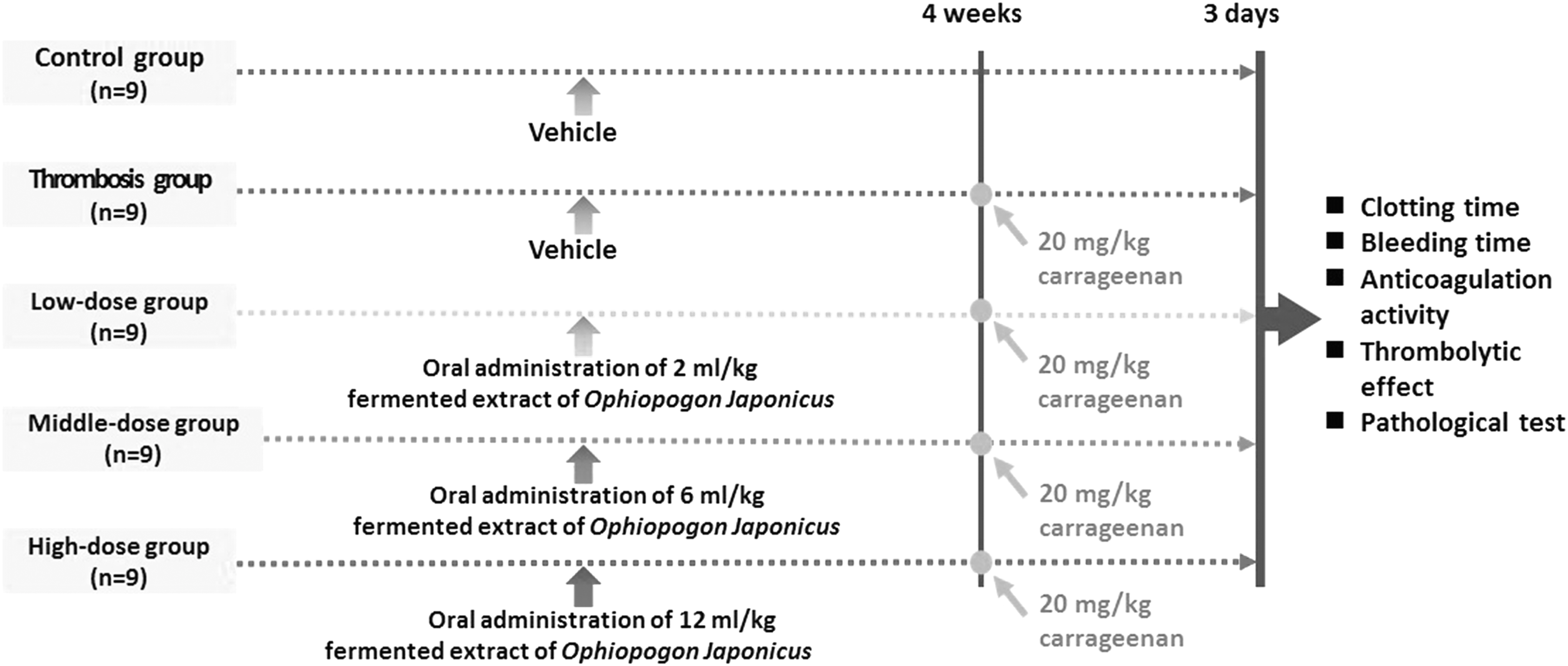
Scheme of experimental processes for thrombosis animal model.
Analysis of weight, blood, and organs
Body weight was measured weekly, at a specified time of the day, during 7 weeks of experiment. After 4 weeks of experimental feeding, animals were anesthetized with Rompun (0.5 mL/kg) and Zoletil 50 (20 mg/kg). After more than 16 h of fasting, blood was collected using antithrombotic syringe from abdominal aorta. Blood was centrifuged at 1006 g for 15 min to obtain plasma. The weight of organs (liver, lung, heart, kidney, and spleen) was measured immediately after extraction of blood and removal of remaining blood with gauze.
Measuring clotting time and bleeding time
Blood (0.3 mL) was drawn by venipuncture of tail vein 72 h after thrombus induction. Blood was placed in a nonheparinized capillary tube and gently mixed. Clotting time (CT) was measured as the time required for sample of blood to coagulate. Bleeding time (BT) was recorded after making a 2 mm cut on the tip of tail, 72 h after thrombus induction.
Measuring anticoagulation activity and thrombolytic effect
To verify the anticoagulation activity and thrombolytic effect of FEOJ, APTT, PT, and FBG were investigated. Automated coagulation analyzer (Coapresta 2000, Tokyo, Japan) was used to analyze the APTT in the intrinsic pathway of activation of clotting inhibition; PT in the extrinsic pathway; and FBG in coagulation effect. Tube containing the sample was placed in ice for about 1 h, followed by centrifugation at 1006 g (4°C) for 15 min. The separated plasma was stored in a refrigerator and analyzed within 1 day.
Pathological test
The liver and lung tissues were dehydrated after fixing with 10% neutral formalin. The tissues were embedded in paraffin and subjected to microsectioning (5 μm). The microsections were stained with H&E, and clotting conditions of capillary vessels, small arteries, and veins were examined microscopically (magnification × 200).
Statistical analysis
The experimental results of this study were analyzed using SPSS ver. 21.0 (SPSS). The results from each group are expressed as mean ± standard error. One-way analysis of variance (ANOVA) was performed to assess significance of differences among experimental groups. Duncan's multiple range test was performed as a post-hoc test. Statistical significance was verified at P < .05.
Results
Weight of body and organs
The mean body weight of rats in all groups was 206.9 g at the beginning of experiment, and 371.2 g at the end of the study. The increase in mean body weight was calculated to be 149.7 g. There were no differences in mean body weights of rats among the different groups at the beginning and end of experiment or in the increases in body weights during the experiment. A similar pattern was observed in the organ weight, suggesting that there was no significant difference in the weights of liver, lung, heart, kidney, and spleen among groups (Table 1).
Values are presented as mean ± SE (n = 9).
CON, control group; FEOJ, fermented extract of Ophiopogon japonicus; HOJ, high-dose FEOJ; LOJ, low-dose FEOJ group; MOJ, middle-dose FEOJ; NS, not significant; SE, standard error; TMB, thrombosis group.
Effect of FEOJ on CT and BT
CT was found to be 85.3, 89.3, 89.5, 96.8, and 94.3 sec in CON, TMB, LOJ, MOJ, and HOJ groups, respectively, showing delayed CT was statistically significant only in MOJ and HOJ groups compared with that in the CON group. BT was found to be 426.7, 363.3, 593.3, 625.0, and 653.3 sec in CON, TMB, LOJ, MOJ, and HOJ groups, respectively. Thus, indicating that BT was effectively delayed in a dose-dependent manner in all FEOJ groups compared with that in CON and TMB groups (P < .05) (Figs. 2 and 3).
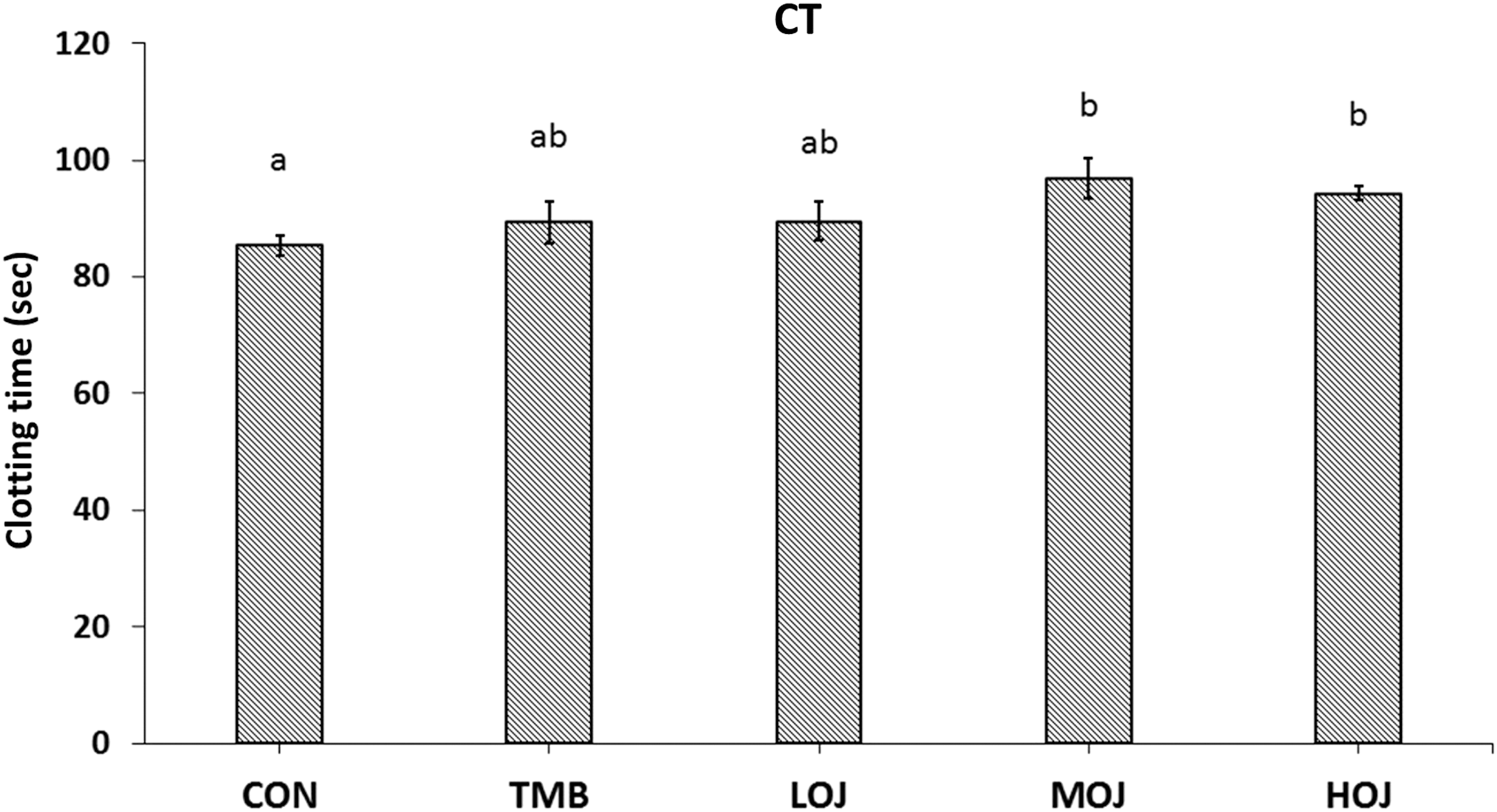
Change of CT when FEOJ was administered to thrombosis-induced rats. Values are presented as mean ± SE (n = 9). Significance was tested by one-way ANOVA followed by Duncan's multiple range test. Values sharing the same superscript letters differ significantly at P < .05. ANOVA, analysis of variance; CON, control group; CT, clotting time; FEOJ, fermented extract of Ophiopogon japonicus; HOJ, high-dose FEOJ; LOJ, low-dose FEOJ group; MOJ, middledose FEOJ; TMB, thrombosis group.
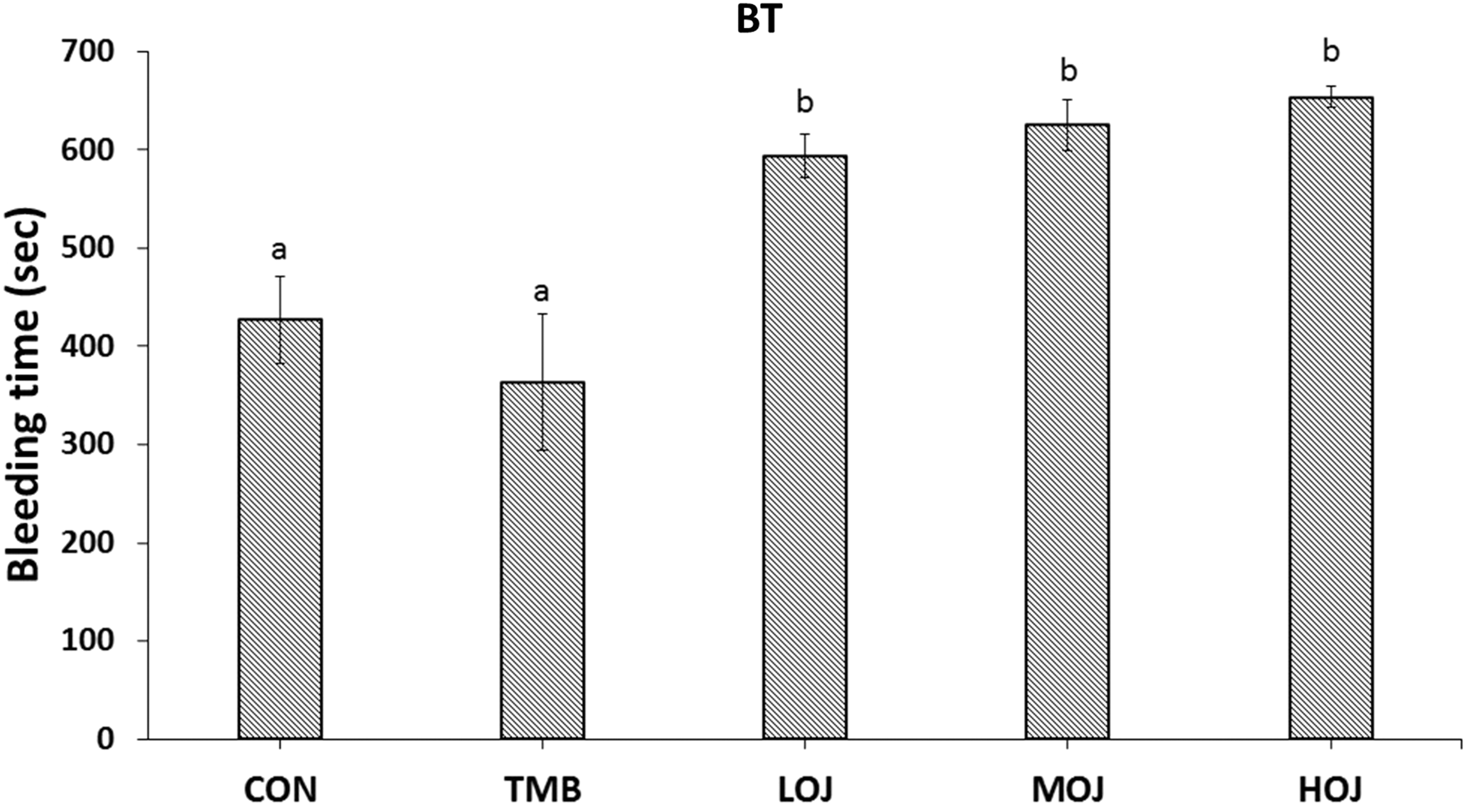
Change of BT when FEOJ was administered to thrombosis-induced rats. Values are presented as mean ± SE (n = 9). Significance was tested by one-way ANOVA followed by Duncan's multiple range test. Values sharing the same superscript letters differ significantly at P < .05. BT, bleeding time.
Impact of FEOJ on indicators of anticoagulation activity and thrombolytic effect in serum
Furthermore, the effect of FEOJ administration on the clotting pathway in rats administered with thrombus-inducing substance (Fig. 4) was investigated. PT in the extrinsic pathway was observed to be 48.4, 30.3, 36.8, 39.4, and 38.5 sec in CON, TMB, LOJ, MOJ, and HOJ groups, respectively, indicating significantly delayed PT in FEOJ groups compared with that in TMB group. APTT in the intrinsic pathway was found to be 33.2, 31.1, 28.7, 38.8, and 42.0 sec in CON, TMB, LOJ, MOJ, and HOJ, respectively. This result suggests that APTT was significantly delayed only in MOJ and HOJ groups compared with that in CON and TMB groups.
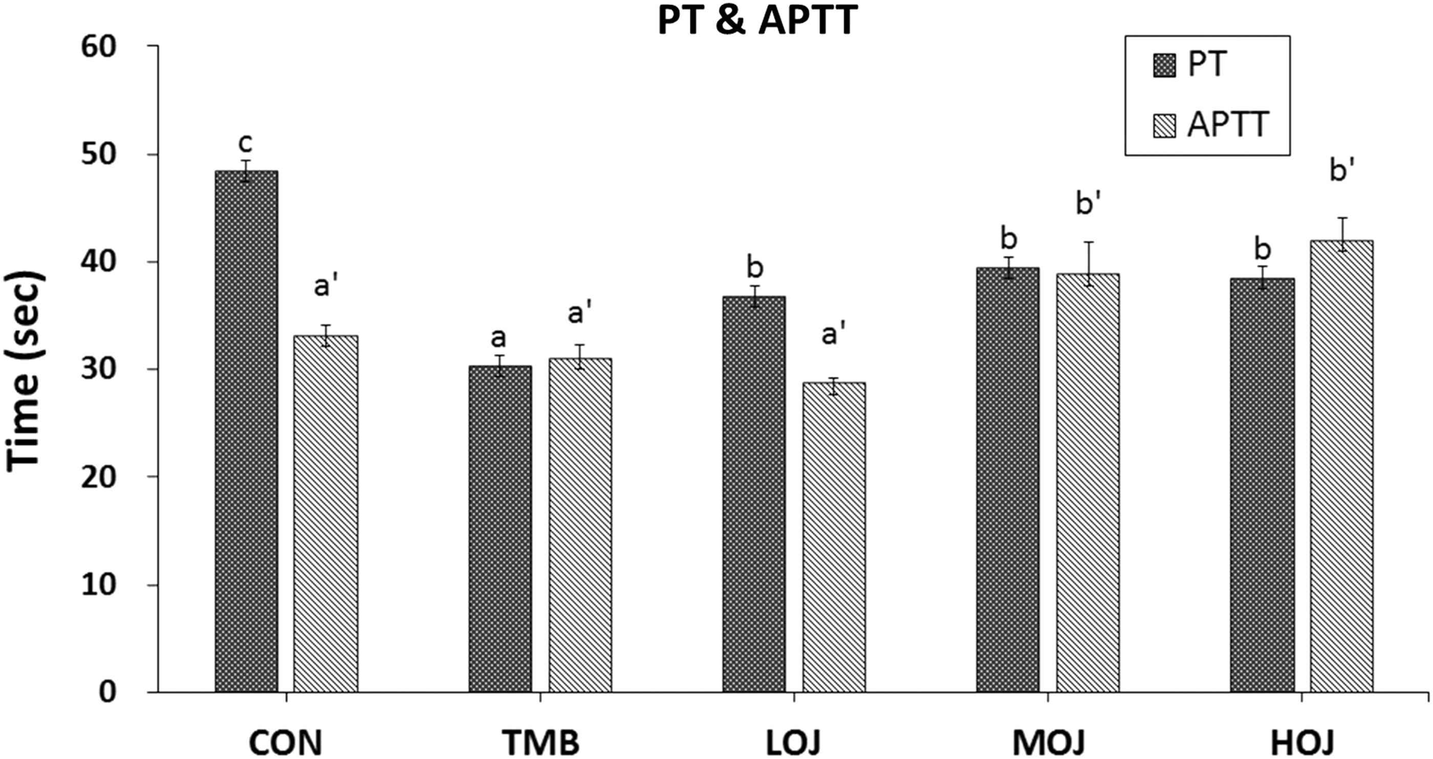
Change of PT and APTT when FEOJ was administered to thrombosis-induced rats. Values are presented as mean ± SE (n = 9). Significance was tested by one-way ANOVA followed by Duncan's multiple range test. Values sharing the same superscript letters differ significantly at P < .05. APTT, activated partial thromboplastin time; PT, prothrombin time.
FBG was found to be 191.7, 195.0, 196.7, 185.5, and 177.4 mg/dL in CON, TMB, LOJ, MOJ, and HOJ groups, respectively. Although, the tendency of decreased concentration of FBG in MOJ and HOJ groups has been observed, it was not statistically significant (Fig. 5).
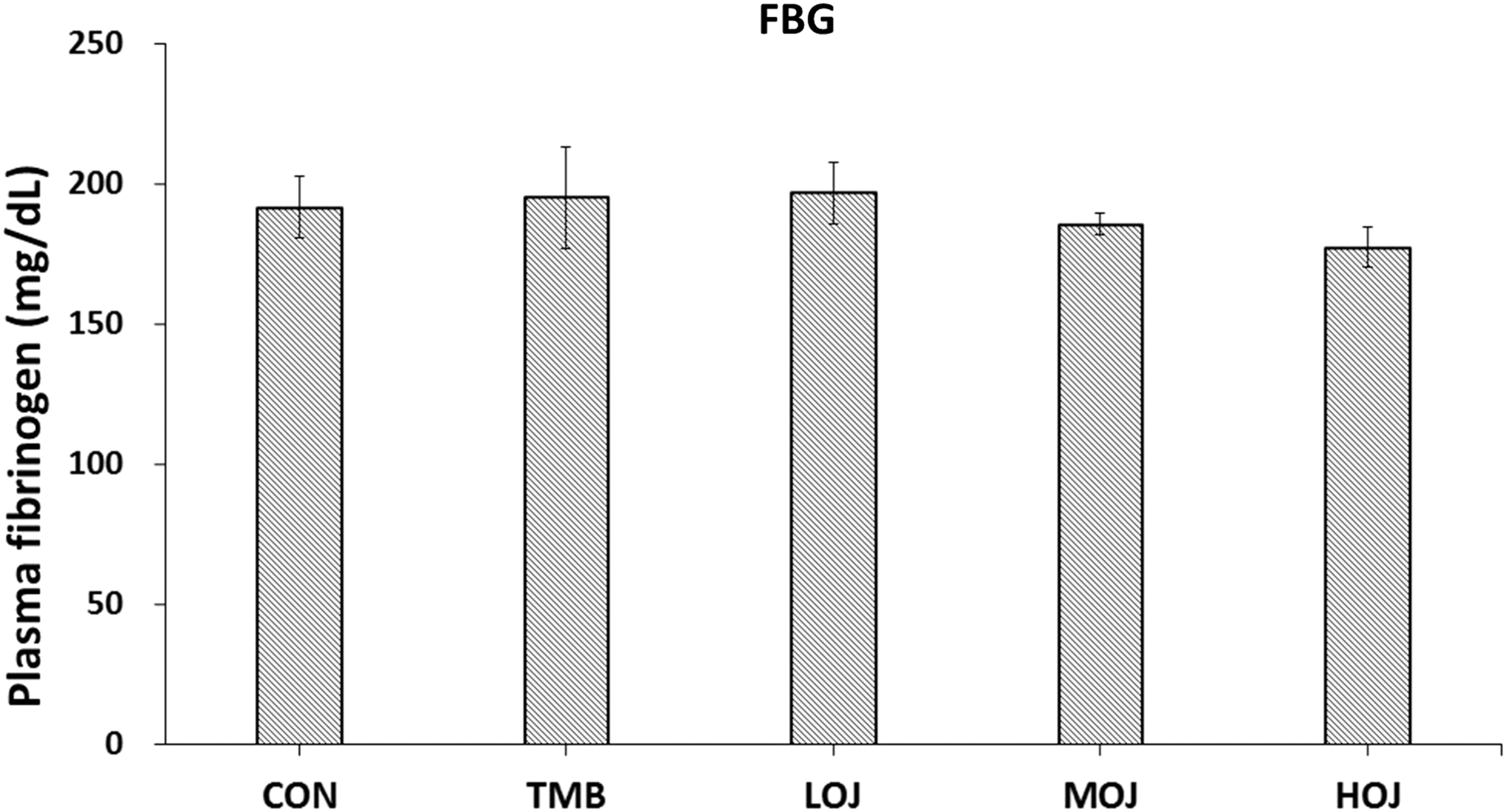
Change in plasma FBG when FEOJ was administered to thrombosis-induced rats. Values are presented as mean
Effect of FEOJ on histopathology of rat tissues
Liver and lung tissues extracted at 72 h after the administration of the thrombus-inducing substance carrageenan were stained with H&E stain to observe the coagulation status in capillaries, small arteries, and veins. Thrombosis, inflammatory foci, congestion, hemorrhage, and necrosis were observed in a section of liver tissue in TMB group. Thrombosis in TMB group was more prominent in lung tissue compared with that in liver tissue. Although, similar observations were made in FEOJ groups, thrombosis was partially inhibited by FEOJ administration (Figs. 6 and 7).
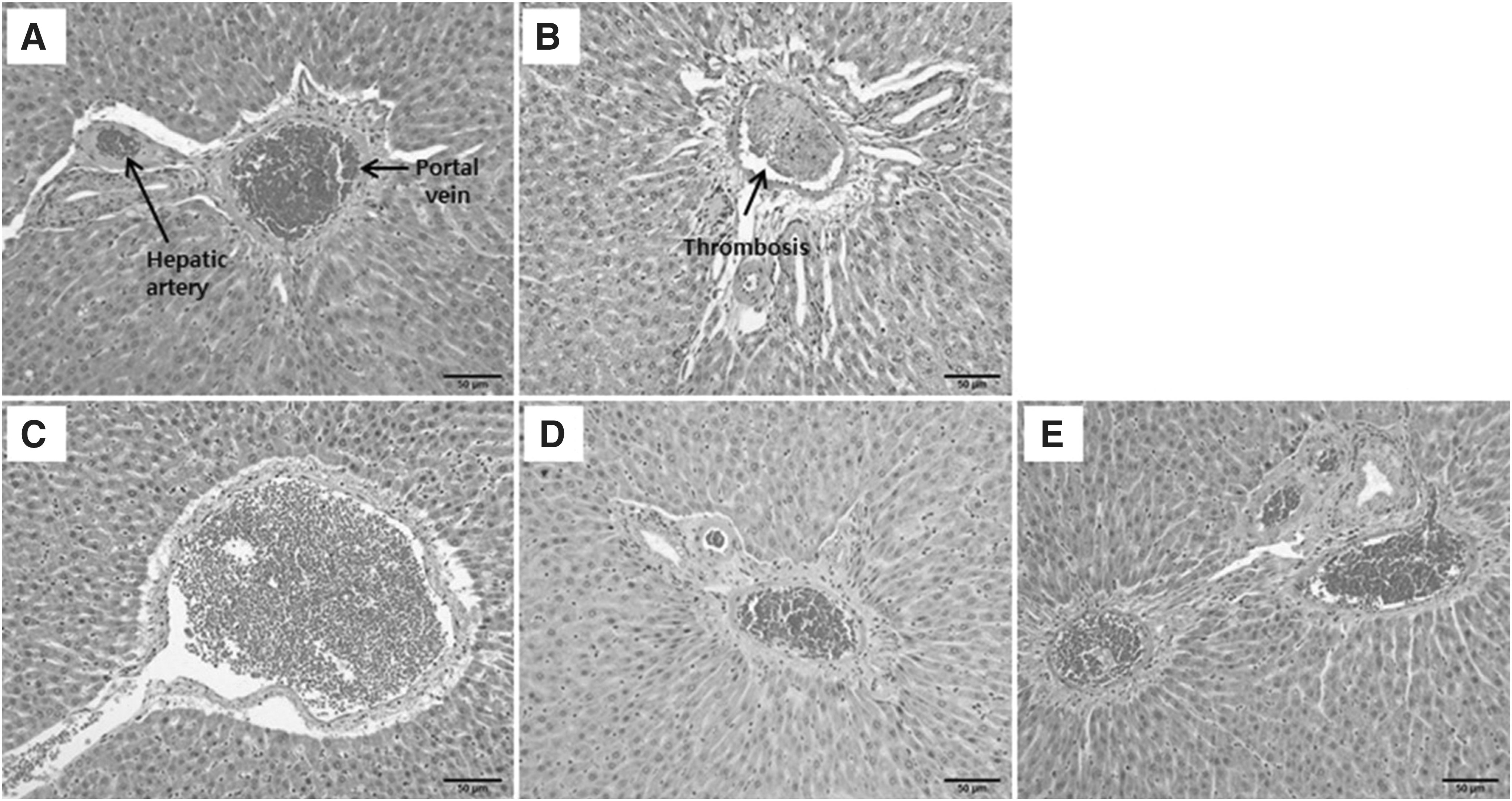
Histopathological changes in liver tissue when FEOJ was administered to thrombosis-induced rats. Thrombus formation in small blood vessels was indicated:
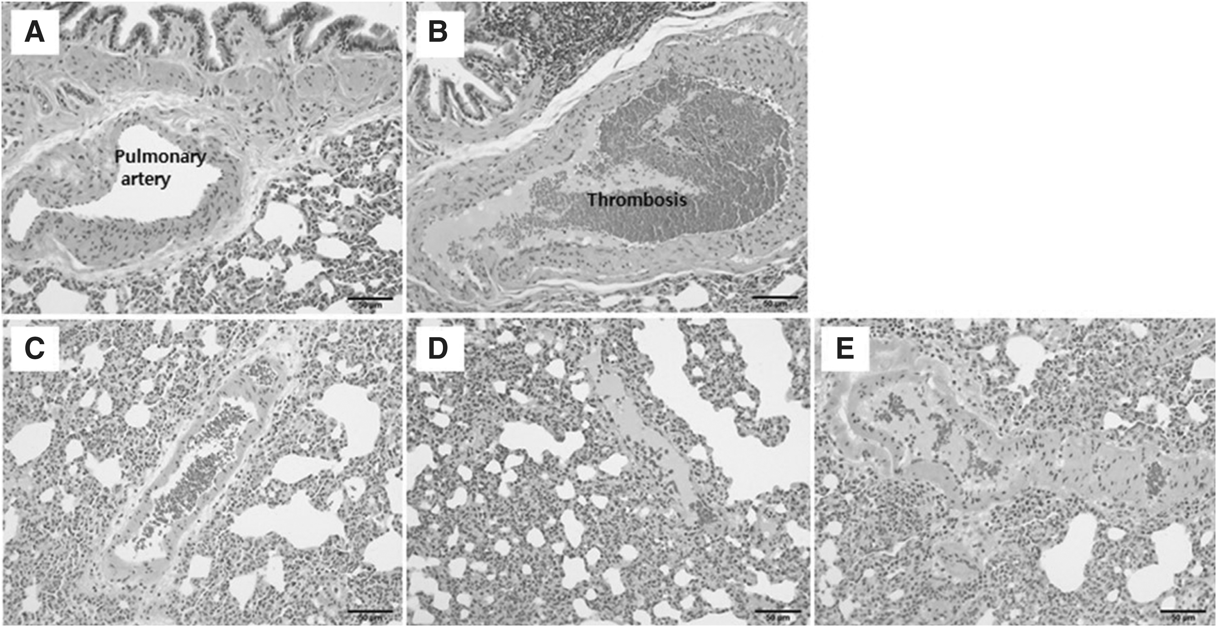
Histopathological changes in the lung tissue when FEOJ was administered in thrombosis-induced rats. Thrombus formation in small blood vessels was indicated:
Discussion
In this study, FEOJ was administered to SD male rats for 4 weeks to verify antithrombotic and thrombolytic effects of twice fermented extract of OJ. The increase in body weight, at the end of the study, exhibited no difference among the different groups. Similarly, organ weight of rats among different groups before and after the experiment remained unaltered (Table 1). The body weight of experimental animals is a good indicator to assess the change in a diseased condition. The administration of FEOJ used in this study did not affect the growth and maintenance of rats. Kim et al. 27 measured the effect of Liriope muscari extract on diabetes mellitus and obesity, by oral administering to OLETF rats. A similar pattern was observed in that study, which showed no significant difference in body weight between L. muscari extract and vehicle groups.
CT tended to be delayed in MOJ (6 mL/kg) and HOJ (12 mL/kg) compared with that in CON (P < .05) (Fig. 2). However, BT was dose-dependently delayed in all FEOJ groups compared with that in CON and TMB (P < .05) (Fig. 3). BT is an important measurement variable of antithrombotic activation studies. Prolonged BT indicates not only that blood smoothly circulates in blood vessels without interruption of blood flow by a clot or other substances, but also that blood is clean without impure substances in the vessels. 28 The result of this study showed that oral administration of FEOJ exhibited antithrombotic effects. Thrombin is activated through extrinsic and intrinsic pathways, converts FBG to fibrin, and produces a solid thrombus through a cross-linking process. 29 In the present study, indicators such as PT, APTT, and FBG were analyzed to investigate the effect of FEOJ on activation of thrombin. PT was observed to be shorter in FEOJ groups compared with that in CON. However, PT was significantly delayed in FEOJ groups compared with that in TMB, indicating prolonged CT. APTT was significantly delayed only in MOJ and HOJ compared with that in CON and TMB. No significant difference was observed in LOJ in comparison with CON and TMB (Fig. 4). Although there was a dose-dependent decrease in concentration of FBG in FEOJ groups, no statistically significant difference was observed among groups, including CON and TMB (Fig. 5).
The early stage of thrombus formation begins with the adhesion, activation, and aggregation of platelets in response to vascular endothelial injuries in a wound. This process is stimulated by the activation of coagulation pathway. Platelets release adenosine diphosphate (ADP) and thromboxane A2 (TXA2), activating more platelets. Platelet membrane receptor, GPIIb/IIIa, binds to FBG, thereby triggering platelet aggregation. 30 Activated platelets release serotonin, Ca2+, and TXA2, amplifying platelet aggregation and forming clots by reacting with coagulation factors in plasma. Thrombin, produced by the activation of intrinsic and extrinsic coagulation process, converts FBG to fibrin. 31 Considering that platelet aggregation and clotting occur rapidly in the same region, PT is the time until a fibrin clot is formed in the clotting cascade of extrinsic pathway, including II, VII, IX, and X factors. APTT is the time until fibrin clot is formed in the clotting cascade of intrinsic pathway, including XII, XI, IX, X, and II factors. 30,32 Under normal conditions, activated plasminogen (plasmin) degrades fibrin and FBG, dissolving thrombus, resulting in recovery of normal blood vessel. Thrombosis is caused under abnormal conditions. Therefore, it was speculated that FEOJ used in this study had an inhibitory effect on coagulation system, and affected the intrinsic and extrinsic pathways. Kou et al. 33 measured thrombus weight, PT, and APTT after inducing thrombus and administering the ethanol extract from OJ in animal model. They reported that thrombus weight significantly decreased in the extract from Radix OJ compared with positive control group (warfarin), but PT and APTT were not significantly delayed. These observations could be due to different components of nonfermented OJ, unlike FEOJ used in this study.
Kou et al. 6 have reported that ADP-induced platelet aggregation was significantly inhibited after distilled water extract of Radix OJ was orally administered to thrombus-induced rats. Cho et al. 34 have reported that the active component of C. militaris, cordycepin, had significant antiplatelet aggregation effect. Although platelets are activated through various pathways, it is known that GPIIb/IIIa activation is necessary to cause platelet aggregation at the last step of coagulation. Currently, thienopyridine drug, clopidogrel, is the most commonly used among antiplatelet agents, except aspirin. 29 Clopidogrel inhibits GPIIb/IIIa activation and affects platelet aggregation by blocking ADP binding to P2Y12 receptor of a platelet. Therefore, further studies are required to understand the anticoagulation effect of FEOJ, which specifically alters platelet aggregation.
The clotting status of capillaries, small arteries, and veins of liver and lung tissues were observed by H&E staining. Although thrombosis was observed in TMB and FEOJ groups, it was partially improved by FEOJ administration (Figs. 6 and 7). Adhesion, proliferation, and migration of WBCs in vascular epithelium were observed in liver tissue. Decreases in vascular epithelial cells and WBC deposits were also observed.
Alternatively, an inflammatory lesion and hyperemia were improved in FEOJ groups. From the results of this study, it is inferred that FEOJ could prevent thrombosis by inhibiting WBC adhesion as well as by protecting the vascular epithelial cells. It has been reported that vascular epithelial cells had important metabolic function in preventing arteriosclerosis. It played various roles such as primary hemostasis control, blood clotting, fibrinolysis, and interaction with WBCs. 35,36 Kou et al. 33 have reported that ethanol extract from OJ protected epithelial cells of a wound under anaerobic condition and alleviates inflammation of vascular walls. Histological analysis after administration of the extract in rats revealed these results.
Ruscogenin and cordycepin are the major active components of FEOJ responsible for antithrombotic and thrombolytic effects. Cho et al. 9 reported that collagen-induced platelet aggregation could be inhibited by using 500 μM cordycepin. Kou et al. 6 reported that 0.7 and 1.0 mg/kg ruscogenin had remarkable antiplatelet aggregation and antithrombotic effects. Fermentation has been suggested as a traditional process for enhancing the pharmacological effect of herbal medicine based on Oriental Medicine theory. Fermentation increases the production of active components in medicinal herbs, which beneficially influences health through health-promoting and disease-preventing effects. 37 Ahn et al. 38 indicated that fermented C. militaris had greater protein content and higher fibrinolytic activity than ordinary C. militaris. Song et al. 25 reported that FEOJ inhibited thrombin-induced growth of smooth muscle cells and inhibited signal transduction in the nucleus. In a previous study, our team measured the change in the content of active component of OJ and C. militaris before and after the second fermentation. Although cordycepin was not detected at all before fermentation, high cordycepin was detected as fermentation began. The content of ruscogenin increased about 2.8 times on the 10th day of fermentation compared with before fermentation. This result suggests that the active components of OJ and C. militaris rapidly increase through the second fermentation. This is the first report, to our knowledge, to demonstrate that FEOJ processed using two sequential fermentations with Lactobacillus and C. militaris would demonstrate greater antithrombotic and thrombolytic effects in thrombosis-induced rat models, compared with OJ or C. militaris alone.
In conclusion, the results of this study suggest that FEOJ developed through a two-step fermentation process with Lactobacillus and C. militaris can partially ameliorate experimental thrombosis in rat models, which supported its therapeutic applications. However, further studies are required, for defining a discrete dose, development of best administration route, and safety evaluation, to show optimal antithrombotic and thrombolytic effects.
Footnotes
Acknowledgments
The present study was supported by the “Food Functionality Evaluation Program” under the Ministry of Agriculture, Food and Rural Affairs and, in part, by the Korea Food Research Institute (Grant No. 20140114). This research was supported by the Chung-Ang University Research Scholarship Grants in 2015.
Author Disclosure Statement
No competing financial interests exist.