
Abstract
The aim of this study was to evaluate the wrinkle improving effect of hyaluronic acid intakes. Wrinkles were induced by exposing the skin of hairless mice to ultraviolet B (UVB) irradiation for 14 weeks. Hyaluronic acid was administered to the mice for 14 weeks including 4 weeks before experiments. Skin tissue was assayed by enzyme-linked immunosorbent assay to determine protein expression of wrinkle-related markers. The group supplemented with high concentrations of hyaluronic acid appeared significantly better than control group for collagen, matrix metalloproteinase 1, interleukin (IL)-1β, and IL-6 assay. Transforming growth factor-β1 (TGF-β1) and hyaluronic acid synthase 2 (HAS-2) were not shown to be significantly different. In conclusion, hyaluronic acid administration regulated expression levels of proteins associated with skin integrity, and improved the wrinkle level in skin subjected to UVB irradiation.
Introduction
U
Hyaluronic acid is a high molecular compound that consists of N-acetyl-
This research aimed to investigate the effect of consuming hyaluronic acid for preventing or reversing wrinkles in photoaged skin in a mouse model. Furthermore, through the enzyme-linked immunosorbent assay (ELISA), we confirmed protein expression levels that are wrinkle-related markers.
Materials and Methods
Preparation of hyaluronic acid
Hyaluronic acid was prepared as follows: Streptococcus zooepidemicus was cultured in an incubator of 37°C, pH 7, for 16–18 h. The medium consists of glucose, soy peptone, yeast, and KH2PO4. After the culturing was completed, the medium was sterilized and filtered and the low molecular weight byproduct was separated by ultrafiltration. After treating with sodium acetate, sedimentation was performed to yield hyaluronic acid and then ethyl alcohol was added for drying.
Animal experiments
This study was conducted with reference to previous experiments. 20 For the experiment, female SKH-1 hairless mice were purchased from DAEHAN Biolink of Korea. Mice were provided standard chow diets for 2 weeks, and then separated into six groups as follows: normal group, wrinkle-induced group (control group), wrinkle-induced group treated with 0.01% retinoic acid (positive control group), and three wrinkle-induced groups fed hyaluronic acid at different concentrations (10, 40, and 160 mg/kg). All six groups were observed for 14 weeks. The animals were kept in environmentally controlled conditions (temperature: 22°C ± 2°C, humidity: 50% ± 10%, lighting: 12-h light/12-h dark cycles, illumination: 200–300 lux, ventilation: 10–15 times/h). Feed was sterilized at 121°C for 15 min, before drying. Animal had free access to drink distilled water. Animal procedures were approved by the EBO animal care (Approval No.: EBOA-2015-06) and use committee and conformed to the guidelines of the Ministry of Food and Drug Safety.
Wrinkle induction
Except for the normal group, all other groups were exposed to UVB irradiation three times per week. First, after investigation, we determined that a standardized 1 minimal erythema dose (MED) consisted of 150 mJ/cm2, which produced the minimal detectable erythema after 24 h of UVB exposure. In the first week, mice have exposed to UV radiation for 1 MED. The next week, mice received illumination of about 2 MED of UV rays. The amount of UV exposure was 3 MED per mouse on the third week, and during weeks 4–10, each mouse received a UV dose of 4 MED. The amount of illumination was measured using a UV radiometer.
Dosage and duration of administration of test substances
Retinoic acid (100 μL) was used and administered three times per week as a positive control. The treatment group was administered hyaluronic acid orally for 4 weeks before UVB irradiation at 10 mL/kg, once a day for 14 weeks, which including the survey period of 10 weeks. Dose concentration of hyaluronic acid powder was dissolved with distilled water and diluted. Diluted samples were fed orally to mice.
Protein extraction of skin tissues
Skin tissues were obtained from the mice using the method described by Song et al. 21 Skin tissues obtained from sacrificed mice were fixed to use in 10% formalin solution, for over 24 h. Skin was prepared as follows: washing, dehydration, transparency, and infiltration. And then tissues were embedded in paraffin, and then sectioned to a thickness of 5 μm. Tissue slices were frozen in a deep freezer and stored until used. One hundred milligrams of separated and disrupted skin tissue was inserted into 1 mL PBS and frozen in liquid nitrogen, and thawed in a 37°C water bath. This process was repeated three times for total protein extraction. Extracted protein was quantified using the bicinchoninic acid (BCA) assay kit (Lot.PD202972; Pierce, USA), and each sample protein was determined using phosphate-buffered saline.
Wrinkle marker analysis
Experiments measuring proteins using ELISA kit for analysis of wrinkle-related factors followed the manufacturer's instructions 22 –25 for each assay (Table 1).
HAS-2, hyaluronic acid synthase 2; IL-1β, interleukin-1β; MMP-1, matrix metalloproteinase 1; TGF-β1, transforming growth factor-β1.
Statistical analysis
Results of all experiments were expressed as average and standard deviation and statistically analyzed using SPSS software (SPSS, Inc., ver 21.0). Significant differences were verified by one-way analysis of variation and was confirmed by Dunnet's t-test (significance level P < .05) and Duncan's multiple range test (significance level P < .05 and P < .01).
Results
As shown in Figure 1, UVB irradiation significantly decreased collagen levels in mouse tissues (normal group: 17.9 ng/mL, control group: 8.4 ng/mL). The hyaluronic acid-fed groups showed dose-dependent increases for collagen synthesis and were significant in the 160 mg/kg administration group at 14.5 ng/mL. Other hyaluronic acid-fed groups, 10 and 40 mg/kg, were not significantly different from the control group. Retinoic acid of 0.01% concentration group also did not show significant difference from the control group.

Effect of hyaluronic acid on the expression of collagen. Data are expressed as mean ± SD (n = 7). Nor.: normal group; UVB: UVB treated group; 10: HA 10 mg/kg treated group; 40: 40 mg/kg treated group; 160: HA 160 mg/kg treated group; RA: 0.01% retinoic acid treated group. *Significant differences from UVB group, *P < .05, **P < .01. SD, standard deviation; UVB, ultraviolet B.
The normal group and control group exhibited MMP-1 concentrations of 17.2 and 33.4 μg/mL (Fig. 2). As indicated in Figure 2, increased MMP-1 was induced by UVB irradiation. In the test groups, the group receiving 160 mg/kg of hyaluronic acid exhibited significantly lower MMP-1 levels. However, there was no significant difference in the groups receiving less hyaluronic acid (10 mg/kg: 28.0 μg/mL, 40 mg/kg: 21.7 μg/mL). The retinoic acid group exhibited decreased amount of MMP-1 synthesis, which was comparable to that of the control group at 20.5 μg/mL.
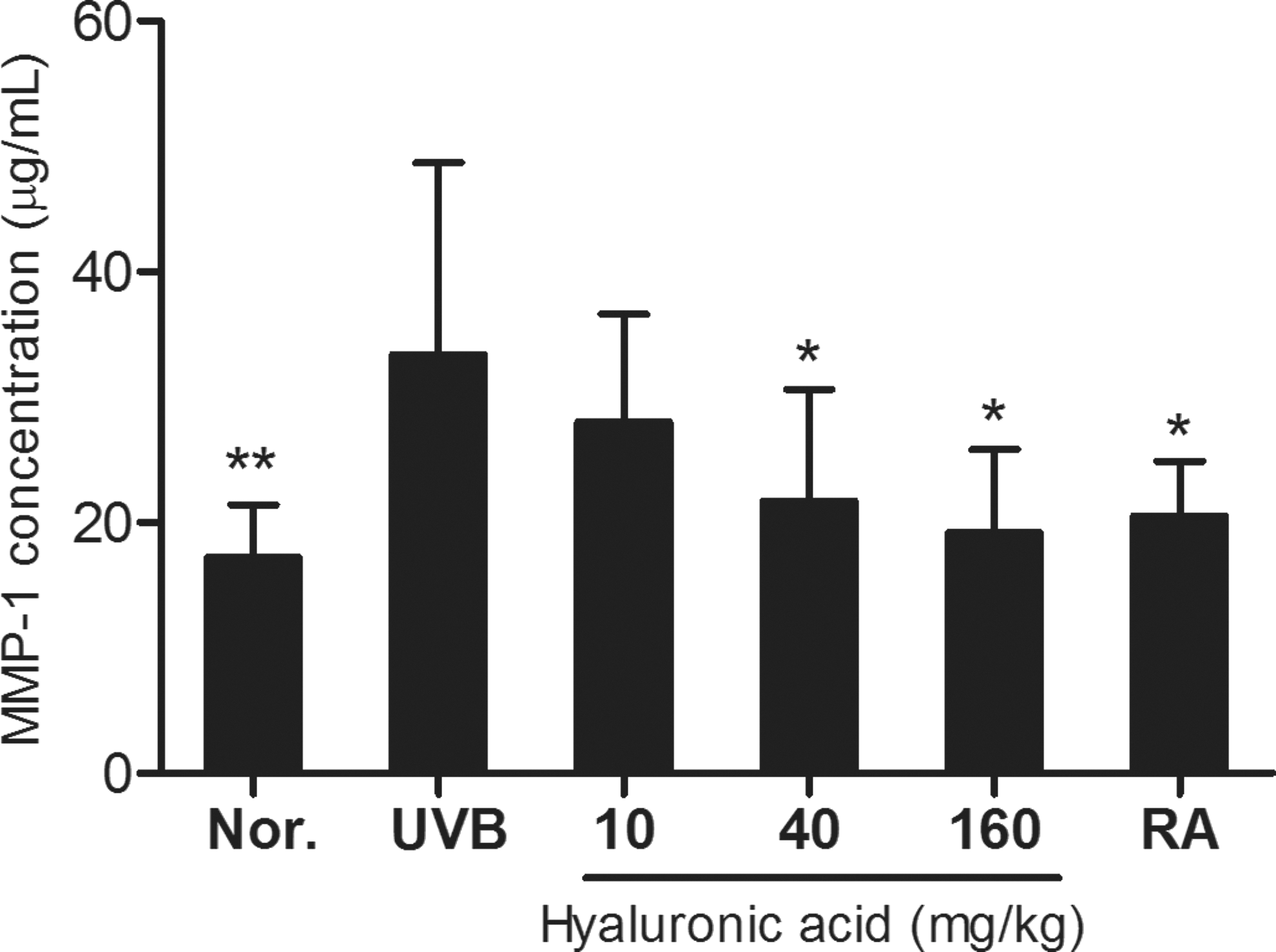
Effect of hyaluronic acid on the expression of MMP-1. Data are expressed as mean ± SD (n = 7). *Significant differences from UVB group, *P < .05, **P < .01. MMP-1, matrix metalloproteinase 1.
Next, we measured IL-1β expression levels in the mouse skin. As shown in Figure 3, the IL-1β expression level was increased in the wrinkle group (920.5 pg/mL) induced by UVB irradiation compared with that in the normal group (453.7 pg/mL). In the three test groups, only the 160 mg/kg capacity group decreased IL-1β levels as 411.5 pg/mL. When comparing the retinoic acid group and control group, the retinoic acid group was not significantly different from the control group.
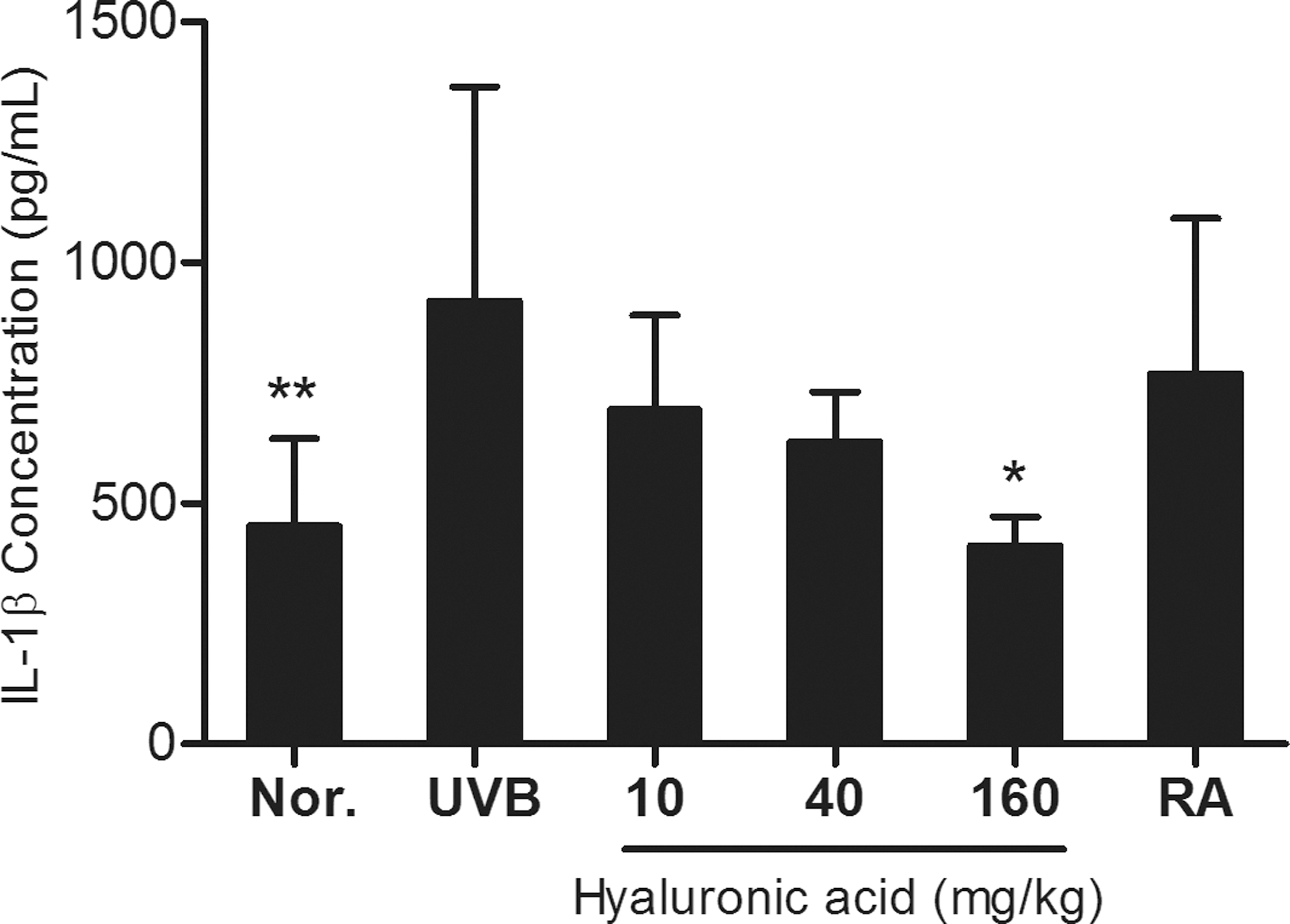
Effect of hyaluronic acid on the expression of IL-1β. Data are expressed as mean ± SD (n = 7). *Significant differences from UVB group, *P < .05, **P < .01. IL, interleukin.
We also performed ELISA for IL-6 analysis. IL-6 was elevated in the wrinkle group compared with that in the normal group (Fig. 4). The groups fed hyaluronic acid tended to dose-dependently decrease in IL-6: 40 and 160 mg/kg were 262.3 and 241.0 pg/mL, respectively. Mouse cells that were treated with retinoic acid had lower amounts of IL-6 synthesis (250.6 pg/mL) than the wrinkle group (321.0 pg/mL).
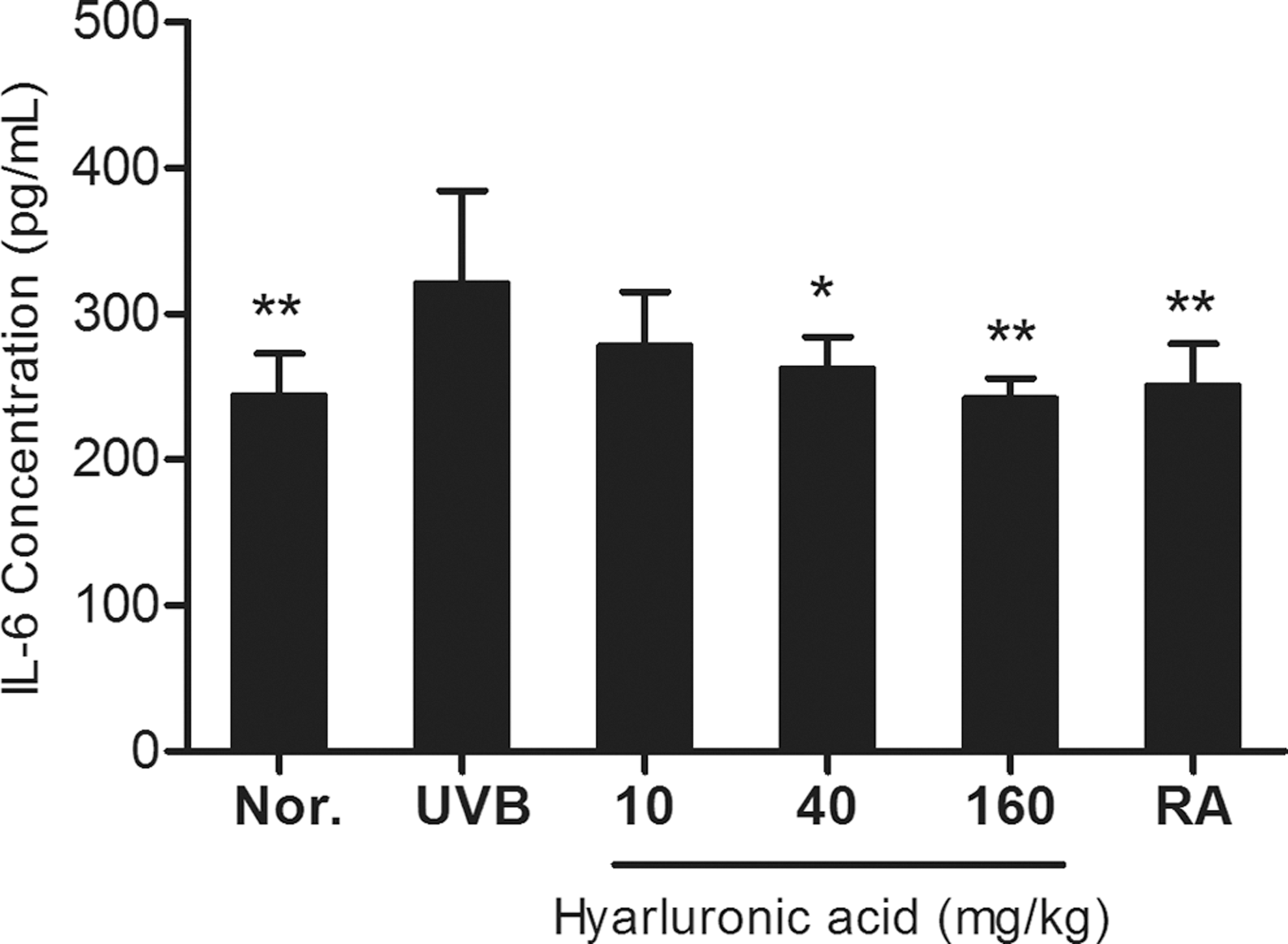
Effect of hyaluronic acid on the expression of IL-6. Data are expressed as mean ± SD (n = 7). *Significant differences from UVB group, *P < .05, **P < .01.
Transforming growth factor-β1 (TGF-β1) in the normal and wrinkle groups was not statistically different as 530.1 and 466.2 pg/mL (Fig. 5). Hyaluronic acid-treated groups tended to have lower levels than the control group, but none of the results were significantly different from the control group. Retinoic acid treatment also made no significant difference.
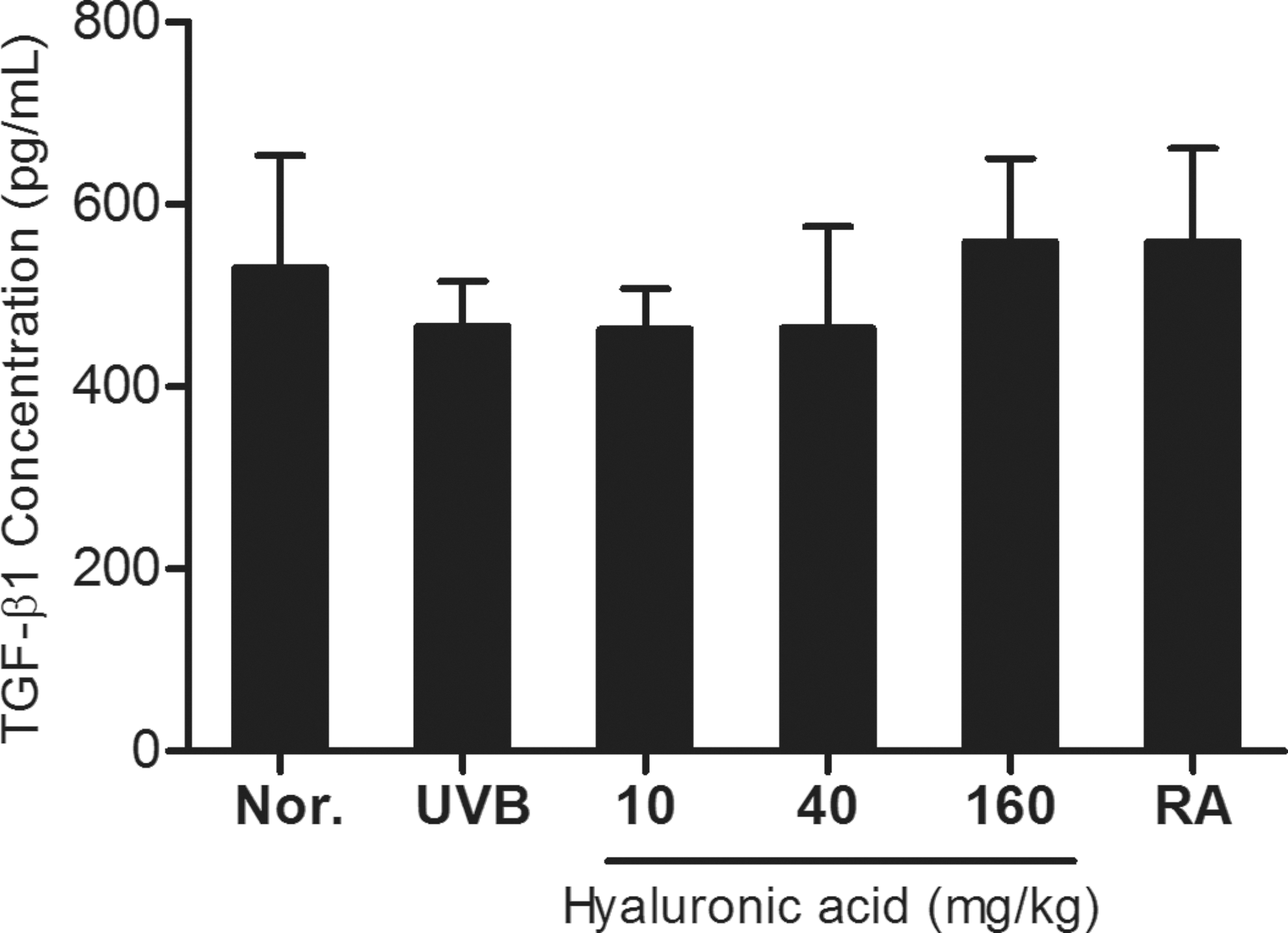
Effect of hyaluronic acid on the expression of TGF-β1. Data are expressed as mean ± SD (n = 7). TGF-β1, transforming growth factor-β1.
As shown in Figure 6, HAS-2 levels in the normal group and wrinkle group were not statistically different at 3946.7 and 2885.1 pg/mL, respectively. There were also no significant differences in the skin tissues of mice treated with hyaluronic acid, regardless of concentration, from those of the control group. The positive control group had 2944.0 pg/mL of HAS-2, which was not significantly different from that of the other groups.
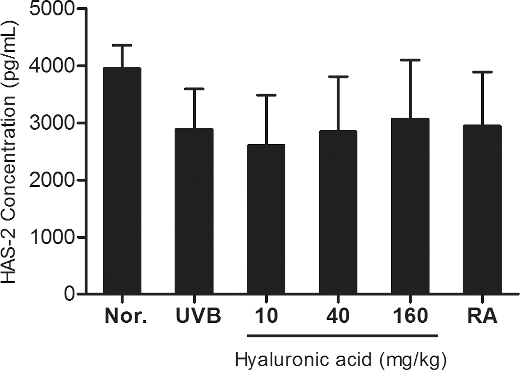
Effect of hyaluronic acid on the expression of HAS-2. Data are expressed as mean ± SD (n = 7). HAS-2, hyaluronic acid synthase 2.
Discussion
To evaluate the antiwrinkle effect of hyaluronic acid intake, skin tissues of hairless mice that were exposed to UVB radiation were evaluated for change in the protein expressions of factors related to skin wrinkles. This study demonstrated that collagen, MMP-1, IL-1β, and IL-6 protein expression level were significantly affected by high-concentration-treated sample, especially 160 mg/kg. However, HAS-2 and TGF-β1 protein expressions were not significantly different. These results indicated that administering hyaluronic acid improves photoaged skin induced by UVB irradiation. After hyaluronic acid treatment, exposure to UVB irradiation results in CD44 and RHAMM (receptor for hyaluronic acid-mediated motility) expressions to be specifically inhibited. A similar mechanism leads to inhibition of hyaluronic acid synthesis. ROS produced by UV rays leads to cytokines, such as TGF-β1, 26,27 keratinocyte growth factor, 28 IL-1β; and negatively affects the regulation and expression of hyaluronic acid metabolizing enzymes. Skin damaged by UVB radiation has produced many fragmented hyaluronic acid molecules and has increased hyaluronidase and decreased hyaluronan synthases, CD44, and RHAMM. These results indicate that skin has limited ability to protect itself from UVB irradiation, which inhibited hyaluronic acid synthesis-induced inflammation. 29,30 Hyaluronic acid binding to CD44 and hyaluronidase in the cytoplasm hydrolyzes hyaluronic acid into small oligosaccharide fragments. These fragments transform NLRP3 cryopyrin into pro-IL-1β and convert it to active IL-1β. 31 CD44 binding with hyaluronic acid activates p38 MAPK and NF-κB. Then, hyaluronic acid also increased transcription levels of IL-1β and CXCL2 mRNA. 22 Through these mechanisms, hyaluronic acid helped to heal skin injured by UVB exposure. As in vivo tests, hyaluronic acid improved wrinkle indicators and histological changes in hairless mouse skin. Furthermore, hyaluronic acid showed improved elastosis and decreased collagen degradation in the treated group. 20 These results indicate that hyaluronic acid has the potential value as a health-promoting functional food.
Footnotes
Author Disclosure Statement
No competing financial interests exist.