
Abstract
This study aimed to identify the changes in the metabolomics profile of liver damage caused by alcohol consumption and verify the beneficial effect of Prunus mume Sieb. et Zucc extract (PME) in protection of alcohol-induced injury by attenuating the level of identified metabolites. Mice were treated with PME and saline or untreated once daily for 5 days, followed by alcohol injection. The plasma samples were analyzed using liquid chromatography–mass spectrometry-based high-resolution metabolomics followed by a multivariate statistical analysis using MetaboAnalyst 3.0 to obtain significantly expressed metabolites, using a false discovery rate threshold of q = 0.05. Metabolites were annotated using Metlin database and mapped through Kyoto Encyclopedia of Genes and Genomes (KEGG). Among 4999 total features, 101 features were significant among alcohol- and PME-treated mice groups. All the samples cluster showed a clear separation in the heat map, and the scores plot of orthogonal partial least squares-discriminant analysis (OPLS-DA) model discriminated the three groups. Phosphatidylcholine, Saikosaponin BK1, Ganoderiol I, and N-2-[4-(3,3-dimethylallyloxy) phenyl] ethylcinnamide were among the significant compounds with a low intensity in alcohol group compared to PME group, suggesting that these compounds have a relation in the development of PME's protective effect. The study confirms the hepatoprotective, antioxidant, and anti-inflammatory effects of PME against alcohol-induced liver steatosis, inflammation, and apoptosis.
Introduction
T
Alcohol has long been known to be a major risk factor for several types of cancers, including cancers of oropharynx, larynx, esophagus, breast, and colon. HCC is now one of the most frequently diagnosed liver cancers. Although the primary risk factors of HCC are aflatoxin and hepatitis B and C infections, lifestyle-associated risk factors such as heavy alcohol drinking, obesity, diabetes, smoking, and nonalcoholic steatohepatitis should also be considered. 6 Despite the significant improvements in treatment by mainstream medicine, mortality rates have not been significantly improved, and new therapeutic strategies are required to put an end to this frightening disease.
Previously, oxidative stress has been shown as one of the key risk factors in hepatocarcinogenesis. 7 A variety of herbal extracts with antioxidant activity such as Cichorium intybus L, Allium sativum, Taraxacum officinale, Glycyrrhiza glabra, Silybum marianum and Taraxacum officinale have been studied for their beneficial effects on cancer and other hepatic diseases. The use of these natural antioxidants has been confirmed as a promising approach for preventing hepatocarcinogenesis. 8 Prunus mume Sieb. et Zucc. (PM), which originates from southeastern China, is well known as an antioxidative and anti-inflammatory fruit. 9,10 PM extract (PME) has been shown to be a potential therapeutic agent for inflammatory bowel disease, 11 diabetes mellitus, 12 vascular dementia, 13 and cancers, including HCC. 14 In our previous study, we identified that our PME contains 5-O-caffeoylquinic acid (NeoCGA) and 3-O-caffeoylquinic acid (CGA), 15 which were previously reported as major bioactive antioxidant compounds in PM fruits. 9 Furthermore, we observed that alcohol treatment elevated serum and hepatic triglyceride, whereas serum triglyceride levels were reduced with PME treatment, which indicated that PME can prevent alcoholic steatosis in the liver. In addition, we demonstrated that PME decreased cellular oxidative stress by inhibiting mitogen-activated protein kinase (MAPK) activation and p53-mediated apoptotic signaling axis, resulting in the inhibition of hepatic steatosis and apoptosis. In this study, we extended our research using a metabolomics approach and investigated the metabolic changes to understand the molecular mechanism by which PME conferred a hepatoprotective effect, using plasma from mice with alcohol-induced liver injury or protected with PME. 15
Metabolomics is an emerging analytical technique to seek global profiles of metabolites in particular samples, including endogenous and exogenous metabolites. Since the metabolites reflect the status of physiology in the body, the metabolomic analysis can be used as a useful tool for diagnosis of latent diseases. Recent studies have suggested that the metabolites can be the indicators of diseases such as cancer, 16 alcoholic liver disease, 17 or Parkinson's disease. 18 This study aimed to identify the changes in metabolomic profiles involved in liver damage caused by alcohol consumption and finally verify if PME is beneficial in protecting against alcohol-induced liver injury by attenuating the level of identified metabolites.
Materials and Methods
Preparation of PME
PME was prepared as discussed in our previous study. 15 Briefly, 50 grams of each ground, seed-free sample was submerged in 1 L of 75% fermented ethanol and the sample was extracted by shaking for 12 h at room temperature (25°C). The extract was filtered through Whatman No. 41 filter paper (Whatman Int. Ltd., Maidstone, UK), freeze dried using a programmable freeze dryer (Ilshin Lab Co., Yangju, Korea), and then ground and stored.
Animals and experimental design
The care and treatment of experimental animals conformed to a protocol approved by the Institutional Animal Care and Use Committee of Korea University (Seoul, Korea). C57BL/6J mice (8-week old) were purchased from Central Lab. Animal, Inc. (Seoul, Korea) and were housed in individual cages in a windowless room with a 12-h light–12-h dark cycle. Mice were treated with PME (75% ethanolic extract, 100 mg/kg of body weight) or vehicle (saline) once daily for 5 days. Thirty minutes after the PME treatment, mice received alcohol (1 g/kg of body weight) (Table 1 and Fig. 1). The mice were euthanized by Avertin (2,2,2-tribromoethanol; Sigma-Aldrich, St. Louis, MO, USA) for the collection of blood.
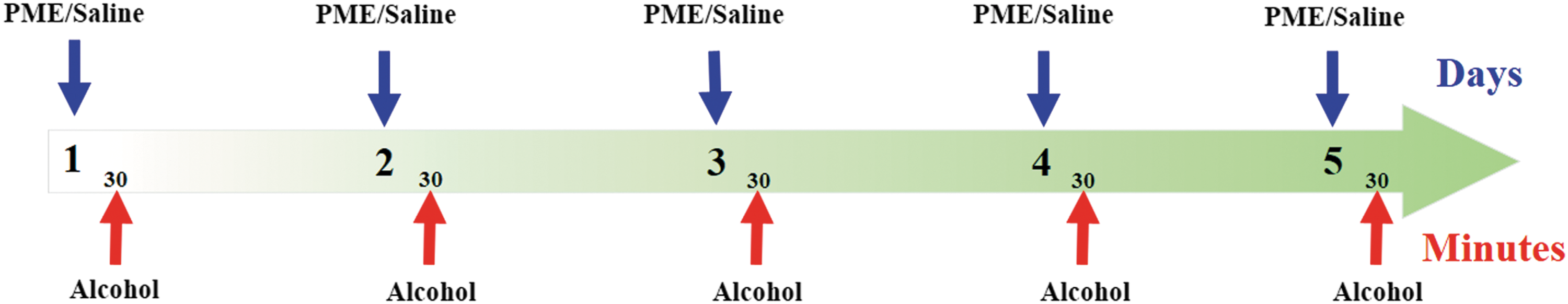
Experimental design of alcohol-induced liver injury mouse model.
IP, intraperitoneal; PME, Prunus mume Sieb. et Zucc extract.
Sample collection
Blood samples were collected by cardiac puncture followed by centrifugation at 2000 g for 20 min at 4°C immediately after collection of blood. Plasma protein was quantified by the Bradford Protein Assay and 3 mg/mL concentration was used for analysis. Fifty microliter aliquots of supernatant samples were treated with 100 μL of acetonitrile and 2.5 μL of internal standard (C18) and then centrifuged at 13,000 g for 10 min at 4°C to precipitate proteins. High-resolution liquid chromatography–mass spectrometry (6530 Q-TOF LC-MS; Agilent Technologies, Santa Clara, CA, USA) was employed to analyze samples. 19 All the samples were run in triplicate in positive mode with 0.1% formic acid and water.
Analysis of metabolites by LC-MS/MS
The chromatographic analytical procedures were performed on an Agilent 1260 Series (Agilent Technologies) LC system equipped with a binary pump, an online degasser, an auto plate-sampler, and a thermostatically controlled column compartment. The columns were maintained at 30°C. The separation was carried out on a Shiseido CapCell PAK C18 column (150 × 4.6 mm, 5 μm), preceded by a C18 guard column (4.0 × 3.0 mm; Phenomenex, Torrance, CA, USA). The binary gradient elution system consisted of 0.1% formic acid in water (solvent A) and 0.1% formic acid in acetonitrile (solvent B), and separation was achieved using the following gradient: 2% B at 0–5 min and 2–95% B at 5–30 min. The composition was held at 95% B for 5 min, then returned to initial conditions, and maintained for 10 min to equilibrate the column. The flow rate was 0.6 mL/min and the injection volume was 10 μL.
Analytes were detected by mass spectrometry using an Agilent 6530 Q-TOF mass spectrometer (Agilent Technologies) equipped with an electrospray ionization interface, and was operated under the same conditions for positive and negative ion mode, with a curtain gas of 40 psi, drying gas temperature of 325°C supplied at 14 mL/min, and sheath gas temperature of 350°C supplied at a flow rate of 10 and 12 mL/min. All samples were run in triplicates and data for each ionization technique were acquired in positive ion mode. Data were collected in centroid mode and the mass range was set at m/z (mass/charge ratio) 50–1000 using the extended dynamic range. The accurate mass capability of the TOF analyzer allowed reliable confirmation of the identity of the detected metabolites, with mass errors below 5 ppm in routine analysis. 20
Metabolic profiling
After running high resolution, the raw LC-MS data were converted into the mzData format using Agilent MassHunter Qualitative Analysis software (version B.05.00; Agilent Technologies) and subsequently processed by software (apLCMS) using Limma R package for quantification of ion intensities. 21 apLCMS provided 11,407 m/z within a range of ions set from 50 to 1000 with 20,000 resolutions. 22 Data were further processed with normalization, scaling, filtering, and statistical analysis using MetaboAnalyst 3.0. 23,24 MetaboAnalyst filtered the noninformative variables such as variables of very small values (close to baseline or detection limit) and variables that are near-constant values by applying an interquantile range filter. In this way, the top 4999 m/z was used in the subsequent analysis. Statistical significance of features was determined among vehicle control, alcohol-treated, and PME-treated mice groups using one-way analysis of variance (ANOVA) with Fisher's LSD test. P < .05 was considered to be statistically significant. Significant features with P < .05 between alcohol and PME groups and/or between alcohol and vehicle groups were identified by Student's t-test. The raw data were log transformed and hierarchically clustered by Pearson correlation and average linkage clustering. Signal intensities of the differentially abundant metabolites were visualized as a heat map and orthogonal partial least squares-discriminant analysis (OPLS-DA) using MetaboAnalyst 3.0. The discriminatory m/z values after OPLS-DA were used to identify metabolites using an online database METLIN, 25 and the feature distribution on Mus musculus (mouse) metabolic pathway was mapped using Kyoto Encyclopedia of Genes and Genomes (KEGG) database. 26 Subsequently, the significant m/z values after OPLS-DA with filtering P-value less than .05 were annotated by MetaboAnalyst 3.0 to create a potential metabolic network model.
Biomarker identification and pathway enrichment analysis
Compounds with significant changes between groups (P-value <.05 and fold-change >2) were selected as biomarkers. The potential biomarkers were identified by an online biochemical database service Metlin Mass Spectrometry Database METLIN. The recorded KEGG numbers were subjected to mouse metabolomics pathway (KEGG) and MetaboAnalyst for further enrichment and a pathway analysis. Detected m/z features that matched known mouse intermediary metabolites were observed as black dots in the map.
Results
Three-way hierarchical cluster analysis
Protective effects of PME treatment for preventing alcohol-induced liver injury in mice such as steatosis, inflammation, and apoptosis were verified using three-way comparisons between the three groups of vehicle control, alcohol, and PME. Figure 2 shows the significant features, which are differentially expressed among vehicle control, alcohol-treated, and PME-treated groups. The Y axis represents the −log10 of the raw P-value between compared groups, while the X axis shows the compounds after normalization, with their m/z values ranging from 50 to 1000. The dashed line represents the false discovery rate (FDR) adjusted q value of 0.05 which separates the significant features (red dots) from other insignificant compounds (green dots). In this way, a total of 223 compounds were detected above the dashed FDR line among total (4999) compounds, and all metabolites (detected 223 features) above the threshold were considered statistically significant between the three groups. Among 4999 total features, 166 features were significantly different among vehicle and alcohol groups, while 101 features were significant among alcohol and PME groups. Furthermore, a three-way hierarchical cluster analysis was performed to organize and cluster samples using significant features obtained from FDR (q = 0.05) using MetaboAnalyst 3.0 as shown in Figure 3. The features under blue, red, and green panels at the top represent vehicle, alcohol, and PME groups, respectively. Sample names are at the bottom and significant m/z values are at the right side. Significant features were used to cluster the samples of vehicle, alcohol, and PME groups. All the samples clusters show a clear separation as shown by blue, red, and green panels at the top. This apparent separation in the heat map suggests that the metabolites are highly differentiated among the three groups. Furthermore, to better define the differential metabolic profile and variations among groups of mice treated with alcohol and those treated with PME followed by alcohol, all observations acquired were analyzed using autoscaled OPLS-DA. As shown in Figure 3b–d, the scores plot of OPLS-DA model discriminated the alcohol group from their corresponding control groups or PME group, which exhibited satisfactory classification.
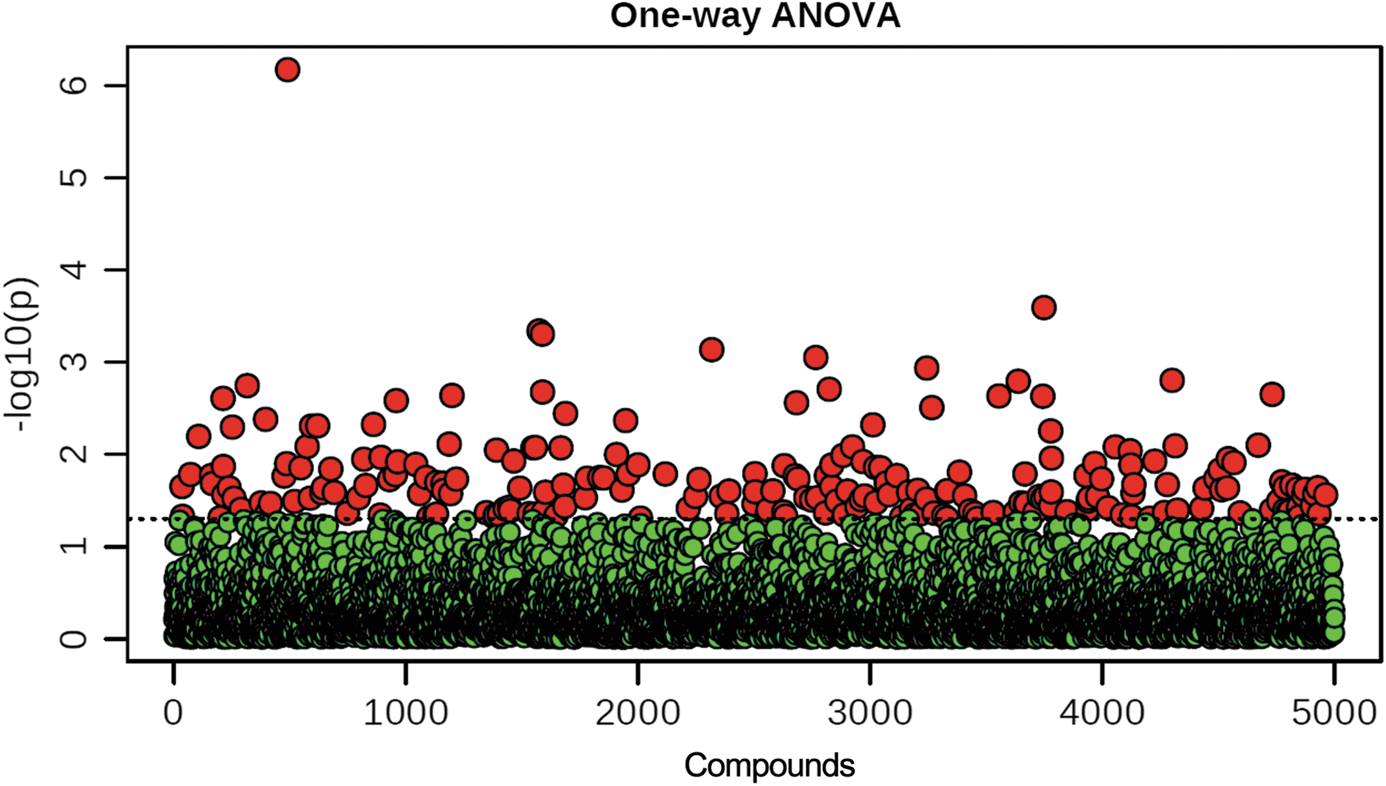
Important features selected by ANOVA plot with a P-value threshold of .05. The Y axis represents the −log10 of the raw P-value between compared groups, while the X axis are the 4999 compounds after normalization of their m/z values ranging from 50 to 1000. The dashed line represents the false discovery rate significant threshold (q = 0.05), which separates the significant features as red dots from other insignificant m/z, the green dots. Fisher's LSD ANOVA test. ANOVA, analysis of variance; m/z, mass/charge ratio.
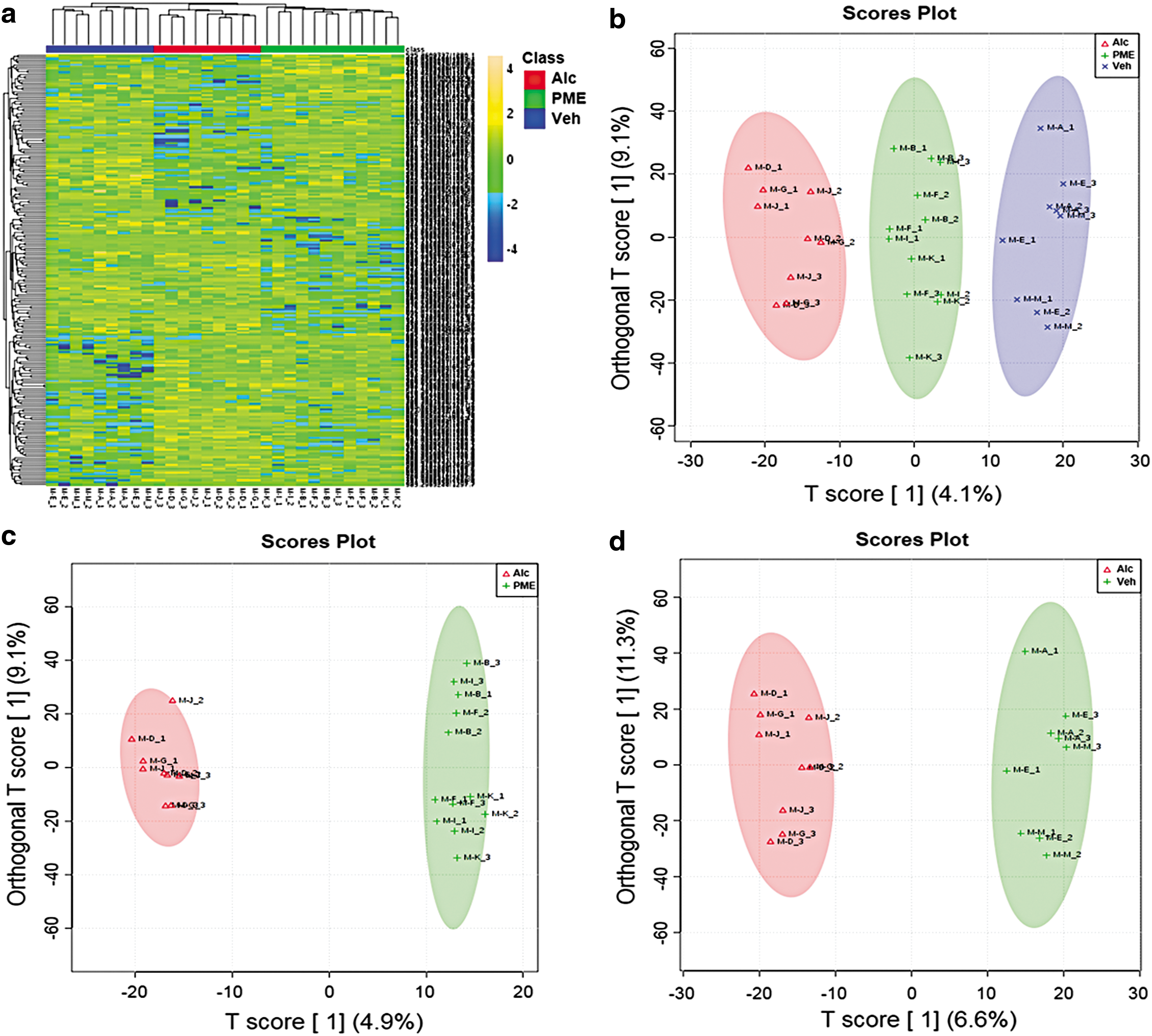
Separation and classification among the metabolites between control, alcohol-treated or PME-treated mice groups.
KEGG pathway analysis
Highly significant features obtained from ANOVA were annotated in Metlin database (
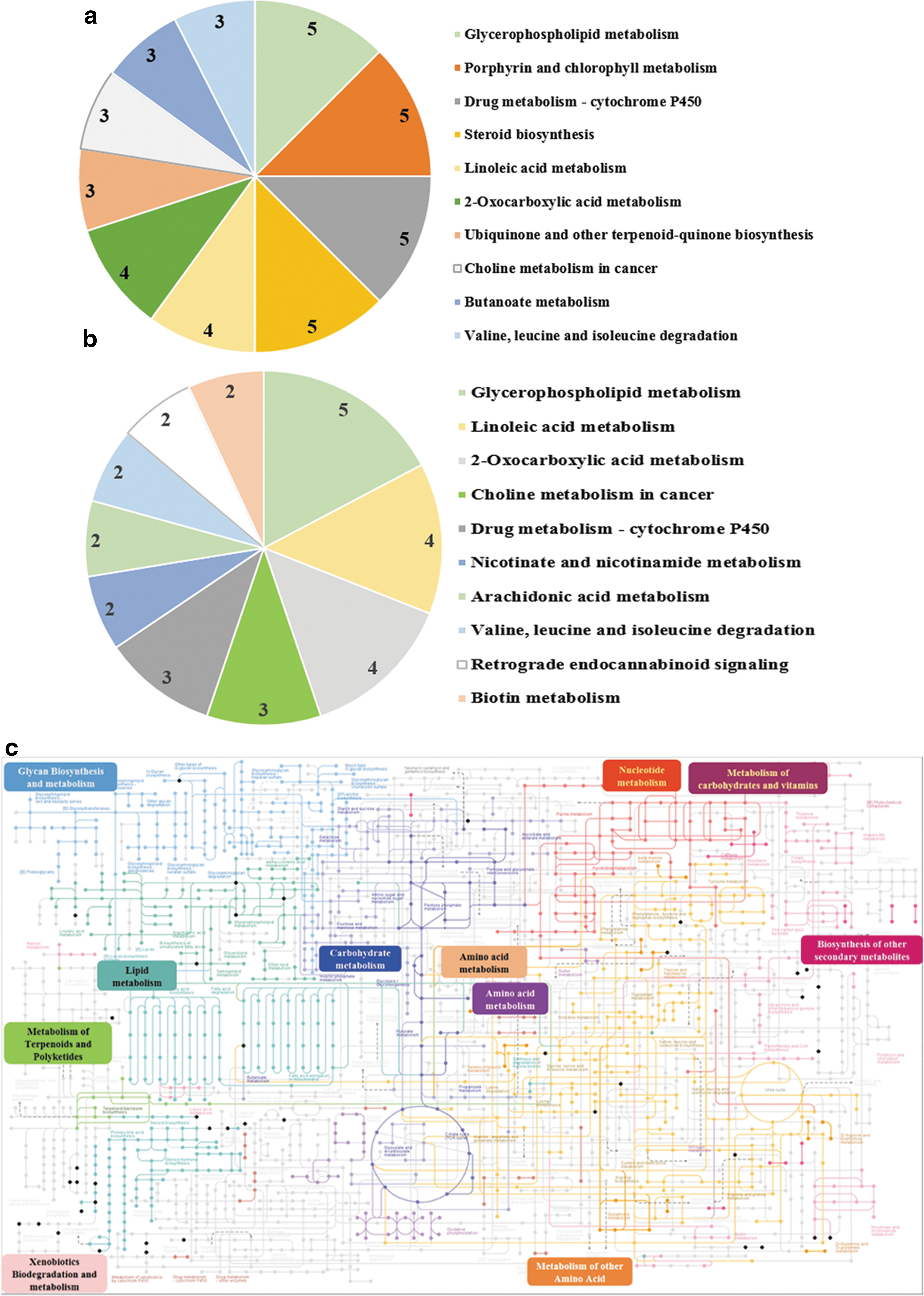
Top 10 KEGG pathways with the greatest number of compound hits.
Effects of PME on molecular pathways of alcohol-induced liver injury
In our previous study, we demonstrated that NeoCGA and CGA in PME decreased cellular oxidative stress, which plays key roles in the activation of MAPK and p53-mediated apoptotic signaling axis, resulting in the inhibition of molecular mechanism involved in the alcoholic liver injury such as hepatic steatosis and apoptosis. 15 In this study, using the metabolomics approach, we aimed to analyze detailed metabolite profiling to elucidate the hepatoprotective mechanisms of PME. We selectively analyzed the 50 highly significant compounds among vehicle, alcohol, and PME groups. Figure 5 shows the relative concentration of the compounds assessed using their respective signal intensities from quadrupole time-of-flight mass spectrometer (QTOF), which are of importance in terms of hepatoprotective effects of PME against alcohol liver injury. Figure 5a shows the relative intensity of phosphatidylcholine (PC), which is observed to be significantly lower in mice treated with alcohol possibly because of alcohol-induced liver damage, in comparison with PME treatment followed by alcohol or vehicle control. PC has been shown to have a vital importance in liver repair in animal models. 27 The increase in the PC concentration with PME treatment to the level of vehicle control mice group supports the hepatoprotective role of PME. Furthermore, we have observed the significantly increased concentration of Saikosaponin BK1 in PME-treated mice in comparison with alcohol alone or vehicle control, as shown in Figure 5b. Saikosaponins isolated from the Bupleurum roots are widely used medicinal herbs in the treatment of chronic liver diseases. 28 This result suggests that PME may contain the Saikosaponin BK1 as one of the active components, which can help reduce the hepatotoxicity caused by consumption of alcohol. Furthermore, as N-2-[4-(3,3-dimethylallyloxy) phenyl] ethylcinnamide obtained from Aegle marmelos (Bael tree) leaves were also documented for hepatoprotective effects in alcohol-induced liver injury in albino rats, interestingly in our study, as shown in Figure 5c, we also have observed a significantly higher concentration of N-2-[4-(3,3-Dimethylallyloxy) phenyl] ethylcinnamide in PME-treated mice compared to control or alcohol-treated groups. 29,30 This result also suggests that PME treatment before alcohol consumption can help reduce the damage of liver caused by alcohol. On the same note, Ganoderma lucidum has a long history of potent hepatoprotective effects on liver injury. 31 –33 In accordance with that in our study, we have observed a higher concentration of Ganoderiol I (an active component of G. lucidum) in PME-treated mice as shown in Figure 5d. This result indicates that PME has an antihepatotoxic activity in mice against the alcohol, which might be exhibited by the presence of Ganoderiols in PME. Furthermore, two more compounds, namely N-(2-hydroxy-hexadecanoyl)-4E,6E-tetradecasphingadienine-1-phosphoethanolamine and a compound with unknown chemical identity m/z: 839.77, were detected significantly higher in alcohol-treated mice in comparison with control group or PME-treated mice group as shown in Figure 6. The role of the unknown compound could not be identified for its relative alcohol-induced toxicity, but N-(2-hydroxy-hexadecanoyl)-4E,6E-tetradecasphingadienine-1-phosphoethanolamine is a subclass of ceramide phosphoethanolamine (CPE), which is shown as the apoptotic bioactive lipid involved in the alcohol-induced neuronal cell loss. 34,35 We suspect that the significantly elevated level of CPE in alcohol-exposed mice is due to the toxicity of alcohol in mouse brains, while with PME treatment, the intensities of these two toxic compounds have returned to baseline, such as in control mice as shown in Figure 6. We could not explain the role of significantly higher intensity of an unknown compound, but we suppose it one of the toxic compounds released as a result of alcoholic toxicity which, like CPE, have returned to the baseline. These results suggest that the treatment of PME before alcohol treatment shielded the toxicity, which may happen due to the undesirable components of alcohol.
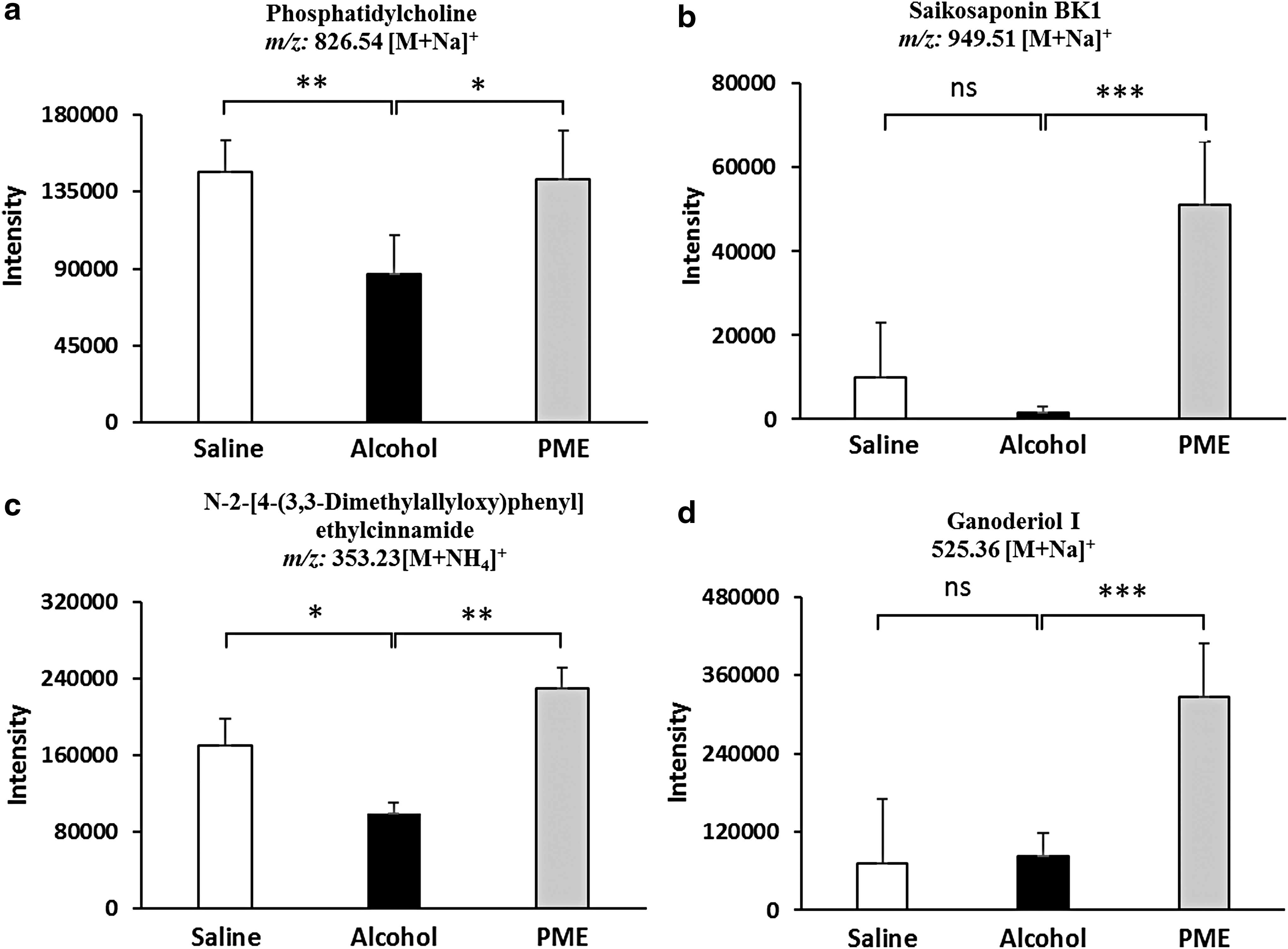
Relative concentrations of significant compounds between control versus alcohol- versus PME-treated mice groups.
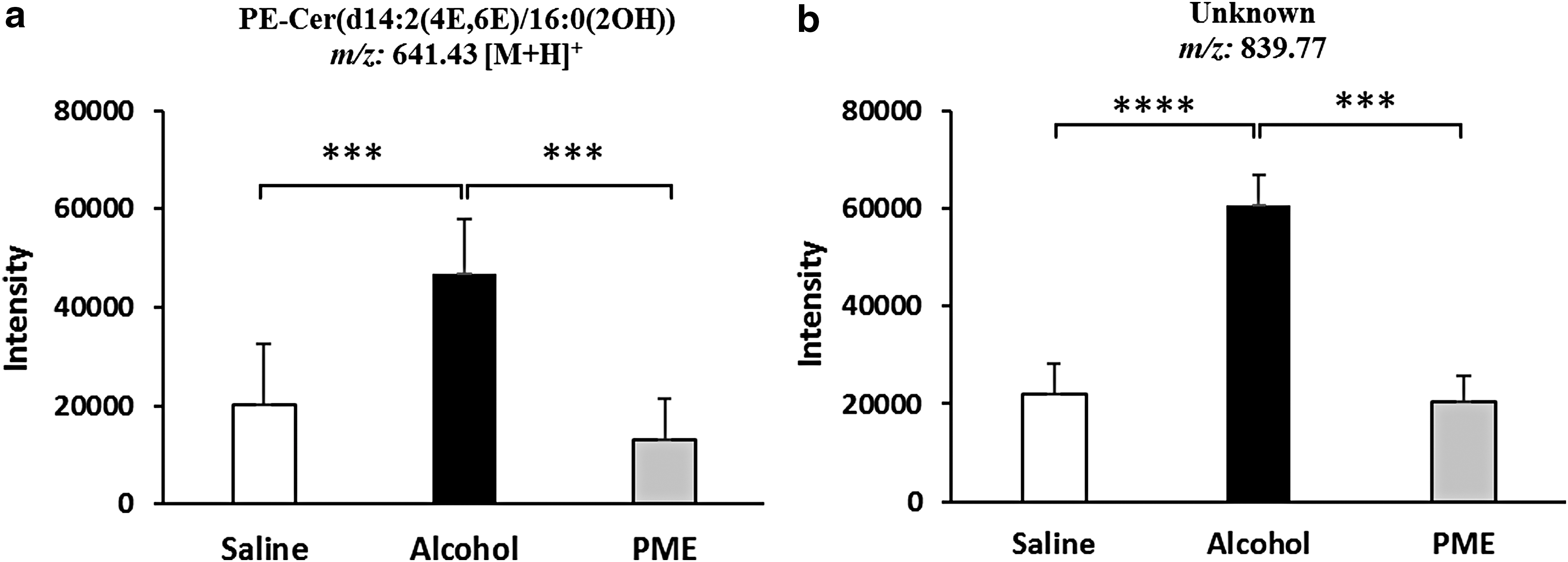
Relative concentrations of significant compounds between control versus alcohol- versus PME-treated mice groups.
Discussion
In the last 2 decades, PME has been investigated extensively for its beneficial effects on various metabolic diseases, including obesity, 36 cancer, 37 and thrombosis. 38 However, to date, little is known about the efficacy of PME for protecting against alcoholic liver injury. In our previous study, we have shown that the PME treatment not only helped lower triglycerides in mouse serum or liver but also inhibited the alcohol-induced apoptosis and ROS-mediated p53 signaling pathway, thereby reducing liver steatosis, inflammation, and apoptosis. In this study, using a metabolomics approach, we have identified the potentially important metabolites generated while treating mice with PME, which can help understand the mechanism involved in the hepatoprotective effect of PME.
PC is the most abundant phospholipid component found in every cell of the human body. Previous studies performed on the importance of PC have demonstrated several important roles of PC, such as PC makes acetylcholine in human brain, 39 and has been shown to be interrelated with several brain-centered conditions such as dementia, 40 Alzheimer's disease, 41 anxiety, and depressive disorders. 42 Heavy alcohol consumption is well documented to have harmful neurologic effects. 43 Elevated levels of ceramides in brain, which are an analogue of sphingolipids, are shown to cause ethanol-induced neurodegeneration such as cell cycle arrest and apoptosis. 44,45 Such an elevated level of CPE in alcohol-exposed mice observed in this study, which in accordance with previous studies, is due to ethanol-induced brain toxicity in mice, while an increase in the level of PC in mice treated with PME has neutralized elevated level of CPE, which is most likely because of protective roles of PC in the brain. In addition, recent studies have examined potential benefits of PC for liver repair. 27 In a mouse model of chronic hepatitis, the PC-fed diet helped in significant reduction of disease severity. 46 Nonetheless, one study has demonstrated a low level of hepatic PC in patients with hepatic steatosis. 47 Interestingly, we also observed a lower level of PC in mice treated with alcohol, possibly because of alcohol-induced hepatic steatosis in mice since alcohol can reduce blood linoleic acid levels and induces essential fatty acid (EFA) deficiency; 47 in this way, a deficiency of EFA could result in a significant reduction of PC in the mice livers. 48,49 However, those mice, which received PME before alcohol treatment, were protected from such an effect, and the intensity PC was observed to be as much as it was in healthy control mice, which were treated with saline. These findings are in support of our previous study that PME is an effective natural substance for the prevention of alcoholic steatosis in the liver.
Traditional herbal medicines have a long history of more than 3000 years. 50 Traditional practice and herbal medicines exist side-by-side with modern medicine because of its popularity for historical and cultural reasons. Herbs and plants are now available in different forms such as herbal dried extract, tablets, syrup, teas, capsules, ointments, tinctures, essential oil, and powdered formulations, and are used for treating cardiovascular disease, prostate problems, depression, inflammation, respiratory syndrome, cancer, and to boost up the immune system. 51 In the United States, a large proportion of patients with liver disorders use complementary and alternative therapies, and many of them now take herbs to treat their liver disease. 52 A number of herbs have been reported with promising activity against several liver diseases such as liver fibrosis, chronic hepatitis B and C, chronic viral hepatitis, alcoholic cirrhosis, and liver failure. 53 Saikosaponins are the major triterpenoid saponins isolated from Radix bupleuri, a widely used natural remedy in traditional herbal medicine, due to its hepatoprotective, antipyretic, analgesic, immunomodulatory, and anti-inflammatory effects. 54,55 Previous studies have demonstrated Saikosaponin A can inhibit proinflammatory cytokines due to its inhibitory effect on the MAPK signaling pathway and blocking effect on NF-κB signaling pathway. 55 It was reported that alcohol consumption can stimulate proinflammatory mediators in the liver by increasing the production of nitric oxide, 56,57 and overproduction of proinflammatory cytokines and excessive ROS production can induce activation of MAPK, 58,59 which results in the development of alcohol-induced liver injury such as hepatocyte dysfunction and apoptosis. 60 Our previous study confirmed a variety of phenotypes representing the reduction of liver injury, apoptosis in tissue and hepatic cells. Furthermore, we showed that PME treatment effectively inhibited the activation of p38 and MAPK due to its antioxidant activity. 15 However, the components of PME responsible for such actions were not identified. In this study, we have identified the chemical compound as Saikosaponin BK1, which is responsible for the hepatoprotective and anti-inflammatory effects of PME that were observed in our previous study in the form of antiapoptosis, and its inhibitory effect on MAPK. 15 Because Saikosaponin BK1 was observed with a higher intensity in mice treated with PME followed by alcohol, it suggests that PME may contain this unidentified amphipathic glycoside compound in its fruit extract, which has protected the mice from alcohol-induced liver injury. However, further study is warranted to confirm the presence of Saikosaponins in PME. Furthermore, similar with Saikosaponin BK1, we also observed a higher concentration of Ganoderiol I in PME-treated mice. Ganoderiol I is an active component of G. lucidum, used in Asia for health promotion for centuries due to its anticancer, anti-inflammatory, antioxidant, antimicrobial, hypoglycemic, and liver-protecting properties. 61 –63 Ganoderiol are the triterpenoids found in several species of Ganoderma, and numerous studies have demonstrated Ganoderma to have a potent hepatoprotective effect on liver injury. 64 The presence of Ganoderiol I in PME in surprisingly interesting, which opens a new futuristic approach in herbal extraction. However, further studies are required to confirm our findings. Furthermore, N-2-[4-(3,3-dimethylallyloxy) phenyl] ethylcinnamide or marmeline is an alkaloid present in the unripe fruits of A. marmelos (Bael tree). 65 In an animal study of alcohol intoxication, Bael extract treatment was reported to have a hepatoprotective effect against alcohol-induced liver injury in albino rats. 30 However, the bioactive component responsible for such a hepatoprotection activity is yet to be identified. In our study, concomitants with Saikosaponin BK1or Ganoderiol I, we have observed a higher concentration of N-2-[4-(3,3-Dimethylallyloxy) phenyl] ethylcinnamide in PME-treated mice compared to control or alcohol-treated groups. Our results suggest that N-2-[4-(3,3-Dimethylallyloxy) phenyl] ethylcinnamide, found in Bael extract, might also be present in the fruit of PME, which can explain why mice were protected against alcohol-induced liver injury using Bael extract and PME as shown in our previous study. 15
In conclusion, using a metabolomic approach, our study dissected the hepatoprotective phenomena of PME treatment against alcohol-induced liver injury in mice by exploring their metabolic profiles. There is a clear separation and satisfactory classification among the metabolites between alcohol-treated or PME-treated mice groups. The presence of hepatoprotective, antioxidant, and anti-inflammatory components of PME in PME-treated mouse plasma provides an insight into the mechanism underlying the protection against alcohol-induced liver steatosis, inflammation, and apoptosis by PME. The significantly higher intensities of these hepatoprotective agents in PME such as PC, Saikosaponin BK1, Ganoderiol I, and marmeline makes PME as an excellent candidate for future research and clinical trials. Further studies should be focused on isolation and confirmation of in-vivo pharmacological activity of bioactive components of PME, especially Saikosaponin BK1 and Ganoderiol I.
Footnotes
Acknowledgments
We thank Korea Health Industry Development Institute grant (HI14C2686) of Korea University, Ministry of Trade, Industry and Energy (MOTIE), and Korea Institute for Advancement of Technology (KIAT) for financially supporting this research. We thank Carl Medriano for providing different insights on the statistical analysis. This research was supported by Korea Health Industry Development Institute (Grant No. HI14C2686), Ministry of Trade, Industry and Energy (MOTIE), and Korea Institute for Advancement of Technology (KIAT) through the Research and Development for Regional Industry (Grant No. R0004235).
AUTHORS' CONTRIBUTIONS
Y.H.P., A.K., J.H.P., and S.C. carried out the metabolomics experiments and comprehensive data analysis. J.H.P., S.L. and Y.J.K. organized the animal study. Y.H.P. and A.K. prepared the article. All authors read and approved the final article.
Author Disclosure Statement
No competing financial interests exist.