
Abstract
Obesity is the largest and fastest growing public health catastrophe in the world affecting both adults and children with a prevalence impacting more than one-third of United States (US) adult population. Although the long-term solution lies in lifestyle changes in the form of dieting and exercise, intervention is required for those who are already obese. Unfortunately, treatment options remain quite limited due to associated side effects of conventional therapeutics. As a natural alternative, in this study we describe the beneficial effect of a standardized composition (UP603) comprised of extracts from Morus alba, Ilex paraguariensis, and Rosmarinus officinalis in improving metabolic disorders in high fat diet (HFD) and high fat & high fructose diet (HFFD) induced obese C57BL/6J mice. Mice treated with UP603 showed dose-correlated decrease in body weight gains compared to vehicle treated HFFD group. Following 7 weeks of treatment, the changes in body weight gains from baseline were found as 6.4%, 27.3%, 2.0%, 3.1%, 0.4%, and −2.9% for normal control diet, HFFD, Orlistat, 450, 650, and 850 mg/kg UP603 treated animals, respectively. Reductions of 7.9–21.1% in total cholesterol, 25.4–44.6% in triglyceride, and 22.5–38.2% in low-density lipoprotein were observed for mice treated with 450–850 mg/kg of UP603. In a dual energy X-ray absorptiometry scan, percentage body fat of 18.9%, 47.8%, 46.1%, and 40.4% were found for mice treated with normal control, HFD, Orlistat, and UP603, respectively. Reductions of 65.5% and 16.4% in insulin and leptin, respectively, and 2.1-fold increase in ghrelin level were also observed for the UP603 group. Statistically significant improvements in nonalcoholic steatohepatitis scores were also observed from liver histology for mice treated with UP603. Hence, UP603, a standardized botanical composition from M. alba, I. paraguariensis, and R. officinalis could potentially be considered as a natural alternative to maintain healthy body weight and to manage metabolic syndrome.
Introduction
O
Although the long-term solution lies on lifestyle changes in the form of dieting and exercising, medical intervention is required for those who are already obese. Unfortunately, treatment options remain quite limited due to associated side effects of conventional weight loss therapeutics. For instance, the withdrawal of sibutramine as a result of cardiovascular risk and rimonabant as a result of serious psychiatric problems are recent memories in this arena. 4 As a result, in some patients, as an alternative to pharmacotherapy, psychological therapies, such as cognitive behavioral therapy and bariatric surgery, have been used with various degrees of success. 5
Moreover, after 13 years of Orlistat, the recent US Food and Drug Administration (FDA) approval of lorcaserin (Belviq) and phentermine/topiramate (Qnexa) for weight management despite enough evidence of associated risks further strengthen the argument that there still is a desperate need for therapeutic agents to moderate the ever growing worldwide obesity epidemic. 6,7
As a result, given limited options, significant portions of obese and/or overweight population are turning to complementary and alternative medicines to bridge the gap. In this context, it is not uncommon to find countless weight loss related natural herbal medicines or dietary supplement products currently being marketed as slimming magic with little or no scientific evidence to lure desperate patients, even some adulterated with conventional pharmaceutical drugs. 8
Significant preclinical and some clinical efficacy of Morus alba, Ilex paraguariensis, and Rosmarinus officinalis extracts in managing blood glucose and improving metabolic risk factors has been reported separately. For instance, (1) the major bioactive component of rosemary leaf extract, carnosol and carnosic acid, has showed a wide range of anti-obesity related activities, which include adipogenesis inhibition, 9,10 glucose and lipid metabolism regulation, 11 weight gain reduction, 12,13 cholesterol level and glycemia improvement, 13 liver steatosis improvement, 12 and gastric lipase inhibition yielding body weight and plasma lipid management. 8
(2) Similarly, a number of animal studies have demonstrated that I. paraguariensis (Yerba matẻ) leaf extract alleviates weight gain and improves plasma glucose and lipid profiles. A reduction in preadipocyte differentiation, decrease in adipocyte lipid accumulation, a decrease in food intake, a decrease in body weight, reductions in serum cholesterol, serum triglycerides, and blood glucose concentrations, 14 –17 reduction in insulin and leptin levels, 18 and adipogenesis gene regulation 18 of I. paraguariensis (Yerba matẻ) were reported.
Due to the proliferation of many scientifically unverified dietary supplements for weight management in the last decade, multiple reviews have been done to address their safety and efficacy concerns. After a systematic review of data from double-blind randomized controlled trials of a number of commonly used herbal supplements, it was suggested as a merit of further investigation of Yerba matẻ for the use of weight loss. 19
(3) The root bark of M. alba has been traditionally used as a natural remedy for various types of human ailments. To put its legendary folk medicine benefit into research based scientific evidence, significant amount of reports has been published to demonstrate its value on in vivo and in vitro models. In animal studies, a long-term administration of Morus root bark extract was found to decrease body weight and adiposity and regulate hepatic lipid accumulation in DIO mice. 20 Similarly, improved insulin resistance, 21 decreased expression of white muscle adipocytokines in mice, 22 and decreased blood glucose level in alloxan diabetic mice 23 were reported from in vivo studies as a result of oral administration of root bark extracts from Morus plant. In vitro, increased adiponectin from murine adipocyte, 24 increased glucose uptake and GLUT4 translocation in rat adipocytes, 24 and inhibition of α-glucosidase activity 25 were reported.
As described above, the use of extracts from M. alba, Rosmarinus officinalis, and I. paraguariensis for blood glucose and/or lipid management has been reported separately. To the best of our knowledge, this is the first report of its kind to formulate standardized bioactive extracts from all these three traditionally well attested plants together for weight management and metabolic disorder related evaluations. Therefore, the current study was designed to assess efficacy of a specific composition of these three standardized extracts to suppress visceral fat accumulation and to ameliorate a number of obesity-related phenotypic and biochemical markers in a HFD/HFFD-induced mouse model of obesity.
Materials and Methods
Material preparation
Composition UP603 is a proprietary blend of standardized extracts of M. alba root bark, R. officinalis leaves, and I. paraguariensis leaves.
M. alba root bark extract was extracted with 70% ethanol, followed by ethyl acetate to give an extract with a yield of 30:1 containing not less than 20% bioflavonoids quantified as total amount of morusin, kuwanon G, and albanin G. The super critical CO2 extract of dried Rosemary leaf was prepared at a ratio of 8:1 with standardization of at least 13% of carnosol and carnosic acid by weight. I. paraguariensis leaves were extracted with water and standardized caffeine in a range of 4–10% in an extract ratio of 4–5:1.
Composition UP603 was prepared by mixing the standardized M. alba root bark extract, R. officinalis leaf extract, and I. paraguariensis leaf extract to yield a composition containing not less than 2% bioflavonoids, 4% carnosol and carnosic acid, and 2% caffeine.
Induction and intervention
(A) Study-1: A high fat diet (HFD) induced obese animal model was developed and utilized. Methodologies of the obesity induction and consequential outcomes have been described by Hariri N and Thibault L in the Journal of Nutrition Research Reviews. 26 In brief, obesity was induced in male C57BL/6J mice (5 weeks old, 18–24 g average body weight, obtained from Korea Research Institute of Bioscience & Biotechnology) when fed a HFD of 60% Kcal in fat (Research diet D12492; Doo Yeol Biotech) for 8 weeks. During induction period, body weights were taken once per week for 8 weeks where at the end a 38.8% increase in average body weight gain was observed. Once induction was confirmed, mice were randomized according to body weight into four groups of (1) normal control + vehicle (N = 10, Normal diet, Research diet D12450B), (2) HFD + vehicle (N = 10), (3) HFD + Orlistat (N = 10, 40 mg/kg/day), and (4) HFD + UP603 (N = 10, 1700 mg/kg/day) and administrations were initiated orally twice per day and sustained for 7 weeks. (B) Study-2: Documenting data from the first study, additional study was designed and executed in High fat & high fructose diet (HFFD) induced obesity model to simulate the western diet. 27 Mice were provided with 60% HFD (Research diet D12492; Doo Yeol Biotech) and 30% fructose by weight (ADM CORNSWEET®, ADM, Lot; AE14032911) in water ad libitum. After 5 weeks on the HFFD a 23% increase in average body weight gain was observed, and deemed mice were ready for randomization according to the body weight for treatment intervention into six groups of (1) normal control + vehicle (N = 7, Normal diet, Research diet D12450B), (2) HFFD + vehicle (N = 7), (3) HFFD + Orlistat (N = 7, 40 mg/kg/day), (4) HFFD + UP603 (N = 7, 450 mg/kg/day), (5) HFFD + UP603 (N = 7, 650 mg/kg/day), and (6) HFFD + UP603 (N = 7, 850 mg/kg/day) and administrations were initiated orally twice per day and sustained for 7 weeks.
The vehicle treated animals received 0.15% xanthan gum +0.5% Tween 80 only in both studies. The positive control Orlistat (Lipidown Cap 120 mg, Lot #12003; Hanmi, Korea), N-Formyl-L-leucine (1S)-1-[[(2S,3S)-3-hexyl-4-oxo-2-ox-etanyl]methyl]dodecyl ester, with a trade name of Xenical or Alli is a US Food and Drug Administration approved human pancreatic lipase inhibitor which inhibits the absorption of approximately one-third of fat from ingested food that would ultimately result in weight reduction in human.
Water was provided ad libitum. Animals were maintained in a temperature and air flow controlled room (22.2°C, and 10–15 filtered air changes per hour, respectively) on a 12-h light/12-h dark cycle with a relative humidity of 50°C ± 10°C. Feed and water consumption were measured twice a week throughout treatment period in both studies. To better estimate caloric-intake by mice, instead of 5 mice per cage, as of week 6, each mouse was placed in an individual cage for the whole treatment duration. All animal experiments were conducted according to institutional guidelines congruent with the guide for the care and use of laboratory animals under Institutional Animal Care and Use Committees (IACUC) Approval No. UIK21407.
Body composition analysis using dual energy X-ray absorptiometry
After 6 weeks of oral treatment of Study-1, sedated mice were subjected to dual energy X-ray absorptiometry (DEXA; Lunar PIXImus, GE, USA) for body composition of fat and lean mass analysis. The system was calibrated according to manufacturer's instructions before the start of the experiment. Software integrated to the scan was used for data analysis. DEXA uses two separate low-dose X-ray exposures to read bone and soft tissue mass with a high degree of precision.
Blood chemistry analysis for liver function and lipid profiles
At necropsy day, mice from Study-2 were fasted for 16 h, and ∼0.6–0.9 mL each of blood samples was collected from abdominal vein. Samples were centrifuged, and serum was transferred to Biotoxtech Co., Ltd. for liver function and lipid profile analysis. Serum level of Alanine aminotransferase (ALT), Aspartate aminotransferase (AST), Total cholesterol (T-Chol), Triglycerides (TG), LDL-cholesterol (LDL-C), and HDL-cholesterol (HDL-C) was measured using Hitachi auto-analyzer (7180; HITACHI, Japan).
Necropsy and tissue collection for histopathology
On the last day of the assay, all animals were exsanguinated and examined for gross pathology. Once the abdominal cavity was opened, organs were subjected to gross examination. Liver and visceral fat pads (epididymal, retroperitoneal, perirenal fat pad, and mesenteric fat) were collected and weighed individually for organ-to-body weight ratio determination then specimens were fixed with 10% buffered neutral formalin, trimmed, processed, embedded in paraffin, sectioned, and liver tissues were stained with hematoxylin & eosin for microscopic nonalcoholic steatohepatitis (NASH) score analysis according to modified scoring system method of Kleiner et al. 28
Biomarkers
At the end of the 7th week of treatment, blood samples of ∼0.6–0.9 mL were collected from abdominal artery of ether anesthetized mice after 16 h of fasting. The blood sample was centrifuged at 704 g for 15 min at 4°C. After centrifugation, serum of each group was pooled, divided, and stored at −70°C until analyzed. Enzyme Linked Immunosorbent Assay (ELISA) Kits were used to determine the levels of leptin (#EZML-82K; Millipore Co.), active ghrelin (#EZRGRA-90K; Millipore Co.), and insulin (#EZRMI-13K; Millipore Co.). Measurements were performed using microplate leader (Victor™ X3; PerkinElmer, Inc.) with software version of PerkinElmer 2030.
Statistical analysis
All nondiscrete data from clinical chemistry, body weights, and food consumptions are represented as mean ± SD and were analyzed using SigmaPlot (Version 11.0). Statistical significance between groups was calculated by means of single factor analysis of variance followed by a paired t-test. P values less or equal to 0.05 (P ≤ .05) were considered as significant. When normality tests failed, data for nonparametric analysis were subjected to Mann–Whitney sum ranks for t-test and Kruskal–Wallis one way analysis of variance on ranks for ANOVA. Interpretations of the results were made based on findings from the in-life body weights and feed consumption data, DEXA scan, serum biomarkers, and NASH score.
Results
Effect of UP603 on body weight
A progressive and stable body weight increase was observed when C57BL/6J mice (5 mice/cage) were provided with a 60% Kcal HFD and 30% fructose in water ad libitum (HFFD) for 5 weeks. Mice were transferred to a “mini-cage” that houses a single mouse per cage as of week-6 (first day of treatment) for the duration of study on the same diet. After 5 weeks on the HFFD, a 23% increase in average body weight gain was observed, and deemed mice were ready for randomization for treatment intervention. As seen in Figure 1, statistically significant rapid drop in body weight was observed for mice treated with 40 mg/kg/day of Orlistat for the first two weeks of oral treatment period followed by a moderate body weight gain compared to vehicle treated HFFD group. In contrast, mice treated with UP603 showed dose correlated and very stable minimal body weight gains throughout the duration of treatment. In particular, mice treated with the high dose of UP603 showed 2.9% reductions in weight gain compared to week-0. Compared to vehicle treated HFFD group, body weight gains for mice treated with UP603 were significantly lower as of week-3 for the 850 mg/kg/day, as of week-4 for the 650 mg/kg/day, and as of week-6 for the 450 mg/kg/day of treatment and these differences remained statistically significant for the rest of treatment duration. The percent change in body weight gain at week-7 compared to week-0 was found to be 6.4%, 27.3%, 2.0%, 3.1%, 0.4%, and −2.9% for normal control diet, HFFD, Orlistat, 450 mg/kg UP603, 650 mg/kg UP603, and 850 mg/kg UP603 treated animals, respectively (Fig. 1). When these changes were computed against the HFFD group, it was found that mice treated with the composition UP603 showed 18.1–23.6% reductions in body weight gains at the end of the seven week oral treatment period.

Body weight changes observed after 7 weeks of daily oral UP603 treatment in HFFD fed obese C57BL/6J mice. ***P ≤ .0001, **P ≤ .001, *P ≤ .01. HFFD, high fat & high fructose diet.
Similarly, in the HFD study, mice were kept on the HFD for 8 weeks and a statistically significant 38.8% increase in average body weight gain was observed before treatment start. As depicted in Figure 2, as expected, statistically significant rapid drop in body weight post 2 weeks of treatment and modest increase thereafter were observed for the mice treated with 40 mg/kg/day of Orlistat compared to vehicle treated HFD group. In contrast, oral treatment with 1.7 g/kg/day of UP603 showed limited body weight gains throughout the course of treatment. Compared to vehicle treated HFD group, body weight gains for mice treated with UP603 were significantly lower after two weeks of oral treatment and these differences remained statistically significant for the rest of the treatment duration. The changes in body weight gains at week-7 from baseline were found to be 2.42 ± 1.5, 6.59 ± 1.5, 2.35 ± 3.5, and 0.85 ± 3.4 g for normal control diet, HFD, Orlistat, and UP603 treated animals, respectively (Fig. 2). At the end of study (week-7), 12.8% and 9.3% reductions in body weight gain were observed for mice treated with 1.7 g/kg of UP603 and Orlistat, respectively, compared to vehicle treated HFD.
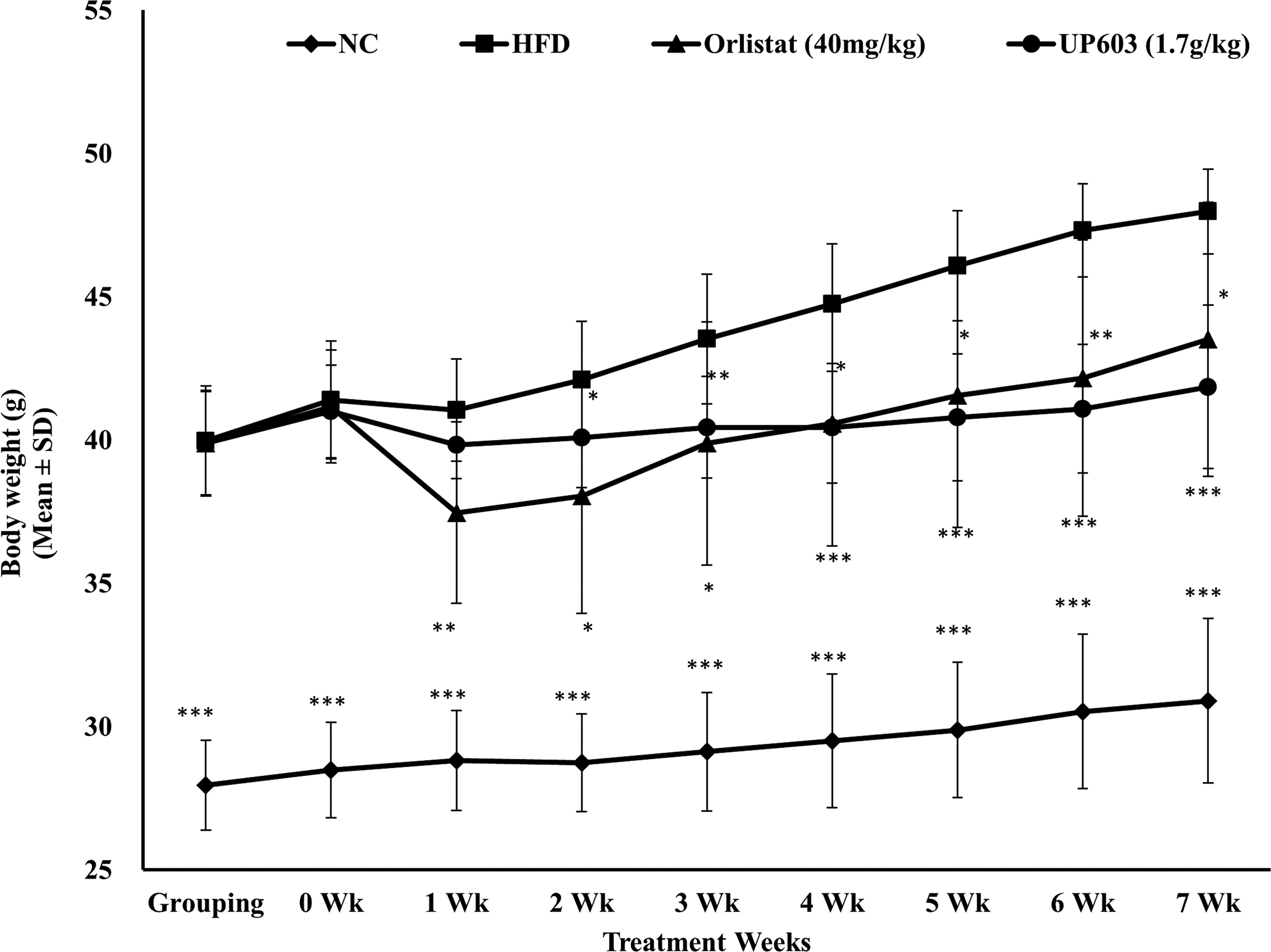
Body weight changes observed after 7 weeks of daily oral UP603 treatment in HFD fed obese C57BL/6J mice. ***P ≤ .0001, **P ≤ .001, *P ≤ .01. HFD, high fat diet.
Effect of UP603 on calorie intake
To better understand the possible mechanism(s) underlying homeostatic control mechanisms of energy intake to expenditure and the subsequent weight gain, feed intake and water consumptions were monitored twice a week for the course of treatment. As described in Table 1, mice treated with Orlistat consumed similar amount of calorie intake for the first week of treatment followed by a significant increase in calorie intake compared to that of the HFFD vehicle group, and such weight gains of Orlistat treated obese animals are parallel to the HFFD vehicle group lasted for the rest of treatment duration. In contrast, mice treated with UP603 at both dosages showed statistically significant decreases in calorie intake for the first 2 weeks of treatment and remained lower for the duration of treatment period compared to vehicle treated HFFD vehicle group (Table 1).
P ≤ .05, ** P ≤ .0001, † P ≤ .001.
Effect of UP603 on liver and lipid biomarkers
Dyslipidemia is the major component of metabolic disorder of obese and metabolic syndrome population that could occur as a result of consumption of high calorie diets with the elevated levels of LDL and total cholesterol with direct correlation of cardiovascular risk. To evaluate this association, and impact of daily oral administration of Orlistat and UP603 to mice, serum samples were collected, and level of liver enzymes, as well as lipid biomarkers, was determined. As illustrated in Table 2, the composition UP603 restored altered metabolic disturbances as demonstrated by reduced serum liver enzymes and lipid panel levels toward near normal. While a 44.6% reduction in serum triglyceride was observed for mice treated with UP603 at a dose of 450 mg/kg/day, a 38.2% reduction in LDL was observed for mice treated with UP603 at a dose of 850 mg/kg/day compared to vehicle treated HFFD group. Similarly, 11.2%, 7.9%, and 21.1% reductions in total cholesterol were observed for mice treated with the composition UP603 at oral doses of 450 mg/kg, 650 mg/kg, and 850 mg/kg for 7 weeks, respectively. In contrast, mice treated with Orlistat showed a 56.9% increase in serum triglyceride compared to vehicle treated HFFD. Significantly low levels of liver enzymes (AST = 25.6–31.1% decrease and ALT = 58.3–64.4% decrease) were also observed for mice treated with UP603 compared to vehicle treated HFFD obese mice; similar reductions were also observed by the regular diet fed normal control mice (Table 2). Mice treated with Orlistat showed 53.0%, 21.3%, 17.9%, and 43.8% reductions in ALT, AST, total cholesterol, and LDL, respectively, compared to vehicle treated HFFD obese mice.
P ≤ .001, * P ≤ .01.
ALT, alanine aminotransferase; AST, aspartate aminotransferase; TG, triglycerides.
Effect of UP603 on body composition and relative organ weight
DEXA scan was performed to determine the lean body and fat mass distribution of sedated mice (Fig. 3). After 7 weeks of daily oral treatment, no difference was observed for the lean body mass between groups. However, statistically significant decreases in fat mass were observed for mice treated with UP603 or Orlistat compared to vehicle treated HFD mice (Fig. 4). In relative to their body weight, the percentage fat was found to be 18.9%, 47.8%, 46.1%, and 40.4% for mice treated with normal control, HFD vehicle, Orlistat, and UP603, respectively (Fig. 3). In agreement with the DEXA scan data, a decrease in wet tissue visceral fat weight was observed for mice treated with UP603 compared to vehicle treated HFD mice where most of the reductions were contributed by low mass in the mesenteric and perirenal fat (Table 3). Mice treated with Orlistat showed a statistically significant decrease in the level of fat mass at perirenal and mesenteric fat; however, the total fat mass was not different from vehicle treated HFD mice (Table 3). Similarly, statistically significant increase in relative liver weight was observed for obese mice fed HFD and treated with vehicle (Table 3). Substantiating Study 1 data, in the second study that utilized HFFD-induced obese mouse model, regardless of the dosage used, mice treated with UP603 showed statistically significant reductions in epididymal, retroperitoneal, perirenal, and visceral fat deposits compared to vehicle treated HFFD group (Table 4). In this study, the positive control Orlistat also showed similar significance in reductions of fat deposits to that of UP603 groups (Table 4).
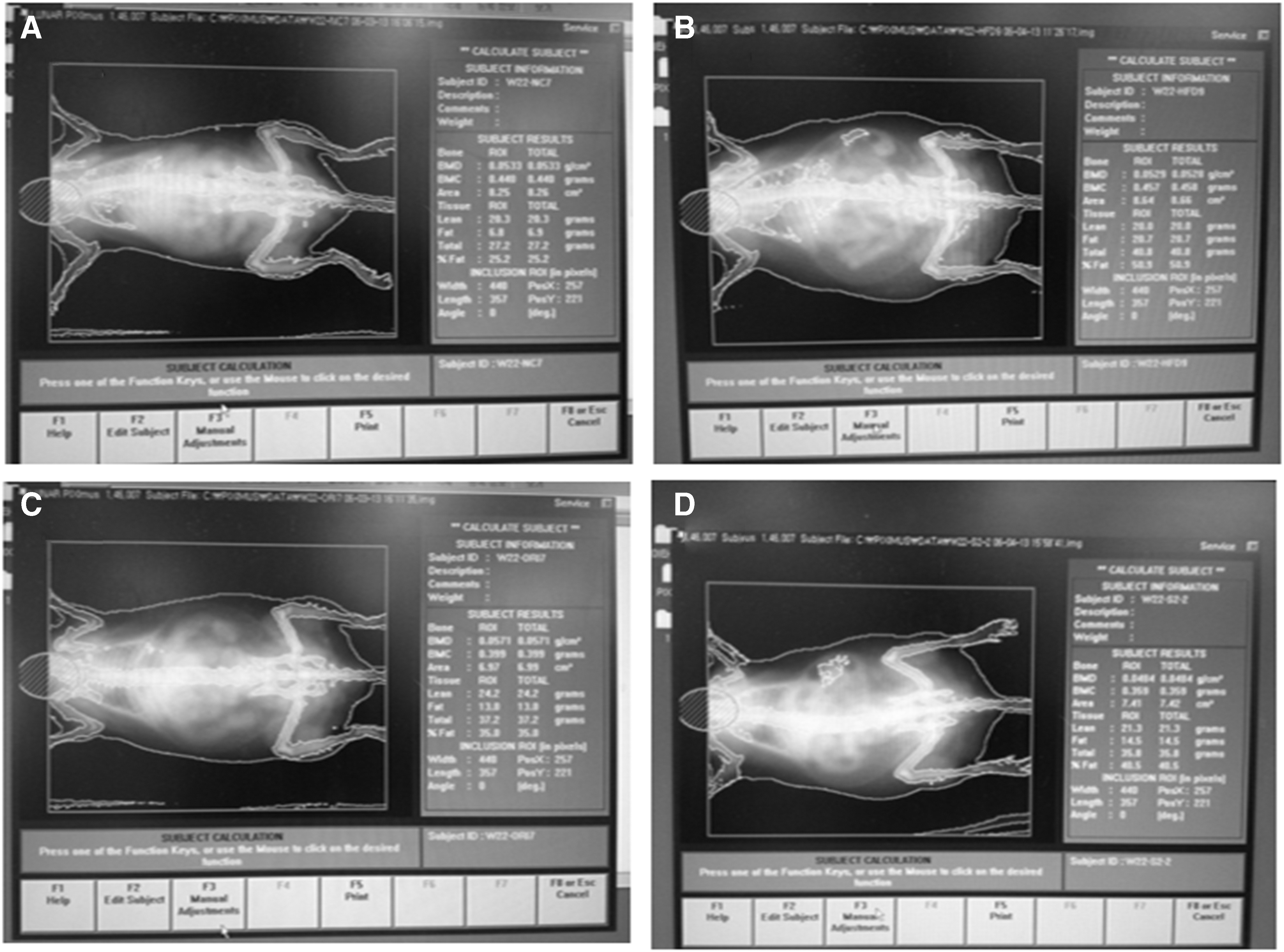
DEXA scan images for mice in the HFD-induced obesity group.

DEXA scan measurements for lean body and fat mass.
P values are compared to HFD.
P ≤ .05, ** P ≤ .0001, † P ≤ .001, ‡ P ≤ .00001.
HFD, high fat diet.
P values are compared to HFFD.
P ≤ .05, ** P ≤ .0001, † P ≤ .001, ‡ P ≤ .00001.
Effect of UP603 on liver histopathology
A rapid onset of lipid accumulation in the liver with both macro- and micro-vacuolation is a common phenomenon when mice are fed with HFFD, which becomes more severe when untreated with increasing duration on HFFD. Histological analysis of liver sections from the HFFD groups of obese mice demonstrated enlargement of hepatocytes and steatosis represented by the vacuolation in hepatocytes of the vehicle treated HFFD mice in comparison with those in the normal diet lean control mice. As the histopathology data showed, after 7 weeks of daily oral treatment, significantly improved NASH scores in steatosis, lobular inflammation, and hepatocellular ballooning were observed for mice treated with UP603 or Orlistat compared to vehicle treated HFFD group (Figs. 5 and 6). There was no obvious difference among the normal control and high dose groups of UP603 with regard to liver histology.
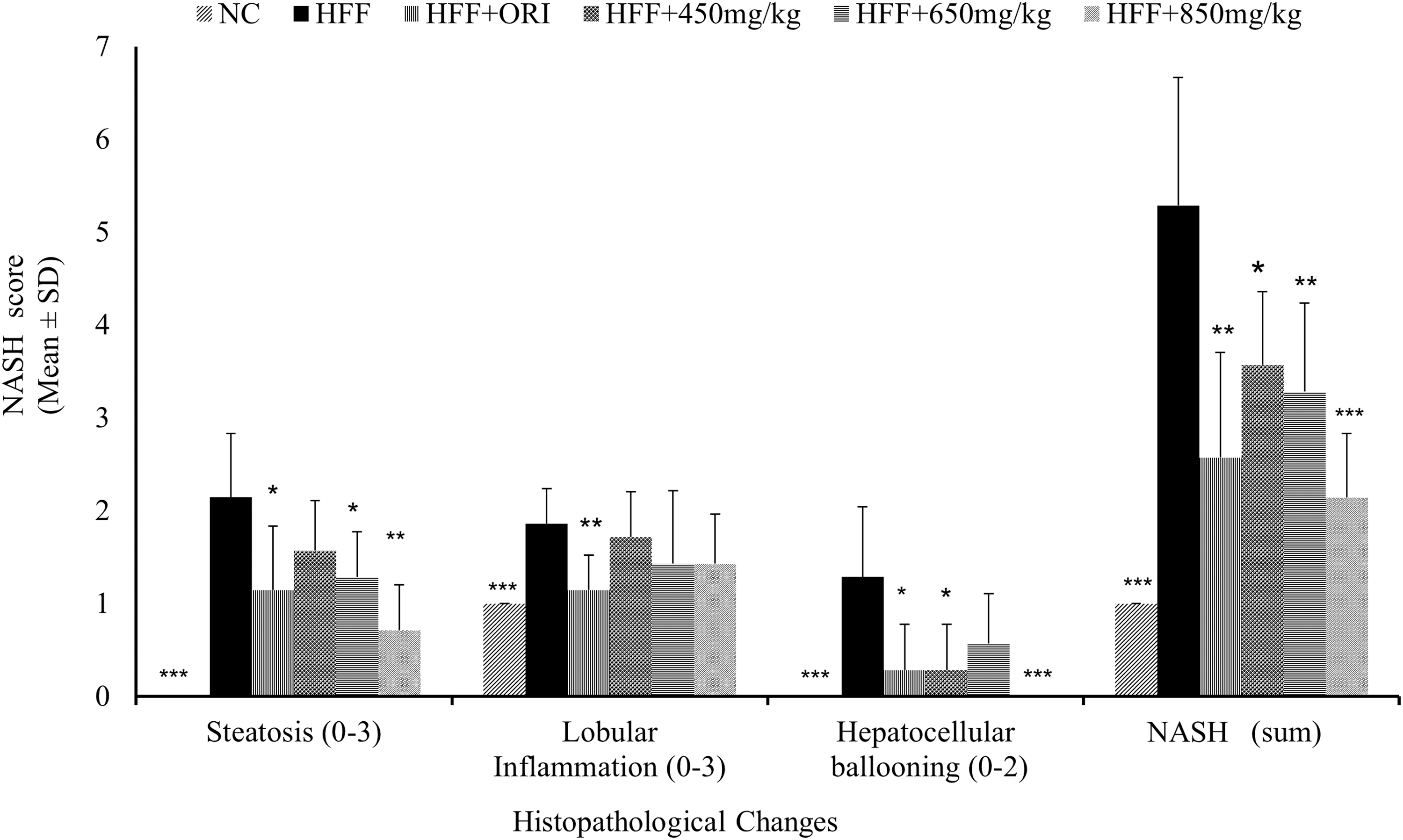
Nonalcoholic Steatohepatitis Scores for mice in the HFFD study group treated with Orlistat and UP603 at doses of 450, 650, and 850 mg/kg/day. *P ≤ .05, **P ≤ .001, ***P ≤ .0001.

Hematoxylin & eosin staining of liver tissue. 200 × magnification.
Effect of UP603 on the metabolic biomarkers in the DIO model
Serum level of insulin, leptin, and ghrelin was measured for mice treated with UP603 at oral dose of 1.7 g/kg/day for 7 weeks in the HFD study. As depicted in Figure 7, obese mice treated with vehicle showed almost six-fold and three-fold increase in serum insulin and leptin levels, respectively, and six-fold decrease in active ghrelin level indicating the presence of altered metabolism. Mice in the UP603 oral treatment group showed 65.5% decrease in insulin, 16.4% decrease in leptin, and 2.1-fold increase in ghrelin level compared to the vehicle treated obese mice. The reference compound 40 mg/kg/day of Orlistat showed 34.5% increase in insulin, 7.6% decrease in leptin, and two-fold increase in ghrelin compared to the vehicle treated obese mice.
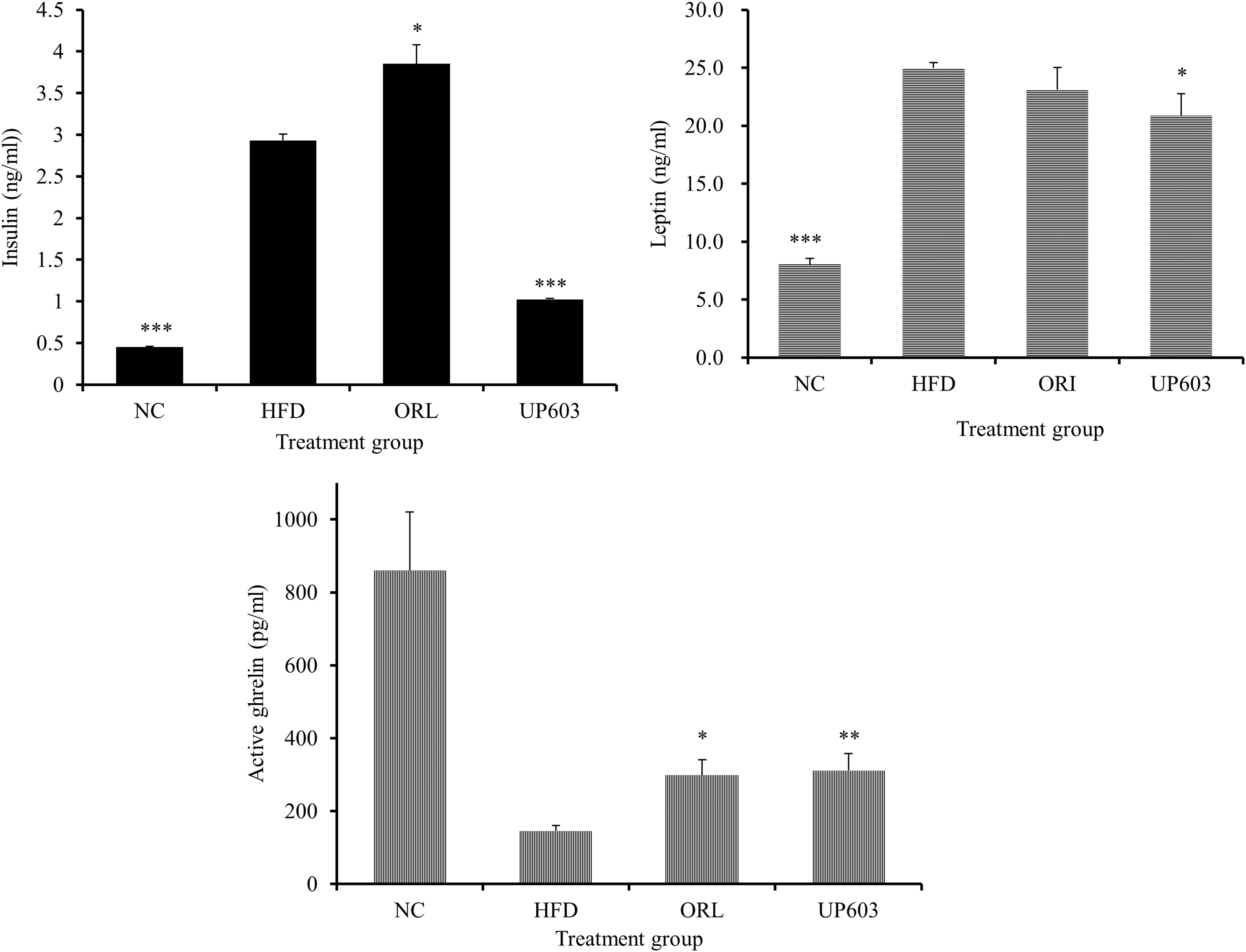
Insulin, leptin, and ghrelin level of obese mice treated with UP603 at oral doses of 1.7 g/kg/day for 7 weeks. *P ≤ .001, **P ≤ .0001, ***P ≤ .00001.
Discussion
Obesity, in association with insulin resistance and type-2 diabetes, is the most prevalent risk factor for cardiovascular diseases worldwide. 3 The long-term solution for weight management lies primarily on lifestyle changes in the form of dieting and exercising, which are difficult to achieve, and hence, there will always be a need for intervention especially for those patients who already are obese. In reality, treatment options remain quite limited due to the associated side effects of conventional therapeutics. As a result, given limited options, significant portions of obese and/or overweight population are turning to dietary supplements as an alternative.
In the current study, the biochemical and biological activity of a well-defined composition, UP603, from standardized extracts of three (M. alba, I. paraguariensis, and R. officinalis) historically very well-known plants was evaluated in two very well established animal obesity models that mimic human metabolic disturbances that frequently exhibit in western diets.
As an individual entity and as evidenced in multiple animal models, these plant extracts contain bioactive compounds that possess metabolic disease modifying activities contributed to the beneficial effects observed in the present study. For example, rosemary leaf extract that was standardized to carnosol and carnosic acid had showed moderation in weight gain, glucose levels, and lipid homeostasis. 12,13 In these studies, the authors reported a decrease in body weight gain, fasting blood glucose, and epididymal fat mass and increased fecal fat excretion without altering feed consumption in vivo and pancreatic lipase inhibition and PPAR-γ agonist activity in vitro. Additional studies have also reported that the lipid and glucose metabolism effects of rosemary extract could potentially be through activation of AMP-kinase; 11 by selectively modulating cecum, inhibiting β-glucosidase activity, increasing fiber, and decreasing short chain fatty acid fecal excretions; 29 and/or by stimulating glucose uptake in skeletal muscle cells through a PME-1/PP2A/PKB signaling axis. 30 It has also been reported that the active constituent to significantly improve glucose tolerance and reduce hepatic triglyceride accumulation is by decreasing hepatic lipogenic related gene (L-FABP, SCD1, and FAS) expression and increasing lipolysis related gene (CPT1) expression after 4 weeks of oral delivery to mice. 31 The beneficial moderation in body weight and metabolic markers observed in the current study could partially be explained by the inherent activities of rosemary extract contributed to the composition as described in this summary.
Augmented studies have also demonstrated that I. paraguariensis extracts rich in polyphenols (chlorogenic acid), xanthines (caffeine and theobromine), purine alkaloids (caffeic acid, 3,4-dicaffeoylquinic acid, and 3,5-dicaffeoylquinic acid), and flavonoids (quercetin, kaempferol, and rutin) alleviate weight gain and improve plasma glucose and lipid profiles in animals. The impacts of I. paraguariensis on obesity and insulin resistance in animals and humans in association with its potential mechanisms of actions have been well reviewed by Gambero and Ribeiro. 32 Arcari et al., have reported a marked attenuation of weight gain, adiposity, a decrease in epididymal fat pad weight, and restoration of the serum levels of cholesterol, triglycerides, LDL cholesterol, and glucose in HFD-induced obese mice treated orally with I. paraguariensis extract for 8 weeks at a dose of 1 g/kg. 15,16 These results were identical to what were observed in our study. Recently, it has also been reported that HFD- induced mice treated with I. paraguariensis extract for 8 weeks exhibited a reduction in body weight and epididymal fat. 16 Following gene expression study, the authors also reported the ability of the extract to downregulate the expression of genes that regulate adipogenesis and upregulated the expression of genes related to the inhibition of adipogenesis suggesting the importance of the extract in obesity management. In this study, again, the composition UP603, might have assumed to adapt the suggested mechanisms of actions and activities of I. paraguariensis as shown by data depicted in the current study.
Various studies have also reported the merit of M. alba root bark extracts in metabolic disorders like diabetes and obesity. Hypoglycemic activity in diabetes patients, 33,34 improving hyperglycemia and associated complications in the diabetic rats, 35 decreasing body weight and adiposity, regulating hepatic lipid accumulation in diet induced obese mice, 20 and decreasing expression of white muscle adipocytokines in mice 22 and blood glucose level in alloxan diabetic mice 23 were some of the beneficial effects documented for this plant. Previously it has also been reported that mulberry juice in combination with blueberry inhibited body weight gain, decreased serum cholesterol, reduced insulin resistance, attenuated lipid accumulation, and decreased leptin secretion in HFD mice administered orally for 12 weeks. 36 Some have suggested that these anti-obesity effects of morus extract were partially explained by increasing adiponectin level of adipocytes, 24 increasing glucose uptake and GLUT4 translocation in adipocytes, 24 inhibition of α-glucosidase activity, 25 and inhibition of human and rat intestinal disaccharidase. 37 It has also been reported that extract from morus ameliorated LXR alpha-mediated lipogenesis and upregulated lipolysis-associated markers such as lipoprotein lipase in the HFD-fed mice. 38 Inhibition of preadipocyte proliferation and differentiation on 3T3-L1 cells has also been reported indicating the association of morus extract and adipocyte deposition. 39 In support of the other two components of the composition, M. alba root bark extract also reenforces the potential of UP603 to yield a boosted efficacy in weight management as a result of collective mechanism of actions contributed by all the three components.
Given these individual previous reports of each component of the composition, it is possible to postulate the composition to have similar characteristics in moderating metabolic disorders signifying the merit of their combination for weight management. However, demonstrating efficacy of the composition in multiple animal models is pragmatic and a necessity. Indeed, the hypothesis was tested, and beneficial weight management activities were confirmed by orally administering the composition to HFD and HFFD-induced obesity models for 7 weeks. UP603 effectively and dose dependently attenuates body weight gain and counteracts metabolic disorders, especially obesity-associated liver damage, induced by a HFD and/or HFFD in mice. Data from two consecutive studies were compiled in this report. In the second study (Study-2 which was carried out in the HFFD-induced group) we used a reduced dosage of the composition (i.e., maximum of 850 mg/kg/day half of the 1.7 g/kg/day used in Study-1 conducted using HFD-induced obese mice) for dose correlation effects of the composition to extrapolate a feasible human consumption daily dosage. Both the HFD and HFFD studies showed significantly reduced body weight compared to their respective HFD or HFFD control.
Substantiating the above individual findings from M. alba, I. paraguariensis, and R. officinalis extracts, in this study, the anti-obesity and metabolic modulation activity of the standardized composition, UP603, was demonstrated by a reduced body weight gain and reduced calorie intake in both HFD and HFFD-induced obese mice models administered orally for 7 weeks. These body weight reductions were analyzed and were found to be 12.8% in the HFD study and 18.1–23.6% for HFFD-induced obese mice treated with UP603 at doses of 450–850 mg/kg compared to vehicle treated HFD/HFFD groups at week-7. Body weight was significantly decreased by UP603 treatment in comparison to the HFD or HFFD group. Especially in the HFFD study, almost complete prevention of body weight gain was observed in the group of obese mice receiving higher dose of UP603. Compared to week-0 (treatment start), the lower daily dosages (450 and 650 mg/kg) of the composition maintained the initial treatment start body weight for the duration of the treatment group; the highest dose (850 mg/kg/day) of UP603 resulted in a 2.9% reduction from the initial body weight of treatment start compared to the end of treatment period. For comparison, the HFFD vehicle group gained a 27.3% and the Orlistat group gained 2% of their treatment start initial body weight. At the time of induction, accelerated increase in body weight gain was observed for all the mice provided with 30% fructose in the drinking water in addition to the 60% kcal HFD. However at the course of treatment, the calorie intake from food and water for mice treated with UP603 was significantly reduced for the first few days and remained low for the duration of study suggesting that the composition may have additional benefit in suppression of appetite and increasing metabolic efficiency.
The beneficial metabolic management effects observed from UP603 were very comparable or better than those found with the Food and Drug Administration approved obesity drug-Orlistat. We have found, however, that treatment with UP603 did not produce lipase inhibitor associated side effects usually exhibited in this class of drugs such as a transient drop in body weight, increase in calorie intake after few weeks of treatment, and steatorrhea.
As evidenced by the liver histology and serum biochemical data, UP603 treatment prevented hepatic steatosis induced by the HFD/HFFD diet. The liver weights of UP603-treated mice were significantly lower compared with high calorie diet fed mice. Treatments of HFFD-induced obese mice with UP603 significantly reduced triglyceride, total cholesterol, and LDL levels which agree with the findings for individual plants as mentioned in the body of the articles above. Thus, UP603 appears to possess the beneficial effects in regulation of dyslipidemia. Furthermore, mice treated with UP603 showed significantly improved NASH scores in steatosis, lobular inflammation, and hepatocellular ballooning without affecting the liver enzymes (AST and ALT) suggesting that the composition may have an indication in nonalcoholic fatty liver disease without associated adverse effects. In addition to what has been described above for each component of the composition, in agreement with our study, it has been reported that extracts from Morus lead to significantly lower body weight gain, plasma triglycerides, and lipid peroxidation levels with concomitantly suppressing hepatic fat accumulation and reducing epididymal adipocyte size after the 12-week treatment period in HFD-induced obese mice. 40 The other constituent of the composition, rosemary extract, has also showed significantly reduced body weight gain, percent of fat, plasma ALT, AST, glucose, insulin levels, liver weight, liver triglyceride, and free fatty acid levels when HFD induced mice supplemented with rosemary extract were enriched with carnosic acid for 16 weeks. In their study the authors also reported decreased lipid accumulation in the hepatocytes and enhanced fecal lipid excretion indicating the inhibition of lipid absorption in comparison with the mice fed with a HFD without rosemary extract supplement. 41 Matching observations were also reported for Yerba maté. When HFD induced C57BL/6J mice were treated with Yerba maté extract at a daily dose of 0.5–2 g/kg for 4 weeks, a significant reduction in the differentiation of preadipocyte, a decrease in the accumulation of lipids in adipocytes, a decrease in food intake, and reductions in serum cholesterol, serum triglycerides, and glucose concentrations were reported. 14 Therefore, the data depicted in the current study solidify the merit of combining these plant extracts for a better outcome targeting multiple pathways that are indicated in various aspects of metabolic disorders.
Mounting evidences have been reported to show the strong association of abdominal obesity with metabolic disorders in humans. 42 Accumulation of fat in the visceral depot can cause increased secretion of pro-inflammatory mediators and decrease secretion of adiponectin 43 which subsequently prone subjects to insulin resistance and metabolic complications. It was found that the 7 weeks oral UP603 treatment significantly changed body composition of obese animals by decreasing total fat, percent body fat, and visceral fat weight. As depicted in the DEXA scan data, obese mice treated orally with UP603 showed a decrease in fat mass with no change in lean body mass, which magnifies that the decrease in body weight gain observed in these mice was mainly due to the result of reduced visceral fat accumulation. In agreement with this occurrence, the relative organ weight data also demonstrated statistically significant lower degree perirenal and mesenteric fat deposits. Similarly, in the HFFD-induced obese mice study, the weights of the mesenteric, epididymal, perirenal, and retroperitoneal fat depots were significantly decreased by UP603, with the larger decrease occurring in the mesenteric depot which was 62.4% compared to vehicle treated HFFD obese mice.
Insulin resistance, the fundamental etiology of type 2 diabetes, is one of the primary complications of obesity. 44 Untreated, it can result in deleterious consequences characterized under metabolic syndrome that includes hypertension, hyperlipidemia, and atherosclerosis. 45 Patients with chronic metabolic imbalances experience high level of insulin to compensate high calorie intakes. This phenomenon was observed in the vehicle treated HFD group in the current study. However, the surge of insulin was reduced to the level of the normal control mice as a result of the 7-week oral treatment of UP603.
The anorexigenic hormone, leptin, and the orexigenic hormone, ghrelin, are two hormones that have been recognized to have a major influence on energy balance and obesity. While leptin mediates the long-term regulation of energy balance by suppressing food intake, ghrelin seems to play a role in meal initiation. Clinically, obese subjects have increased level of circulating leptin and decreased level of ghrelin 46 due to leptin resistance developed under long times of overloading of high calorie diets. Again, these phenomena were observed in the vehicle treated HFD group in the current study. In contrast, these abnormal levels of leptin and ghrelin were reversed significantly toward the normal level due to UP603 treatment. These findings further give strong support to the efficacy of UP603 in moderation of calorie intake and metabolic disorders.
In summary we have standardized and demonstrated a novel and efficacious weight management composition UP603 from historically well known plants, M. alba, I. paraguariensis, and R. officinalis. Even though preclinical to clinical data translation requires human clinical trial validation, the data depicted in this study and safe historical usage of individual components of the composition suggest that UP603 could potentially be considered as a natural alternative to maintain healthy body weight, body composition, and metabolism management.
Footnotes
Acknowledgments
The authors express their best gratitude to Drs. Ed Cannon, Wenwen Ma, Doug Bradley, Min Chu, and Eujin Hyun and Unigen team for their incalculable support for the completion of this research. The authors also extend their utmost gratitude to Mr. Bill Lee, the owner of Econet/Unigen, Inc., who supported the entire project described in this article.
Author Disclosure Statement
All authors are current Unigen employees, therefore have competing financial interests.