
Abstract
We investigated the cytotoxic and antitumor effects of nine leaf extracts from Artemisia dracunculus (Tarragon). Five extracts were obtained using different organic solvents and four by supercritical CO2. The cytotoxic effects were expressed as IC50 in 100, 80, 80, 100, and 80 μg/mL by respective solvents: hexane, ethyl acetate, acetone, ethanol, and acetonitrile in L5178Y lymphoma cells. For supercritical CO2 extract A, IC50 was 100 μg/mL; for extracts C and D, IC50 was 150 μg/mL. The antitumor activity was assessed through a tumor growth inhibition test that measured ascites fluid volume and tumor cell counts of BALB/c mice (2 × 10⁴ cells L5178Y i.p.). Twenty-four hours after inoculation, mice were treated with 100 mg/kg of acetonitrile extract or extract SF-A daily for 15 days in independent groups of five mice, using two administration routes. We observed tumor evolution with and without treatment. Without treatment, tumor evolution was 17,969 × 106 ± 5485 L5178Y cells in 2.6 mL ascites volume, whereas the orally treated acetonitrile extract group showed 0.1 × 10⁶ ± 0.07 L5178Y cells (P < .05). The oral SF-A group showed 12.9 × 10⁶ ± 243 L5178Y cells, and intraperitoneal (i.p.)-treated SF-A group showed 0.1 × 10⁶ ± 0.05 L5178Y cells (P < .05) without any ascites volume development. The acetonitrile extract contains abundant polyphenols and possibly a flavone with antioxidant activity. The SF-A contains abundant alkamides. Both extracts are complexes and the identity of the compounds responsible for observed antitumor activity remains unknown.
Introduction
W
Artemisia dracunculus is a perennial herb in the Asteraceae family that is highly popular in European cuisine. Studies of the chemical composition of A. dracunculus extracts have revealed the presence of essential oils, phenylpropanoids, alkamides, flavonoids, and coumarins. 8,9 Certain extracts and compounds isolated from this herb have many associated pharmacological activities, such as antibacterial, antifungal, anti-inflammatory, antidiabetic, hepatoprotective, gastroprotective, and cytotoxic effects. 10,11
In this work, total extracts from A. dracunculus were obtained by maceration in solvents of different polarities or through supercritical CO2 extraction. These extracts were analyzed by thin layer chromatography and their biological activity was assessed by cytotoxicity assays in L5178Y lymphoblast and antitumor effects against murine lymphoma L5178Y in BALB/c mice.
Materials and Methods
A. dracunculus
The dried and crushed leaves of A. dracunculus used in this work were purchased from a distributor (Dry Fruits of Quality S.A. C.V.). The leaves were pulverized and classified by their particle size; using 50 mesh (0.297 mm) and 100 mesh (0.149 mm) screens, they were then packaged in plastic bags and stored at room temperature in a cool place protected from light until use.
Organic solvents and supercritical CO2 extraction
Five different solvent extracts [hexane (Hx), ethyl acetate (EA), acetone (DMK), acetonitrile (ACN), and ethanol (EtOH)] were obtained by maceration of 75 g of A. dracunculus leaves in light-protective vessels with 300 mL of respective solvent. The supernatant of each extract was filtered through Whatman N. 4 paper, and the solvent was renewed in the vegetable material every week for 6 weeks. The combined supernatants from each extract were evaporated to dryness using a rotavapor Büchi R-200 and heating bath B-490 (BÜCHI Labortechnik AG, Switzerland), then weighed to determine the yield (percentage). Four extracts were obtained with 75 g of A. dracunculus leaves at different conditions of pressure, temperature, and particle size (Table 1) using an SFE Bio-Botanical Extraction System SFE-500 MR (Waters®, Mildford, MA, USA). We used USP grade CO2 (INFRA®, México) for extraction at 25 g/min. At the end of each extraction, the weight of the extract obtained was recorded.
SF-A, SF-B, SF-C, and SF-D extracts obtained with supercritical CO2.
Cytotoxic activity
The cytotoxic effects of A. dracunculus extracts were evaluated in vitro using L5178Y cells. Cultures of 50,000 cells per well were seeded in 96-well plates at a total volume of 200 μL. Cells were incubated in presence or absence of various concentrations of A. dracunculus extracts (10, 50, 80, 100, and 150 μg/mL) or cyclophosphamide (11 μg/mL), which served as a positive control. Cells were cultured in RPMI (Gibco® by Life Technologies™, USA) medium, pH 7; supplemented with previously inactivated 10% fetal bovine serum (Corning®, Manassas, VA, USA), 5000 IU/mL penicillin, and 5000 μg/mL streptomycin Pen Strip (Gibco by life technologies, USA) with an incubation time of 24 h at 37°C under 5% CO2 and 95% humidity in a cell incubator (Nuaire NU4750). MTT [3-(4,5-dimethylthiazol-2-yl −2,5-diphenyltetrazolium bromide] (Sigma-Aldrich, St Louis, MO, USA) assay staining was used to assess cell viability; optical density was measured at 570 nm with a Microplate reader Model 680 (Bio-Rad Laboratories, Inc., USA). The extracts from each process exhibiting the greatest cytotoxic effects were evaluated in the antitumor model in vivo in BALB/c mice inoculated with L5178Y lymphoblast.
Animals
Male BALB/c mice (6 to 8 weeks old, 20–24 g) were provided by the Centro de Investigación Biomédica de Occidente (CIBO-IMSS). Animals were maintained in a specific pathogen-free animal care facility in a regulated environment (22 ± 1°C, 50–60% relative humidity and 12-h light cycle). All mice were fed a commercial diet Lab Chow (Purina Lab Chow–México) with autoclaved tap water ad libitum. For this study, all procedures involving animals were performed according to protocols approved by the Research Center's animal care committee and in compliance with national guidelines on animal welfare (NOM-062-ZOO-1999). 12
Antitumor effects
The antitumor effects of A. dracunculus extracts exhibiting the greatest cytotoxic effects were evaluated in BALB/c mice. On day 0, a suspension of 100 μL 2 × 10⁴ L5178Y lymphoblast (in isotonic saline solution) was infiltrated i.p. in all mice. Twenty-four hours later, eight randomized groups of five mice were performed. One group without treatment was considered the negative control group (L51); two groups received 100 μL/15 days isotonic saline:dimethyl sulfoxide (DMSO) solution (1:1 v/v), orally (DMSO-o) or i.p. (DMSO-i.p.); to evaluate the solvent effects, the remaining groups were individually treated with either (1) cyclophosphamide (CY) 75 mg/(kg ·3 days) i.p., (2) ACN 100 mg/(kg ·15 days) orally (ACN-o), other group i.p. (ACN-i.p.), or (3) SF-A 100 mg/(kg ·15 days) orally (SF-A-o) and other group i.p. (SF-A-i.p.). Extracts were dissolved in isotonic saline:DMSO (1:1 v/v) solution.
On day 17, mice were euthanized by ether inhalation and ascites were collected from the intraperitoneal cavity of each mouse. The ascites fluid was recovered in a graduated centrifuge tube to measure the volume, and a cell count was performed in a Neubauer's chamber of viable lymphoblast with trypan blue (Sigma-Aldrich). In mice groups without ascites accumulation, an intraperitoneal wash with 1 mL physiological saline solution was performed to obtain lymphoblast for counting. The body weight of the mice was monitored in the course of treatment, because weight variations also reflect tumor development.
Thin layer chromatography analysis
Four microliters of 20 mg/mL extracts from A. dracunculus, or kaempferol, quercetin, catechin, diosgenin, berberine, and cholesterol as reference standards, was applied to 20 cm × 10 cm HPTLC Silica gel plates 60 F254 (Merck-Millipore, Germany) with automated LINOMAT 5 (CAMAG, Switzerland) equipment using flow air at 100 μL/min. Extracts were applied in bands of 7 mm at a distance of 5 mm from the lower edge and 14.5 mm from each side of the plate, with a separation of 17 mm between the lanes; the plates were developed with a 1:1 mobile phase of toluene and ether saturated with 5% acetic acid. The chromatoplates were analyzed at 254 and 365 nm, then plates were developed with phosphomolybdic acid, Dragendorff reagent (followed by 10% NaNO2), polyphenol developer, and 2,2-diphenyl-1-picrylhydrazyl (DPPH), and the retention factor (Rf) of each band of all extracts was recorded. Moreover, a densitometric analysis in percentage of area under the curve (% AUC) of bands at 300 nm was also carried out using a TLC SCANNER 3 (CAMAG) at Centro de Investigación y Asistencia en Tecnología y Diseño del Estado de Jalisco (CIATEJ) unit in Zapopan, Jalisco.
Statistical analysis
Data are reported as mean ± standard deviation and analyzed using one-way analysis of variance, followed by Dunnett test and corroborated by student t test. When the probability (P) was less than .05, the difference was considered to be significant.
Results
Solvent and supercritical CO2 extraction
Total extract yields from A. dracunculus were 1.6 g Hx, 2.5 g EA, 3.1 DMK, 8.5 g EtOH, and 1.7 g ACN, corresponding to 2.1%, 3.3%, 4.1%, 11.4%, and 2.3%, respectively. The amount of A. dracunculus extract obtained with supercritical CO2 under different conditions of pressure, temperature, and particle size was 0.39 g SF-A, 0.78 g SF-B, 0.97 g SF-C, and 0.62 g SF-D corresponding to 1.66%, 3.33%, 4.15%, and 2.67%, respectively.
Cytotoxic activity
The cytotoxicity of A. dracunculus extracts obtained by organic solvents and supercritical CO2 was evaluated in L5178Y lymphoblast using the MTT assay. All the extracts tested have dose-dependent cytotoxic activities, as shown in Figure 1. The most cytotoxic extracts from each type of extraction were ACN and SF-A, wherein the IC50 was 80 μg/mL and 100 μg/mL, in percentage of inhibition, respectively.
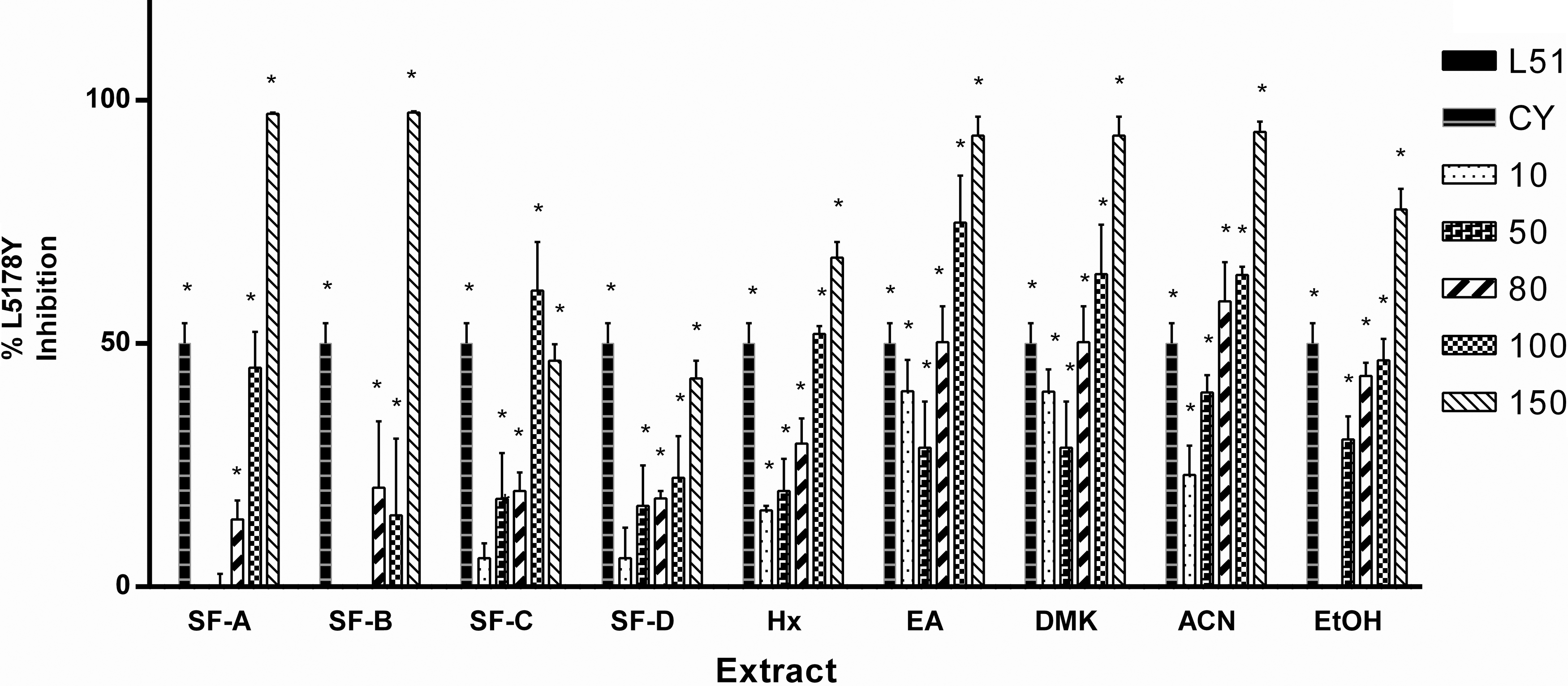
Graphical representation by the inhibition percentage of L5178Y lymphoblast induced by extracts of Artemisia dracunculus obtained by organic solvent or supercritical CO2 extraction. The concentrations of the extracts used were 10, 50, 80, 100, and 150 μg/mL (*P < .05 compared with the untreated group L5178Y). L51, untreated group; CY, 11 μg/mL cyclophosphamide; Hx, hexane; EA, ethyl acetate; DMK, acetone; ACN, acetonitrile; EtOH, ethanol. SF-A, SF-B, SF-C, and SF-D, extracts obtained with supercritical CO2.
Antitumor effect
The ACN and SF-A extracts were evaluated in a murine model with L5178Y lymphoblast to assess the antitumor effects by oral or i.p. administration; the weight changes, ascites volume, and tumor mass of the mice are shown in Table 2. The control group (without treatment) exhibited a tumor evolution of 17,969 × 106 ± 5485 L5178Y cells in 2.6 mL ascites volume. The A. dracunculus extracts ACN and SF-A oral showed antitumor effects of 0.1 × 10⁶ ± 0.07 L5178Y cells and 12.9 × 10⁶ ± 243 L5178Y cells, respectively, and SF-A-i.p. showed 0.1 × 10⁶ ± 0.05 L5178Y cells (P < .05), without accompanying weight gain or ascites development. The least effective extract treatment was i.p. ACN; this group gained 3.7 g and accumulated 962.5 μL ascites containing 39,981.8 ± 17625.1 × 106 cells/mL.
P < .05, with respect to the L51 group.
CY, cyclophosphamide; DMSO, dimethyl sulfoxide; SD, standard deviation.
Thin layer chromatography analysis
The A. dracunculus extracts were analyzed by thin layer chromatography. At 254 nm, 366 nm, and using phosphomolybdic acid, the supercritical CO2 extracts are noticeably more complex, with 25 (SF-A and SF-B) and 24 (SF-C and SF-D) bands, whereas organic solvent extracts are less complex, with 19 (DMK, EtOH, and ACN), 18 (EA), and 14 (Hx) bands.
All the extracts have unsaturated compounds, as evidenced by fluorescence quenching at 254 nm. In almost all extracts, possible coumarin compounds are indicated by the presence of a blue band (Rf ∼0.69) at this wavelength. This band is also seen as a bright blue band at 365 nm on plates developed with natural products reagent. 13 The presence of other polyphenols is also indicated with bands developed with natural products reagent; apparently, the higher number of bands is possibly caused by flavones in the supercritical CO2 extracts (Rf ∼0.0, 0.11, 0.14, 0.20, 0.42, and 0.52) and the EA (Rf ∼0.14, 0.31, and 0.52), DMK (Rf ∼0.14, 0.31, 0.38, and 0.46), EtOH (Rf ∼0.14, 0.31, 0.38, 0.46, 0.52, and 0.63), and ACN extracts (Rf ∼0.14, 0.31, 0.38, and 0.46). In plates sprayed with DPPH to evaluate antioxidant activity, no evidence of antioxidant activity was observed in the SF extracts. 14,15 However, the extracts obtained with organic solvents (except for Hx) showed several bands in the lower part of the plate after DPPH development. In plates developed with Dragendorff reagent, all extracts except Hx show the presence of an orange band, indicating the possible presence of alkamides (Figs. 2 and 3).
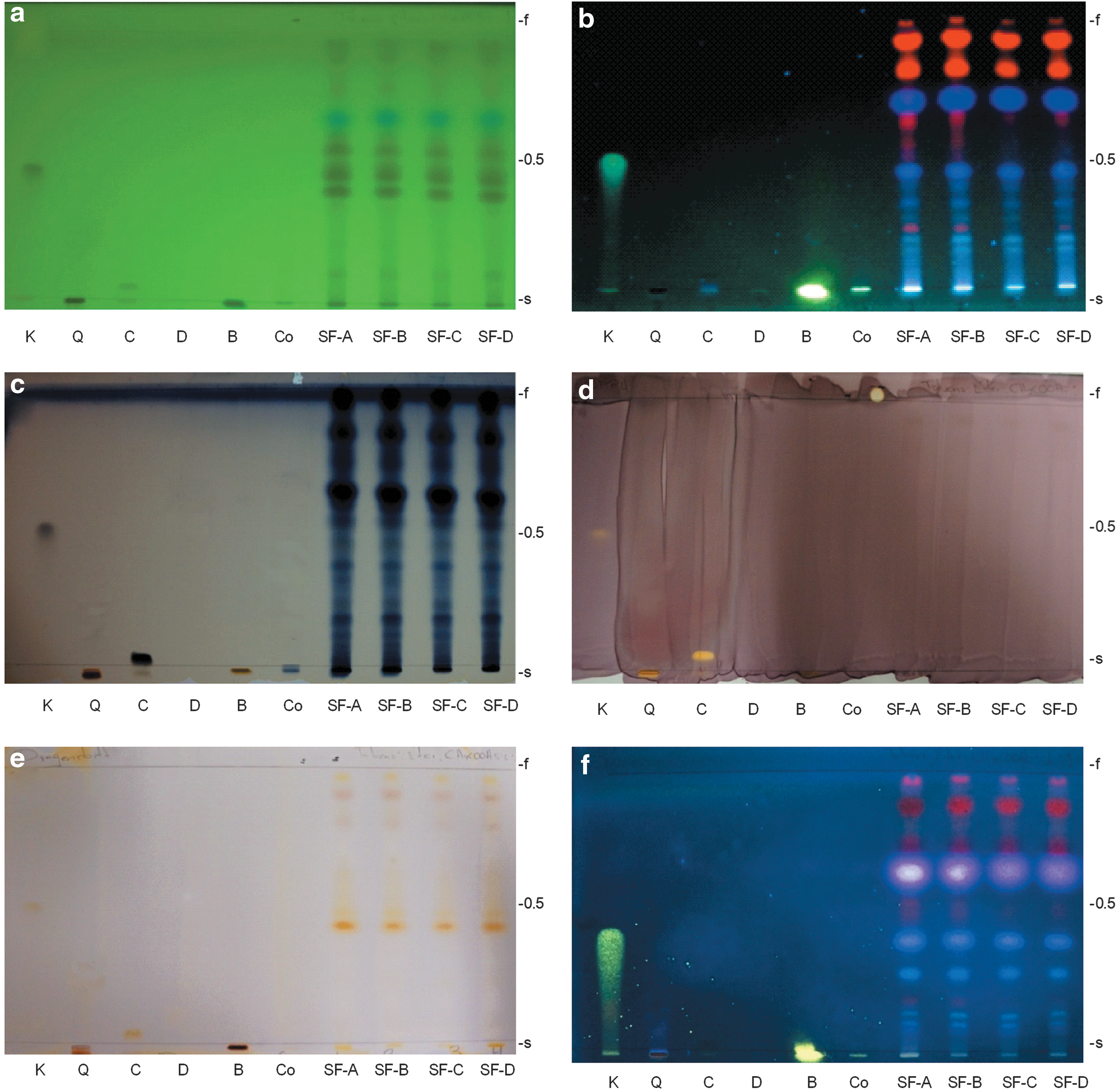
Chromatographs of A. dracunculus extracts obtained with supercritical CO2.
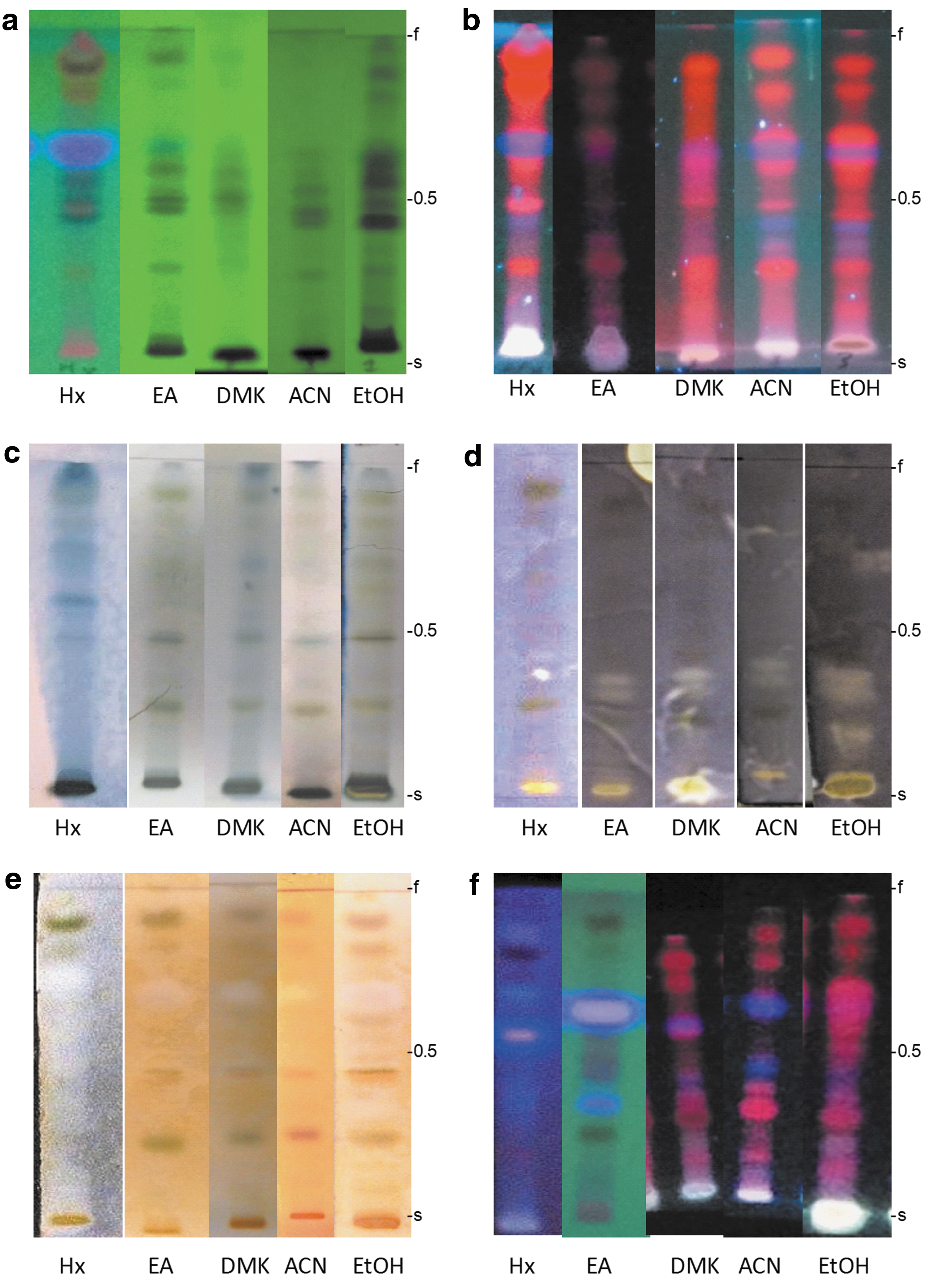
Chromatography of A. dracunculus extracts obtained with organic solvents.
Discussion
A. dracunculus leaf extracts were obtained with organic solvents or supercritical fluids. The solvent extract with the highest yield was the ethanolic extract (11.37%), whereas the hexanic extract was of the lowest yield (2.1%). This difference is likely because of the entrainment of primary metabolism compounds, such as carbohydrates, which are polar in character and they are abundant in plants. 16 In contrast, we need more supercritical CO2 extractions to determine which factors (pressure, temperature, and particle size) are intrinsically ligated on total yield percentage.
Extracts in a range of concentrations from 10 to 150 μg/mL were tested in L5178Y lymphoblast cultures to evaluate their cytotoxic activity using the MTT assay. We observed that the most cytotoxic extracts were ACN (IC
In contrast, the antitumor effects of both extracts were evaluated in BALB/c mice with 100 μL of 2 × 10⁴ L5178Y lymphoblast cells.
The L51 group received no treatment and was considered control of tumor development; they showed 17,969 × 106 ± 5485 L5178Y cells in 2.6 mL ascites. As a positive control, the CY group received 75 mg cyclophosphamide for 3 days, with inhibition of tumor development of 0.4 × 10⁶ ± 0.3 cells without ascites recovered through intraperitoneal lavage; as well as, SF-A-i.p. and ACN-o in 0.1 × 10⁶ ± 0.05 and 0.1 × 10⁶ ± 0.07 L5178Y cells, respectively.
These results show that the best treatment to inhibit tumor development in BALB/c mice inoculated with L5178Y lymphoblast is 100 mg/(kg ·15 days) ACN-o or SF-A-i.p. According to the thin layer chromatography profile and the densitometry analysis at 300 nm (Table 3), the acetonitrile extract contains abundant polyphenols and probably a flavone with antioxidant activity. 19 Similar results were reported by Ju et al 20 and by Hong et al 11 ; however, they did not report in vivo assays. In the chromatographic profile, SF-A contains abundant alkamides, which react with the Dragendorff developer in a weaker way than the alkaloids, giving a tenuous orange coloration. 21 The presence of these types of compounds is congruent with other reports regarding extracts from the leaves of A. dracunculus. 10,22 A future bioassay-guided study may be considered to identify molecules responsible for the antitumor effects of these extracts.
Hx, hexane; EA, ethyl acetate; DMK, acetone; EtOH, ethanol; ACN, acetonitrile; SF-A, SF-B, SF-C, and SF-D, extracts obtained with supercritical CO2.
Conclusions
In vitro assays, ACN was the most cytotoxic extract in L5178Y lymphoblast. In in vivo assays, the treatments with greatest inhibition of tumor development in BALB/c mice with lymphoma L5178Y were the SF-A extract by the intraperitoneal route and the ACN extract orally at 100 mg/(kg·day) for 15 days. These extracts show a greater amount of alkamides, polyphenols, and possible flavones by chromatography analysis.
Footnotes
Acknowledgments
Martha Hilda Navarro-Salcedo acknowledges support from Consejo Nacional de Ciencia y Tecnología (CONACYT) for Scholarship No. 288145/257589.
Author Disclosure Statement
The authors declare there are no conflicts of interest.