
Abstract
Several studies have shown the protective effect of dairy products, especially α-lactalbumin and derived hydrolysates, against induced gastric ulcerative lesions. The mucus strengthening represents an important mechanism in the defense of gastrointestinal mucosa. Previously, a hydrolysate from casein (CNH) and a hydrolysate from whey protein concentrate rich in β-lactoglobulin (WPH) demonstrated a stimulatory activity on mucus production in intestinal goblet cells. The aim of this work was to evaluate the possible antiulcerative activity of these two hydrolysates in an ethanol-induced ulcer model in rats. All tested samples significantly reduced the ulcerative lesions index (ULI), compared with the saline solution, using doses of 300 and 1000 mg kg−1 body weight with decreases up to 66.3% ULI. A dose–response relationship was found for both hydrolysates. The involvement of endogenous sulfhydryl (SH) groups and prostaglandins (PGs) in the antiulcerative activity was evaluated using their blockage. The antiulcerative activity of WPH showed a drastic decrease in presence of N-ethylmaleimide (from 41.4% to 9.2% ULI). However, the CNH antiulcerative properties were not significantly affected. The cytoprotective effect of WPH appears to depend on a PG-mediated mechanism. In conclusion, CNH and WPH demonstrated in vivo antiulcerative properties and represent a promising alternative as protectors of the gastric mucosa.
Introduction
P
It is now accepted that food is not only a supplier of nutrients but also a modulator of different physiological functions in the body. The gastrointestinal tract being in contact with food components and their digestion products, this is the organ where this concept can be directly applied. In fact, many food components of different chemical natures have been found to exert modulation of intestinal functions. 3 Some of them have demonstrated an antiulcerative effect. For instance, flavonoids may act in healing gastric ulcers and have been proposed as new alternatives for suppression or modulation of peptic ulcers associated to H. pylori. 4 Polyphenols, proteins, and especially whey proteins and hydrolysates thereof, have been found to exert antiulcerative properties. Free amino acids consumed with protein hydrolysates are a better source of protein when the repair of tissue damage occurs, rather than the consumption of intact proteins or free amino acids. 5
Several studies have shown the protective effect of α-lactalbumin (α-La) against ulcerative lesions caused by ethanol, stress, or indomethacin. 6,7 Oral administration of this protein increased the level of PGE2 and the mucin contents of both the gastric fluid and the adherent mucus gel layer. 8 This protein also caused PG-independent responses such as an increase of the gastric luminal pH and fluid volume and a delay in gastric emptying. 8
More recently, it has been found that α-La stimulates in cell cultures the synthesis and secretion of mucins. Mucins are the main macromolecular components of mucus. α-La stimulates the thickness of the mucus gel layer in rats, independently of endogenous PGE2. 9 Not only α-La but milk whey protein concentrates (WPC) or whey protein isolates (WPI) containing this protein have also demonstrated this protective effect against ulcerative lesions. These effects were attributed to gastrin, sulfhydryl (SH) substances, and some mechanisms related to mucus production. 10,11 The antiulcerative activity of these WPC was attributed to their α-La content. On the contrary, no antiulcerative effect has been found for β-lactoglobulin (β-Lg). 6
More recently, a cheese WPC was found to protect against dextran sulfate sodium-induced colitis. The effect is attributed to its threonine and cysteine content, given that these amino acids are limited for mucin synthesis under chronic inflammatory bowel disease. 12 A WPC hydrolyzed (WPH) with extracts of Cynara cardunculus has demonstrated a protective effect against ulcerative lesions induced by ethanol. The protective activity of the hydrolysate was attributed to the stimulation of PGs and NO. The effect of its fraction below 3 kDa was partly explained by the SH compounds. 13
With regard to mucus strengthening, certain food peptides have demonstrated a stimulatory activity on intestinal mucin secretion, which could also contribute to their antiulcerative effect. For instance, β-casomorphin-7, a casein-derived opioid peptide, induces the secretion of intestinal mucins by activation of the enteric nervous system and by a direct effect on goblet cells via interaction with μ-opioid receptors. 14,15 This mucin stimulatory effect was also shown on goblet cells with β-Lg hydrolyzed with trypsin 16 and with a peptic casein hydrolysate. 17 Both hydrolysates stimulated mucin secretion and the mucin 5AC gene (MUC5AC) expression in human intestinal cells HT29-MTX.
The aim of this work was to evaluate the antiulcerative activity of these two hydrolysates with previously demonstrated mucin stimulatory effect. The contribution of the SH groups to the gastric protection was assessed by in vivo alkylation. PG protection was also assayed.
Materials and Methods
Production of hydrolysates
Both casein and β-Lg hydrolysates were prepared by Innaves S.A. (Porriño, Pontevedra, Spain). Hydrolysis with food-grade pepsin (Biocatalyst, Cardiff, UK) of commercial casein (Promilk 85, Arras Cedex, France) was carried out to obtain the casein hydrolysate (LowPept®). In brief, casein at 0.06% (w/w) was digested at 37°C and pH acid by pepsin at a ratio of 1:50 (w/w) (enzyme:substrate), with the addition of the enzyme at the beginning of hydrolysis and after 3 h. Hydrolysis was inactivated by increasing pH up to 7 and the supernatant after clarification was collected and spray dried.
The protein content of this hydrolysate determined by the Kjeldahl method was 71.33% ± 1.55% (w/w), as previously assessed. 18 To produce the β-Lg hydrolysate, a WPC rich in β-Lg (Friesland Campina Domo, Zwolle, The Netherlands), containing at least 99.0% of β-Lg of protein content was used. This WPC was hydrolyzed with food-grade trypsin (Biocatalyst), as reported by Anadón et al. 19 In brief, WPC was suspended at 0.05% (w/w) and heated at 90°C for 10 min. After addition of trypsin at a ratio 1:20, hydrolysis was performed at pH 8.0 and 37°C for 3 h and stopped by heat inactivation of trypsin at 95°C for 15 min.
WPH was clarified and spray dried. The protein content for this hydrolysate was 74.07% ± 0.19%.
Animals
Wistar male rats, 250–350 g body weight (bw), were obtained from the Experimental Animal Center (CEMIB) of Campinas University (São Paulo, Brazil). In prior experiments, animals were kept for at least 7 days at 20°C and under alternative light/dark cycles of 12 h, receiving a commercial standard diet (Nuvital Nutrients, Curitiba, Brazil) and water ad libitum. Animals fasted for 12 h before the experiments.
Ethanol-induced gastric ulcer model
The protective effect on rat stomach mucosa was studied using the absolute ethanol ulcerogenesis model. 20 The Ethics Committee for Animal Research of Campinas University approved the experimental protocol (No. 2206-1) in agreement with the ethical principles of The International Association for the Study of Pain (IASP). Animals were divided in groups of five rats, a group per treatment. Sodium chloride solution 0.009% (w/w) at 10 mL kg−1 bw and an antiulcerative drug, carbenoxolone (Sigma, St. Louis, MO, USA), at 200 mg kg−1 bw were used as negative and positive controls, respectively.
Samples were administrated at three different concentrations (100, 300, and 1000 mg kg−1 bw) in a single dose by gastric intubation. One hour after sample or control ingestion, 1 mL of absolute ethanol was provided to each rat. One hour later, animals were sacrificed and their stomachs were extracted and washed with a saline solution for ulcerative lesions analysis.
Ulcerative lesion analysis
The ulcerative lesions were evaluated by visual inspection of rat gastric mucosa. According to Gamberini et al., 21 the ulcerative lesions index (ULI) of each animal was determined by summing the scores associated to several parameters: loss of normal morphology (1 point), mucosa discoloration (1 point), edema (1 point), hemorrhage (1 point), petechial points until 9 mm (2 points), petechial points larger than 10 mm (3 points), ulcers up to 1 mm (n × 2 points), ulcers larger than 1 mm (n × 3 points), and perforated ulcers (n × 4 points), where n is the number of ulcers found.
Percent decrease of the ULI score was calculated by the equation 1:
The effective dose (ED50), defined as the sample concentration that achieves a decrease of 50% ULI, was estimated by using a logarithmic regression equation for ILU values obtained from 100, 300, and 1000 mg kg−1 treatments.
Evaluation of the cytoprotection mechanisms
Evaluation of SH compounds contribution
The contribution of SH in the protection of rat stomach mucosa, against the ulcerogenic activity of absolute ethanol, was evaluated by using N-ethylmaleimide (NEM). NEM has the capacity to block, via alkylation, all active SH groups in the rat body. 22 The experimental protocol was analogous to the one used for the study of the antiulcerative activity of the hydrolysates, except that rats received subcutaneous injection of NEM (10 mg kg−1 bw) 30 min before the intragastric administration of saline solution (negative control) or hydrolysate (single concentration of 500 mg kg−1 bw for CNH and 300 mg kg−1 bw for WPH). The antiulcerative activity degree (% ULI) was calculated using (Eq. 1).
Evaluation of PG contribution
Endogenous PGs in gastric protection analysis was carried out according to Amorim et al. 23 10 mg per kg bw solution of indomethacin (Sigma-Aldrich) was injected subcutaneously to each group (six animals). After 30 min, animals were separated, and one group received CNH (500 mg kg−1) and WPH (500 mg kg−1) dissolved in a saline solution, and the other group received 10 mL per kg bw of saline solution orally. After another 30 min, animals were treated orally with 1 mL of absolute ethanol and sacrificed 1 h later. The following steps were performed as previously described in ethanol-induced gastric ulcer. The antiulcerative activity degree (% ULI) was calculated using Equation 1.
Statistical Analysis
Experimental results were analyzed using a one-way ANOVA method, followed by Tukey's test. GraphPad Prism 4 software was used. Different letters indicate significant differences among dose groups (P < .05). *P < .05, **P < .01 and ***P < .001 express statistical differences between samples and saline control.
Results and Discussion
Evaluation of the antiulcerative properties
The antiulcerative activities of CNH and WPH were studied in a model of ulcer induced by absolute ethanol in rats. By macroscopic observation, the acute ulcer induced by ethanol appeared to have an intense gastric inflammation in red bands, consisting of intense lesions. These signs were observed in a higher degree in control rats that were given only saline solution (negative control), before inducing ulcers by ethanol administration. The damaged gastric mucosa can be easily appreciated with prominent red streaks, with induced macroscopic morphological changes, such as linear hemorrhages and craters ulceration in the mucosal layer (Fig. 1A).

Images of ethanol-induced ulcers in rat stomachs under different treatment: saline solution 10 mL kg−1 bw
In contrast, gastric ulcer induction was strongly suppressed in rats that were given carbenoxolone (positive control) (Fig. 1B). In these cases, the integrity of the mucosa was kept despite the ethanol irritant activity. Rat groups with administration of CNH and WPH present protection of the gastric mucosa. This protective effect was strong when the CNH and WPH doses at 1000 mg kg−1 bw were given (Fig. 1C, D).
The intensity of the gastric ulcer was quantified by the percentage of ULI. CNH and WPH were administrated at three concentrations (100, 300, and 1000 mg kg−1 bw) in one single dose, 1 h before providing absolute ethanol. Figure 2 shows the different treatment groups, ULI scores, and ULI percent decreases compared with the saline solution used as control. All tested samples presented significant decreases in ULI (P < .05), compared with the saline solution.
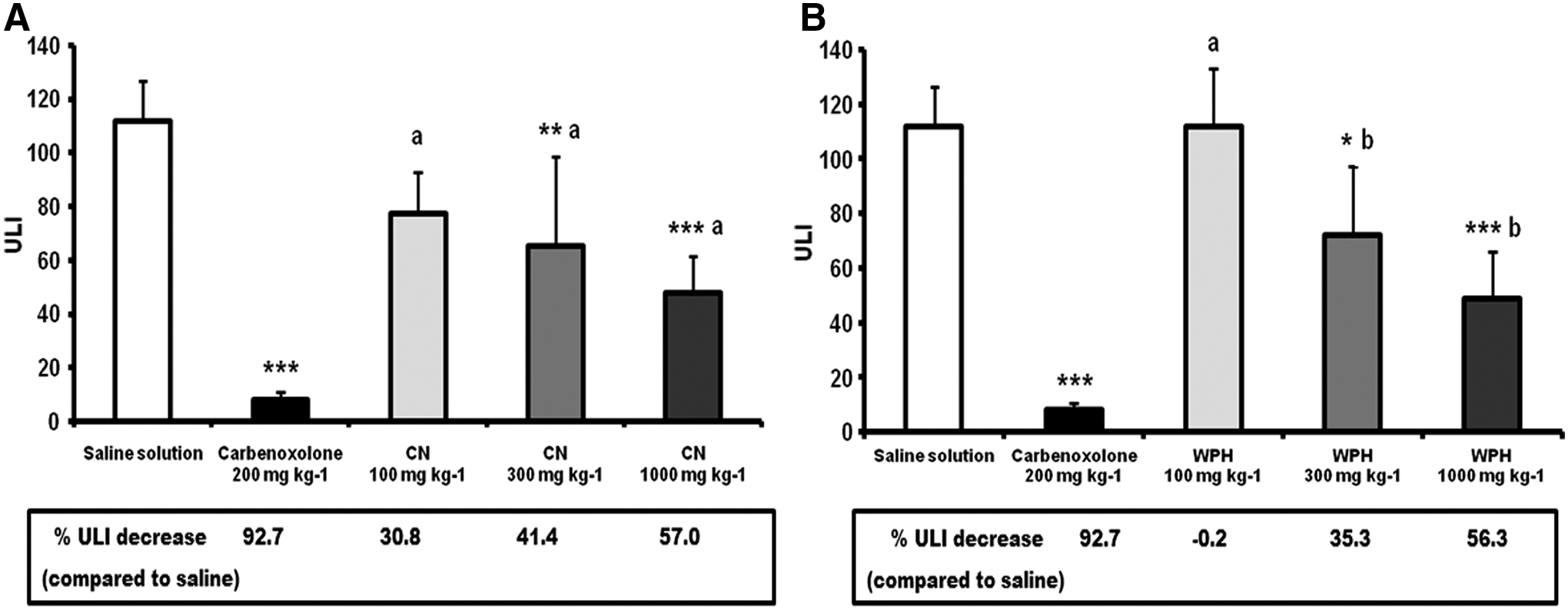
Effect of single dose of commercial CNH
The pretreatment with CNH considerably reduced areas of gastric damage for administration of ethanol and decreased the ULI percentage in a dose-dependent manner (at 100 mg kg−1 bw presented a 28.0% ULI decrease, at 300 mg kg−1 bw presented a 47.0% ULI decrease, and at 1000 mg kg−1 bw presented a 66.3% ULI decrease) (Fig. 2A).
WPH was only active at concentrations of 300 and 1000 mg kg−1 bw with a value of 35.3% ULI decrease and a 56.3% ULI decrease, respectively (Fig. 2B). The effective doses to inhibit 50% of ULI (ED50) were calculated for the two tested samples. The values of ED50 obtained were of 364 mg kg−1 for CNH and 690 mg kg−1 for WPH.
Ethanol acts directly as an irritant agent on gastric mucosa. 20 First, ethanol destroys the protective layer of mucosa, composed of mucus and bicarbonate, and subsequently damages the gastric epithelium, provoking the release of reactive oxygen species, the decrease of cell proliferation, and exacerbated inflammatory responses up to necrosis, edema, and hemorrhage. 24,25 In addition, other ulcerogenic activities are associated to ethanol, such as the stimulation of stomach acid secretion, the decrease of the blood flow in the mucosa, or the reduction in the PG production. 26
The antiulcerative activity has been described for certain food components and represents an alternative or additional therapy in the treatment of peptic ulcers. For example, food-derived polyphenols have demonstrated protective effects against lesions produced by various ulcerogenic agents and H. pylori. 27,28 Dairy products are considered a good source of components with antiulcerative properties, including whole and skimmed bovine milk, which showed activity in different ulcer models. 29
Whey proteins, through WPCs, their hydrolysates, and derived fractions have reported positive influence on gastric mucosa stability. 13,30,31 Based on the research of Matsumoto et al. 6 the protective effect exhibited by WPCs and derivates has been predominantly linked to their content in α-La. Further studies have confirmed the ulcer inhibitory properties of this whey protein. 7,9 Matsumoto et al. 6 found a significant inhibition of ulcer formation (47% damage control) mediated by the ingestion of a WPI, followed by the evaluation of the activity of individual components at representative concentrations, showing that only α-La emulated the observed effect.
Nevertheless, results obtained in our work for WPH differ from the established idea that β-Lg possesses no significant antiulcerative properties. The commercial WPC, which was used in the preparation of our WPH, was enriched in β-Lg up to more than 99% of the protein composition, according to previous analysis. 16 The significant activity observed for WPH with decreases of 35.3% ULI and 56.3% ULI, at doses of 300 and 1000 mg kg−1 bw, respectively, cannot be attributed to α-La or derived fragments but to β-Lg-derived peptides or other nonprotein components. Pacheco et al. 30 reported a comparable protective response for a pancreatic WPH (65.5% ULI decrease after acute 1000 mg kg−1 bw administration). However, it must be considered that the starting WPC contained a considerable α-La concentration. 32 The results exposed in this study are in agreement with the data collected by Matsumoto et al., 6 where low doses of β-Lg (100 mg kg−1 bw) showed a lack of activity against alcohol-induced lesions, as it is the case in the current study.
A more substantial discrepancy was found in the case of casein, since Matsumoto et al. 6 concluded that bovine casein has no antiulcerative properties after administrating 200 mg kg−1 bw (9% decrease ulcer index). To the best of our knowledge, no additional studies have been published about the antiulcerative activity of CNH. In our experiments, a dose as low as 100 mg kg−1 bw of casein achieved a protective effect three times higher than the previous reported activity (30.8% ULI decrease vs. 9%). These results suggest that casein may develop an important protective role versus ulcerative lesions. Such discrepancy may be related to methodological variations, such as, pretreatment time before ethanol administration (30 min vs. 60 min in our study), ethanol concentration (60% vs. absolute ethanol in our study), and the ulcer analysis criteria (the injury length vs. methodology from Gamberini et al., in our study 21 ).
Evaluation of the cytoprotection mechanisms
Evaluation of the contribution of SH groups in antiulcerative activity
To evaluate if the observed antiulcerative activity is mediated by the protective action of endogenous SH groups, an in vivo assay using NEM as inhibitor was performed. The subcutaneous injection of NEM blocks this defensive route via the alkylation of all endogenous active SH groups in the rat body. The role of SH mechanism in the protective activity of both hydrolysates was assessed by acute administration (300 mg kg−1 bw for CNH and 500 mg kg−1 bw for WPH) with or without a previous NEM injection.
Figure 3 shows ULI scores for each treatment group, indicating also ULI reductions in relationship to the reference group of each model. Saline solution presented a value of 81.2 ULI, while the saline solution+NEM presented an increase of ULI with a value of 137.8 ULI, CNH at 300 mg kg−1 bw presented 54.6 ULI, and presented an increase when the sample was treated with NEM with a value of 94.4 ULI.
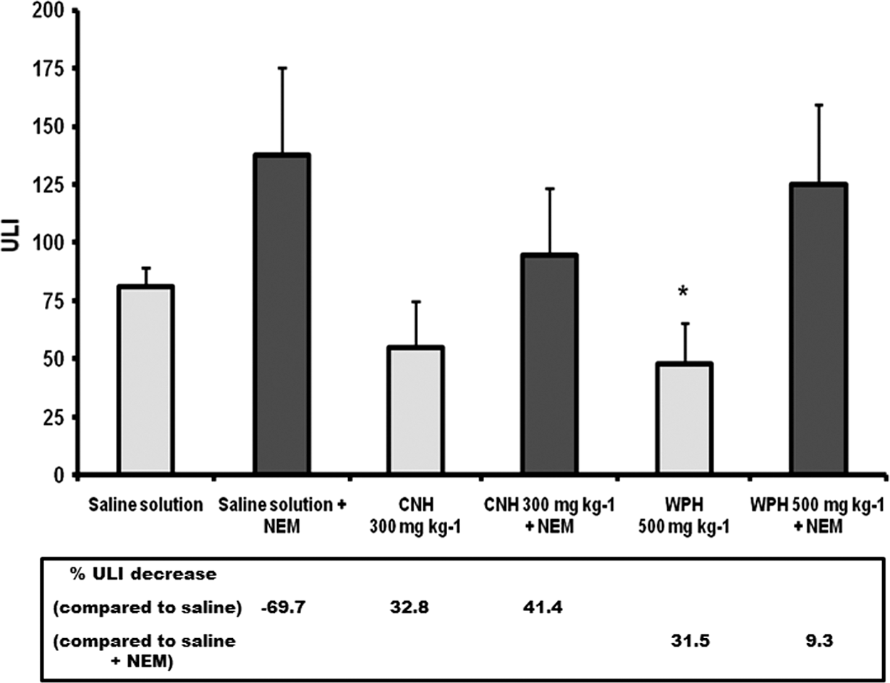
Effect of NEM, administrated by previous subcutaneous injection at 10 mg kg−1 bw, on the protective effect in rat stomachs against absolute ethanol-induced ulcers in treatment with single dose of commercial CNH and WPH. ULI expressed as mean ± standard deviation (n = 5). Data analyzed by a one-way ANOVA and followed by Tukey's test. Significant differences between samples and associated saline solution group as *P < .05. NEM, N-ethylmaleimide.
Similarly, WPH without NEM presented a value of 47.6 ULI, whereas in the presence of NEM, the sample presented an increase of 125 ULI. These ULI increases indicate that SH groups were blocked for NEM. The alkylation of endogenous SH increased the mucosa susceptibility to ethanol-induced lesions. This can be clearly appreciated in the increase of % ULI with a decrease of % ULI previous to the injection of NEM. The CNH sample with antiulcerative activity was not significantly different in both models (with or without NEM) with a value of 32.7% ULI without using NEM and 31.5% ULI using NEM. There was a decrease of % ULI with respect to each control (i.e., saline and saline+NEM) indicating that the protection mechanism was not mediated by SH groups.
On the contrary, the inhibition of ulcer formation when using WPH was drastically decreased (from 41.3% ULI to 9.2% ULI) when rats received NEM. This lack of activity under NEM preinjection supports the involvement of active SH in its action mechanism on mucosa protection. Figure 4 shows the consequences of NEM pretreatment on mucosa integrity after absolute ethanol administration. Figure 4A–C showed the groups of saline solution CNH and WPH without NEM, which can observe less lesions of gastric mucosa. Figures 4D–F show the groups with saline solution and CNH and WPH in presence of NEM, these last pictures show stomachs clearly more damaged, with more lesions.

Images of ethanol-induced ulcers in rat stomachs under different treatment with or without previous injection of NEM at 10 mg kg−1 bw: saline solution 10 mL kg−1 bw
Glutathione (γ-L-glutamyl-L-cysteinyl-glycine, GSH), a hydrophilic tripeptide, represents the main source of endogenous nonprotein SH groups in the gastric mucosa and can operate together with some PGs with a protective role on gastric mucosa. 33
Therefore, GSH constitutes one of the most important cytoprotective agents against peptic ulcer formation. 34 Previous reports have revealed that protective properties associated to whey proteins or their hydrolysates depend on the mechanisms of active SH groups. For example, Rosaneli et al. 31 and Pacheco et al. 30 demonstrated that a WPC and a WPH with pancreatin, respectively, present an important decrease of gastric protection when endogenous SH groups were blocked. Mezzaroba et al. 7 detected a similar prevented inhibition for α-La and two α-La-derived hydrolysate fractions on rats treated with NEM. Interestingly, Tavares et al. 13 found that endogenous active SH groups contributed to a greater extent to an antiulcerative action of low-molecular-weight peptides fractions compared with their whole whey protein-derived hydrolysates.
The stimulation of mucus production represents another important mechanism in the defense of gastric mucosa. The gastric mucus layer covers the gastric mucosa and constitutes a physical barrier between lumen and stomach tissue. Antiulcerogenic activity of kefir was recently reported in rats that received milk preparations after ethanol-induced gastric ulcers. 35 This antiulcer activity was related to a decrease of HCl secretion by gastric H+/K+-ATPase inhibition and the stimulation of mucous secretion. Mucins, high-molecular-weight glycoproteins, are mainly responsible for mucus viscous properties.
Moreover, MUC5AC and MUC6 represent the most abundant gastric secreted mucins, being MUC5AC the main component of mucus layer, whereas MUC6 can develop other activities such as antibiotic functions against H. pylori. 36 Recently, our research group has described enhanced mucin production under the action of the hydrolysates used in this work (CNH and WPH), when they were evaluated in intestinal human mucin-secreting HT29-MTX cells. 16,17 Both hydrolysates increased the mucin secretion and overexpressed the gene MUC5AC, the major secreted mucin gene in this cell line, which exhibits a gastric-like mucus secreting phenotype. CNH improved the mucin secretion up to 210% with respect to controls and upgraded the expression of MUC5AC over 1.8-fold basal level, 17 demonstrating to be slightly more active than WPH, which exhibited a maximum mucin secretion of 152% of controls and a MUC5AC transcription level of 1.52-fold basal level. 16
Each hydrolysate contains peptides with proved or probable μ- or δ-opioid activity whose mucin-producing activity justifies partially the detected activity of the hydrolysate. Fragments of α s1-casein 90–94 (RYLGY), 143–149 (AYFYPEL), and 144–149 (YFYPEL) contained in CNH, besides β-lactorphin (YLLF) in WPH demonstrated effect on mucin secretion when tested individually. 16,17,37 Therefore, it can be hypothesized that food-derived peptides may play a significant role in the mucosa protection via stimulation of mucus secretion. Taking into consideration the results of this work, the mucus strengthening could contribute remarkably to the antiulcerative activity, in particular for CNH whose protector effect seem to be independent from active SH groups. Regardless of opioid interactions, gastric mucin release has been related to PGE2 production. 38 α-La as antiulcerative agent. This association has been initially supported due to the gastric mucosa protection against ulcerative ethanol or indomethacin-induced lesion via concurrent stimulation of PGE2 and mucus increase. 7 However, a later in vitro study suggested that its mucin-producing effect was independent of endogenous PGE2. 9
Castro et al. 10 demonstrated that two porcine and bovine collagen hydrolysates exerted significant gastric mucus increases, being their mucosa protective activity mostly reverted by previous NEM SH-alkylation in an absolute ethanol ulcer model. The mucus-secreting and antiulcerative properties reported for some food-derived agents, together with the results obtained for CNH and WPH, are in agreement with the mucus strengthening as a key mechanism in the protective function against ethanol-induced gastric ulcers. Further studies, focusing on individual peptides with mucin-producing activity, should be carried out in rat ulcer models to identify active components in the gastric mucosa protection.
PG protection
Anti-inflammatory drugs such as the indomethacin drug administered in toxic doses (10 mg kg−1) produce visible gastric ulcers in stomach of rats. Indomethacin is a strong inhibitor of PG biosynthesis enzymes. PG enzymes are known to play an important role in maintaining mucosal integrity. An increase in certain endogenous PGs can enhance gastric mucosal resistance to ulcerogenic agents such as ethanol. The mechanisms involved in PG protection are multiple, among them stimulation of mucus and bicarbonate output, gastric mucosal blood flow, a decreasing gastric motility, an increase in the release of endogenous mediators of gastric injury vasoactive amines, and others. 39,40
Figure 5 shows an increase in ULI related to the saline solution observed in rats that had received indomethacin, which confirms the inhibition of this pathway. In the case of WPH (500 mg kg−1) treated with indomethacin, it was observed that the hydrolysate partially loses the antiulcerative effect. This results in a reduction of only 26.42% ULI compared with the effect hydrolysate (without indomethacin) 29.32% ULI. The protective action may be exerted by this mechanism, but the mechanisms involved in PG protection are yet unknown to this study. On the contrary, WPH presented a value of inhibition of 36.20% ULI without indomethacin and 49.9% ULI with indomethacin. WPH conserved its antiulcerative activity with an increase of its activity. This result suggests that the protection is not affected by the blocking of the PGs.
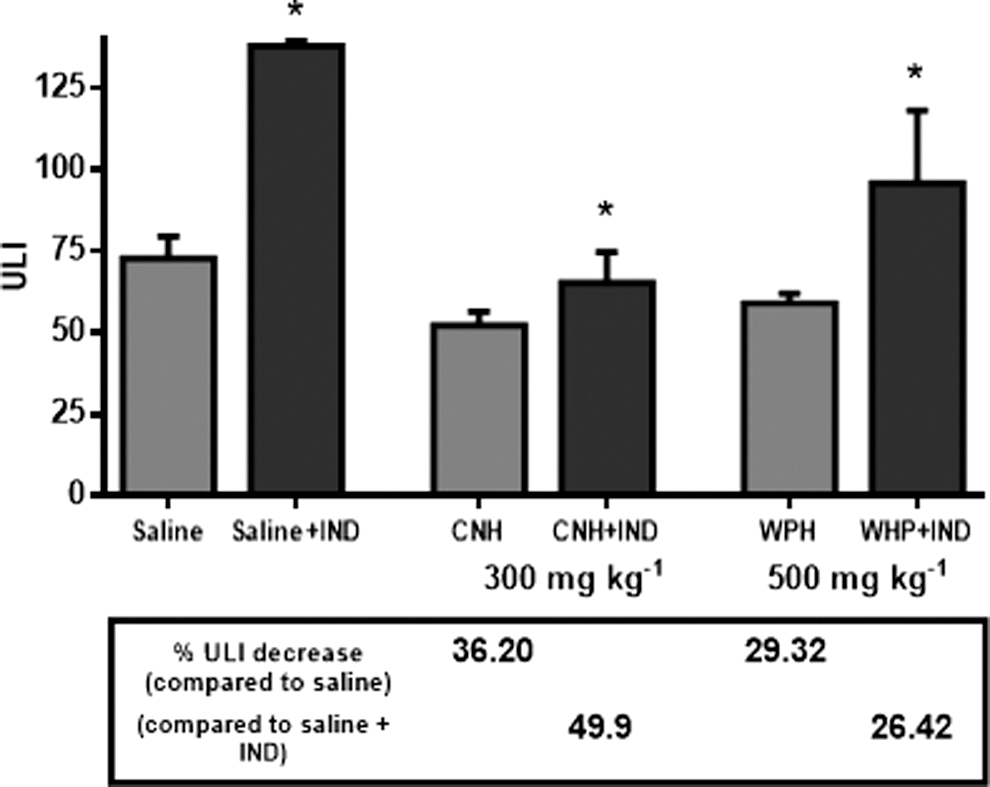
Effect of indomethacin, administrated by previous subcutaneous injection at 10 mg kg−1 bw, on the protective effect in rat stomachs against absolute ethanol-induced ulcers in treatment with single dose of commercial CNH and WPH. ULI expressed as mean−standard deviation (n = 6). Data analyzed by a one-way ANOVA and followed by Tukey's test. Significant differences between samples and associated saline solution group as *P < .05.
The positive results on the CNH and WPH antiulcer activity are indeed promising for further use in the development of functional ingredients for gastric protection. Amorim et al. 23 reported a yeast peptide extract (fraction <3 kDa) with antiulcerative activity in an ulcer induced by absolute ethanol in rats. These authors suggest that the antiulcerative activity was mediated by the PG mechanism. Tavares et al. 13 reported a peptide concentrate PepC obtained from WPC with antiulcerative activity with PG protection, the treatment without indomethacin presented 40.2% ULI, and treatment with indomethacin presented 19.7% ULI. The fraction PepF (below 3 kDa) showed no PG protection. The results of this study are in agreement with this research group, as WPH in this study presented PG protection too.
Summary
Results obtained in this study allow us to conclude that CNH and WPH present antiulcerative protective effects on gastric mucosa of rats in ulcerative lesions caused by ethanol. In the mechanism of protection, WPH implicates SH groups and PGs. In future research, the mechanism involved in PG protection could be identified. Milk protein is a source of most studied bioactive compounds. Caseins and whey proteins are the most used in the elaboration of functional ingredients. CNH and WPH can be used in the future in the elaboration of functional ingredients with antiulcerative activity to prevent and treat chronic diseases such as peptic ulcer.
Footnotes
Acknowledgments
This work has received financial support from the projects CYTED110AC0386 (IBEROFUN), AGL2011-24643, FEDER-INNTERCONECTA-GALICIA (ENVELLEFUN), FP7-SME-2012-315349 (FOFIND), and CNPq (Brasil). The authors are participants in the FA1005 COST Action INFOGEST on food digestion. The English edition has been reviewed by Emilio Labrador.
Author Disclosure Statement
No competing financial interests exist