
Abstract
No specific therapeutics are available for the treatment of sepsis-induced liver dysfunction, a clinical complication strongly associated with the high mortality rate of septic patients. This study investigated the effect of the essential oil of Hyptis crenata (EOHc), a lamiaceae plant used to treat liver disturbances in Brazilian folk medicine, on liver function during early sepsis. Sepsis was induced by the cecal ligation and puncture (CLP) model. Rats were divided into four groups: Sham, Sham+EOHc, CLP, and CLP+EOHc. EOHc (300 mg/kg) was orally administered 12 and 24 h after surgery. The animals were sacrificed for blood collection and liver tissue samples 48 h after surgery. Hepatic function was evaluated by measuring serum bilirubin, alkaline phosphatase (ALP), aspartate aminotransferase, and alanine aminotransferase (ALT) levels. The levels of malondialdehyde and the activity of superoxide dismutase, catalase, and GSH peroxidase (GSH-Px) were measured for assessment of oxidative stress. Liver morphology was analyzed by hematoxylin and eosin staining. EOHc normalized serum ALP, ALT, and bilirubin levels and inhibited morphological changes. In addition, we observed that EOHc inhibited elevation in hepatic lipid peroxidation and reduction of the glutathione peroxidase activity induced by sepsis. Our data show that EOHc plays a protective effect against liver injury induced by sepsis.
Introduction
L
Nowadays, nearly half of the agents used to treat pathological liver disorders are herbal products and their derivatives, including essential oils. 8,9 Previous studies have shown that essential oils extracted from different plant species, such as Rosmarinus officinalis, Pimpinella anisum, and Nigella sativa, exhibited hepatoprotective effects, which are attributed mainly to antioxidant properties of their components. 10 –12
Hyptis crenata Pohl ex Benth is a Lamiaceae plant species commonly found in the north and northeast of Brazil, where it is popularly known as salva-do-marajó and hortelã-do-campo. 13 In folk medicine, teas made with its leaves are used to treat gastrointestinal and liver disturbances. 14 In a previous study performed by our group, we showed that the essential oil of H. crenata (EOHc) mainly comprises camphor (32.78%), 1.8-cineole (18.02%), α-pinene (13.37%), and β-caryophyllene (12.86%) and it reduced gastric mucosa lesions induced by ethanol and indomethacin, supporting its claimed use as gastroprotective agent. 15 However, no experimental studies have investigated its hepatoprotective activity. Thus, the current study set out to evaluate the effect of the EOHc on liver dysfunction induced by polymicrobial sepsis.
Materials and Methods
Plant material
Aerial parts of H. crenata were collected (March, 2016) in the city of São Raimundo das Mangabeiras, Maranhão State, Brazil. The identification of plants was confirmed by Dr. Oriel Herrera Bonilla. A voucher specimen (No. 000106) was deposited in the Marlene Freitas da Silva herbarium. The EOHc was isolated from freshly chopped leaves by steam distillation and analyzed chemically by gas chromatography–mass spectrometry, as described by Diniz et al. 15
Animals
Male Wistar rats (240–300 g) were obtained from the bioscience unit of our institution and housed in standard conditions with free access to standard chow and tap water. All animals used in the current study (n = 60) were kept at room temperature (22°C ± 2°C) with a 12-h light/12-h dark cycle. All procedures described had prior approval from the animal ethics committee of the Universidade Federal de Sergipe (no. 40/15).
Experimental polymicrobial model of sepsis
Sepsis was induced by the cecal ligation and puncture (CLP) model of polymicrobial sepsis, as previously described by Rittirsch et al. 16 Briefly, the animals were anesthetized with intraperitoneal injection of ketamine (15 mg/kg) and xylazine (7.5 mg/kg). After anesthesia, a 3-cm midline incision was made in the anterior abdomen and the cecum was exposed. Fecal content was dragged to the cecum ligation and held below the ileocecal junction pole with a 5/0 Prolene thread (Ethicon), without causing bowel obstruction. The cecum was punctured 10 times with an 18-gauge needle and slightly compressed until a small drop of stool appeared. The cecum was repositioned in the abdomen and incisions in the peritoneal wall and skin were closed. Sham-operated animals underwent an identical laparotomy, but without cecal puncture, and served as controls. Soon after the surgery procedure, 2 mL of 0.9% NaCl solution was administered, subcutaneously, in all animals.
Experimental design
The animals were divided into four groups of seven animals each and treated as follows: Groups Sham and CLP consisted of sham- and CLP-operated animals that received 0.9% NaCl and 0.005% Tween 20 solution (vehicle), orally administered (0.1 mL per 100 mg body weight), every 12 h (i.e., twice daily) for 24 h after sham and CLP operations, respectively. Groups Sham-EOHc and CLP-EOHc consisted of sham- and CLP-operated animals that received 0.9% NaCl solution with 0.005% Tween 20 solution containing EOHc, at a dose of 300 mg/kg, orally administered (0.1 mL per 100 mg body weight) every 12 h for 24 h after sham and CLP operations, respectively. At 48 h after CLP and sham operations, all animals were euthanized by intensive dose of 5% isoflurane. Serum was separated from blood samples and stored at −20°C for posterior biochemical analysis. The livers of the animals were perfused with ice-cold 0.9% NaCl, surgically removed, homogenized, and centrifuged (5369 g for 10 min at 4°C). The supernatants were used for the assays of lipid peroxidation (thiobarbituric acid-reactive substances [TBARSs]), sulfhydryl content and catalase (CAT), superoxide dismutase (SOD), and glutathione peroxidase (GPx) activities (see below). In addition, slices of livers were fixed in 10% formaldehyde for histological processes.
Analytical procedures
Serum makers of hepatic toxicity
Liver function was analyzed by assessing serum levels of total protein, albumin, aspartate aminotransferase (AST), alanine aminotransferase (ALT), total bilirubin, alkaline phosphatase (ALP), and gamma-glutamyl transferase (γ-GT) using analytical diagnostic kits (Labtest Diagnostica SA®, Brazil). All analyses were done in duplicates based on the well-established spectrophotometric methods according to the kits' manuals supplied.
Hepatic oxidative stress analysis
To examine the occurrence of oxidative damage in the liver, malondialdehyde (MDA) and sulfhydryl levels, as well as activities of the antioxidant enzymes, CAT, SOD, and GPx, were assessed in hepatic tissue.
TBARS assay
Lipid peroxidation in liver was assessed by the modified method of the TBARS assay described by Bose et al. 17 Briefly, 50 mg of liver tissue samples, collected at 48 h after CLP-induced sepsis, was homogenized in potassium phosphate buffer (50 mM, pH 7.4) containing butylated hydroxytoluene (12.6 mM). Aliquots of the homogenate were incubated (45°C, 90 min) with thiobarbituric acid (0.37%) in an acid solution (15% trichloroacetic acid and 250 mM hydrochloric acid). After incubation, the homogenates were centrifuged (5 min, 8.000 g) and aliquots of supernatants were extracted with n-butanol, followed by stirring in a vortex for 30 sec and further centrifugation (2 min, 8.000 g). Supernatant absorbance was measured at 535 nm in a microplate reader (with correction by values of absorbance at 572 nm). The results were expressed as nmol of MDA formed per mg of tissue.
CAT assay
CAT activity was measured by the method of Bonaventura et al. 18 Briefly, 5 μg protein from the gastrocnemius muscle was mixed with 2.1 mL of 7.5 mM H2O2 and the reaction was allowed to continue for 10 min at 25°C. The disappearance of peroxide was continuously recorded from the absorbance at 240 nm for the specified period of time. One unit of CAT activity is defined as the amount of enzyme necessary for reducing 1 μmol of H2O2 per minute.
GPx activity
The activity of GPx was determined by Beutler et al. 19 Briefly, solutions of Tris-HCl (1000 mM), EDTA (5 mM) (pH = 8.0), GSH (100 mM), GSH reductase (10 U/mL), nicotinamide adenine dinucleotide phosphate (NADPH) (2 mM), and t-butyl hydroperoxide (7 mM) were incubated with 10 UL hemolysate for 10 min at 37°C. Decrease in NADPH was assessed spectrophotometrically at 340 nm.
SOD activity
The activity of SOD was assayed according to the method of Evans et al. 20 Enzyme activity was measured in an assay mixture containing 2 mL of Tris-HCl (pH 8.2), 2 mL of distilled water, 0.5 mL of tissue homogenate, and 0.5 mL of 2 mM pyrogallol. The resulting color was read immediately at 470 nm at 1-min intervals for 3 min in a spectrophotometer against a blank containing all components except the sample preparation and pyrogallol. The enzyme activity was expressed as units/mg protein.
Liver histological analysis
Slices of rat liver lobe were cleaned, dried, and fixed in a solution of 10% buffered formalin. For histopathological examination, livers were dehydrated with ethanol and embedded in paraffin. The specimen blocks were cross-sectioned at a thickness of 5 μm using an ultramicrotome. Paraffin sections (5.0 mm) were stained with hematoxylin and eosin (H&E) and analyzed using an Olympus CX51 light microscope.
Statistical analysis
The data are expressed as mean ± standard error. The statistical analysis was evaluated by one-way analysis of variance (ANOVA), followed by Bonferroni's multiple range test. Values were considered statistically significant when P < .05.
Results
Effect of EOHc on liver function
Alterations in hepatic function caused by experimental sepsis as well as the effects of EOHc on sepsis-induced liver dysfunction were evaluated by assessment of serum hepatocellular (AST and ALT) and hepatobiliary injury (ALP, serum albumin, urea, blood proteins, γ-GT, and total bilirubin) biomarkers 48 h postsepsis induction. As shown in Figure 1A–C, no significant differences in the serum levels of total proteins, γ-GT and AST, were observed among experimental groups. No significant difference in serum albumin and urea levels was also observed among experimental groups (data not shown). Differently, septic rats (CLP group) showed an increase of serum levels of ALT (40.02 ± 2.36 U/L), ALP (56.84 ± 7.20 U/L), and total bilirubin (1.16 ± 0.19 mg/dL) when compared with the sham group (32.68 ± 2.73 U/L, 38.10 ± 1.00 U/L, and 0.70 ± 0.01 mg/dL, respectively). Oral administration of EOHc (300 mg/kg) inhibited the sepsis-induced increase of serum ALT (34.11 ± 2.28 U/L), ALP (38.30 ± 2.50 U/L), and total bilirubin (0.70 ± 0.06 mg/dL) levels of EOHc-treated septic rats compared with the CLP group (Fig. 1D–F, respectively).
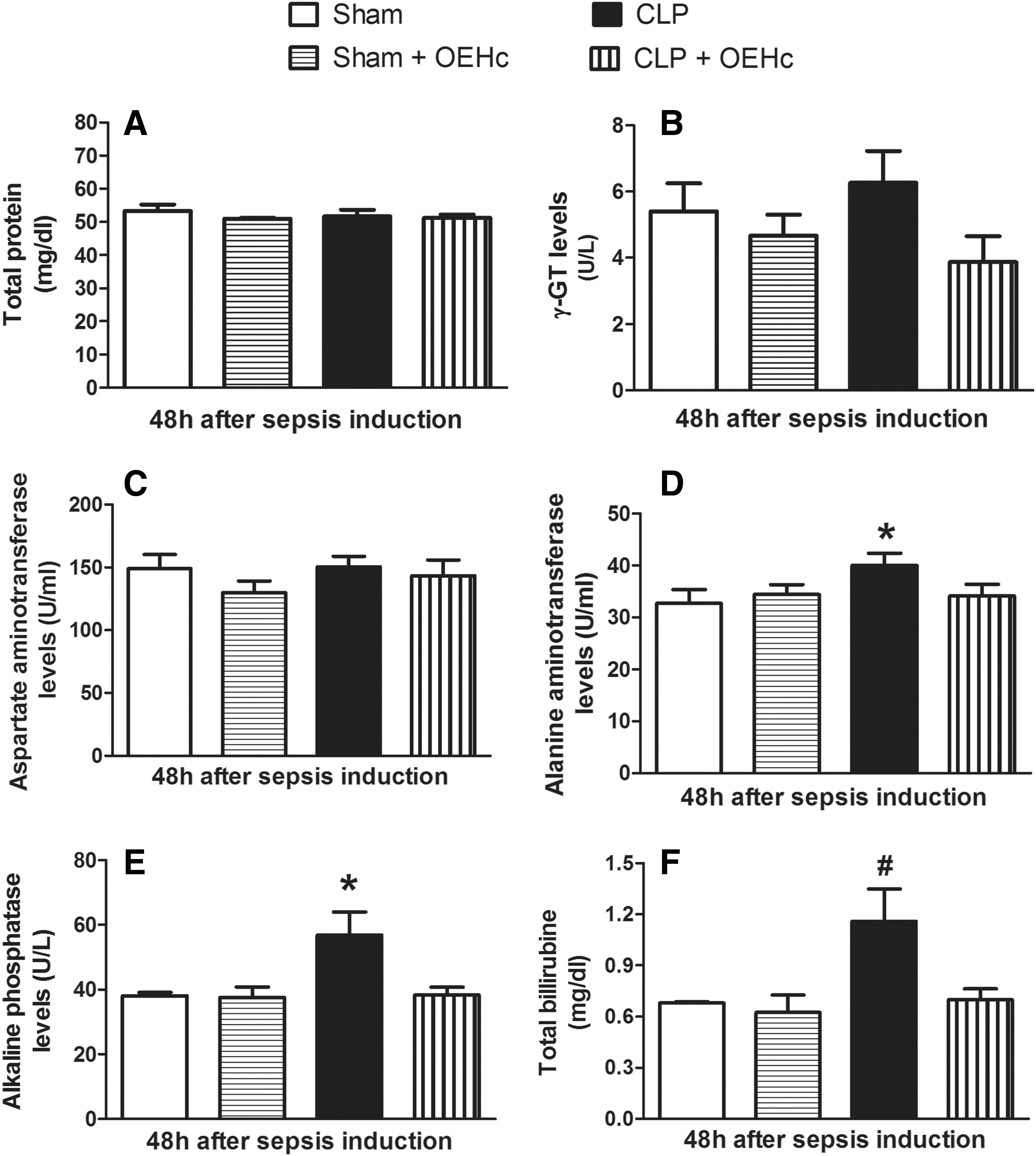
Biomarkers of hepatic function. Serum levels of
Effect of EOHc on hepatic oxidative stress
Figure 2 shows that CLP-induced experimental sepsis did not produce significant alterations in lipid peroxidation, indicated by liver MDA levels, and in the hepatic activities of SOD and CAT (Fig. 2A–C, respectively). However, a decrease in the activity of GPx was observed in CLP-operated rats (58.40 ± 1.72 U/mg protein. min−1 ) compared with sham-operated rats (74.23 ± 3.41 U/mg protein. min−1 ).

Analysis of oxidative stress: Lipid peroxidation and antioxidant enzyme activities. Hepatic tissue levels of
The oral treatment with EOHc (300 mg/kg) did not alter hepatic lipid peroxidation as well as antioxidant enzyme activities of sham-operated rats. Differently, CLP-operated rats treated with EOHc showed a significant (P < .05) decrease in hepatic lipid peroxidation (6.21 ± 0.61 mmol/mg protein) compared with untreated CLP-operated rats (10.33 ± 0.89 mmol/mg protein) (Fig. 2A). In addition, EOHc treatment inhibited the decrease in activity of GPx induced by sepsis, as mentioned above. It was observed that CLP-operated rats treated with EOHc showed glutathione peroxidase activity (72.64 ± 4.62 U/mg protein. min−1 ) compared with sham-operated rats (74.23 ± 3.41 U/mg protein. min−1 ) and CLP-operated rats (58.40 ± 1.72 U/mg protein. min−1 ), as shown in Figure 2D.
Histological examination
Histopathological examination of liver sections of the normal control group (Sham group) showed normal cellular architecture with distinct hepatic cells, sinusoidal spaces, and central vein with no signs of inflammation, necrosis, fibrosis, or cholestasis (Fig. 3A, D, H). Likewise, liver sections of sham-operated rats treated with EOHc (Sham-EOHc group) did not exhibit any significant histological changes (Fig. 3B, E, I). The liver sections of CLP-operated rats (CLP group) showed moderate histoarchitectural changes compared with liver sections of sham-operated rats (Sham group), as indicated by the presence of hydropic degeneration, mainly in the periportal space (red arrows) and increase in the rate of glycogenosis in hepatocytes (white square) (Fig. 3C, F, I). Meanwhile, liver sections of EOHc-treated septic rats (CLP-EOHc group) showed an almost normal pattern of liver tissue, decreased hydropic degeneration, and the presence of glycogenosis (Fig. 3D, G, K).
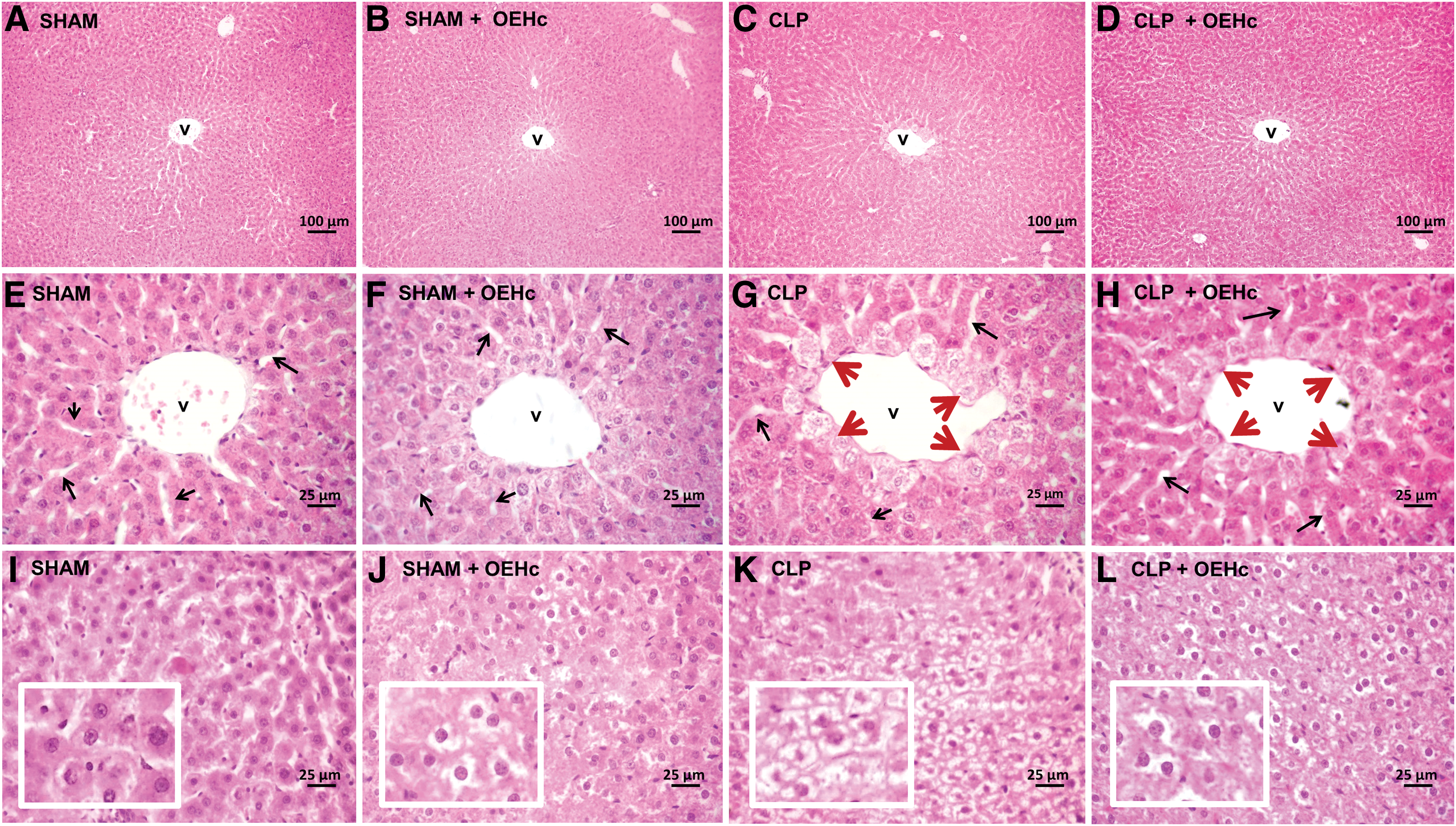
Histological examination of rat livers stained with H&E.
Discussion
Liver dysfunction is a clinical complication strongly associated with impairment of the prognosis of sepsis and it has been used as a powerful predictor of high mortality in ICU patients with severe sepsis and septic shock. 3,21 The present study provides evidences of the potential hepatoprotective effect of the EOHc on liver dysfunction induced by the CLP model in rats. The oral treatment with EOHc (300 mg/kg) significantly attenuated the rises in serum levels of biomarkers of liver injury and hepatic histopathological changes promoted by sepsis. Furthermore, EOHc restored alterations in oxidative stress caused by sepsis.
The sepsis-induced liver dysfunction is usually characterized by hepatocellular injury and impairment of biliary tract cells 22,23 as well as morphological alterations, mainly cholestasis. 24,25 Approximately 11% of patients newly admitted to the ICU exhibit an increase of bilirubin serum levels, which are commonly accompanied by increasing serum ALP, AST, and ALT levels, within 48 h of admission. 24,25 In the current study, we observed a significant increase in serum bilirubin, ALP, and ALT levels (Fig. 1). In addition, moderate morphological changes, as indicated by mitochondrial damage and glycogen storage failure, were also observed in the H&E-stained liver section from CLP-operated rats (Fig. 3). Our results are in agreement with the study performed by Recknagel et al. (2013), in which the authors showed that polymicrobial (CLP model) sepsis produces pronounced hepatocellular excretory dysfunction accompanied by cholestasis and lower degree of cell death compared with cytokine-mediated mechanisms of cellular injury found in endotoxemic rats. 26
The research for new drugs to ameliorate sepsis-associated organ dysfunction and its clinical complications is fundamental to reduce the high mortality caused by sepsis. In this context, Yang et al. (2009) have reported that the ethanol extract of Alpinia katsumadai Hayata seeds improved liver dysfunction of CLP-operated mice. 27 Zhong et al. (2016) have shown that curcumin, a natural plant phenolic food additive, regulated the increase of serum ALT, AST, and ALP levels on sepsis-induced acute liver injury in mice. 28
The hepatoprotective activity produced for different species of genus Hyptis (i.e., H. suaveolens and H. verticillata) 29,30 and the use of H. crenata for the treatment of hepatic disturbances by Brazilian folk medicine 13 –15 led our group to investigate the hepatoprotective activity of EOHc on sepsis-related liver dysfunction. Considering that hepatic dysfunction begins minutes after sepsis induction and may resolve in parallel with the remission of sepsis or remain for a 72-h period after the onset of severe sepsis, 5,21 EOHc treatment was performed by two consecutive oral administrations (300 mg/kg), at 12 and 24 h postonset of sepsis. The liver function was evaluated 48 h postonset of sepsis. The EOHc treatment normalized the rise of ALP, ALT, and total bilirubin observed in nontreated CLP rats (Fig. 1). Moreover, EOHc treatment reduced the presence of hydropic degeneration in the periportal space and glycogenolysis, as illustrated in histological examination of liver tissue stained with H&E in Figure 3.
It is known that reactive oxygen species (ROS) play an important role in the molecular mechanisms of inflammation and cellular damage in the genesis and progression of organic injury induced by sepsis. 26,31 In the present study, we observed a decrease of GPx activity in the liver of CLP-operated rats compared with sham-operated rats (Fig. 2D). A great trend toward increase of lipid peroxidation (Fig. 2A) in the liver of septic rats was also observed, which appears to be linked to inhibition of GPx activity that leads to increase of lipid peroxidation in membranes. 32 Differently, CLP-operated rats treated with EOHc did not show any significant alterations in the oxidative stress parameters evaluated, corroborating with the antioxidant activity produced by EOHc in experimental peptic ulcers induced by ethanol and indomethacin. 15 The antioxidant activities of essential oils from diverse plants, including essential oils obtained from Hyptis rhomboidea, Hyptis brevipes, and Hyptis spicigera, have been credited to components previously identified in the EOHc, such as camphor, 1.8-cineole, α-pinene, and beta-caryophyllene. 33 –35 For example, Ghaffar et al. (2015) have attributed to 1.8-cineole and alpha-pinene the antioxidant activities of essential oils extracted from seven Eucalyptus species. 36 β-caryophyllene and α-pinene normalized the altered hepatic biochemical markers and restored changes in activities of antioxidant enzymes caused by treatment of male mice with cyclophosphamide. 37
Taken together, our data showed that EOHc treatment promotes effective protection of liver dysfunction induced by the CLP model of sepsis, supporting its popular use in the treatment of hepatic disturbance. Furthermore, our results suggest that the hepatoprotective activity of EOHc might be, at least in part, attributed to its antioxidant activity by preventing the increase of ROS and enhancing hepatic antioxidant defenses. Thus, EOHc is a promising therapeutic agent in the research for efficient treatment of liver dysfunction induced by sepsis.
Footnotes
Acknowledgment
The authors are grateful to the staff members of the Institute of Technology and Research for their technical assistance.
Author Disclosure Statement
No competing financial interests exist.