




Abstract
Tropaeolum majus L., popularly known as nasturtium, is a species widely used in the form of infusions and salads. In the last years, the antihypertensive, diuretic, and calcium and potassium sparing activities of T. majus preparations were shown. Moreover, no preclinical 90-day oral toxicity studies were conducted. Thus, this study evaluated the toxicity of the hydroethanolic extract obtained from T. majus (HETM) leaves in female and male mice, rats, and rabbits. Swiss mice and Wistar rats were treated with HETM (75, 375, and 750 mg/kg). The doses of rabbits (30, 150, and 300 mg/kg) were calculated by allometric extrapolation. The control groups received vehicle. The animals were orally treated, daily, for 90 days. At the end, the animals were anesthetized, and body weight gain, relative weight of liver, kidney, and spleen, and histopathological changes were evaluated. Serum hematological and biochemical parameters were also analyzed. No alterations were found in body and organ weights or in histopathological and biochemical evaluation. Hematological analyses revealed small changes in lymphocytes and neutrophil counts in rats after administration of 750 mg/kg of HETM. These results showed that 90-day use of T. majus is safe in rodents and lagomorphs.
Introduction
M
An important medicinal plant native to the Andes region and widely studied in recent years is Tropaeolum majus L. (Tropaeolaceae), popularly known as nasturtium, capuchinha, chaguinha, agrião-do-méxico, mastruço-do-Peru, and flor-de-sangue. 5 Due to its easy adaptation to different climates, this plant has spread rapidly around the world. In Brazil, this species has been used as food, with economic and medicinal importance, being also used as honey-producing plant, natural dye, unconventional vegetable, and ornamental herb. 6
Several preclinical studies conducted with T. majus leaves have demonstrated its powerful therapeutic and pharmacological potential. In fact, antibiotic, 7 anticancer, 8 antithrombotic, 9 diuretic, 10 –13 hypotensive, and antihypertensive 13,14 activities were shown. Despite the evidence of therapeutic effects of T. majus, there are few toxicological data describing the safety of this plant. Lourenço et al. 15,16 demonstrated that T. majus is not completely safe during pregnancy in rats. In another study, no alterations were observed in rats treated with T. majus extract for 28 days. 17 Moreover, no preclinical 90-day oral toxicity studies were conducted. Thus, the aim of this study was to evaluate the preclinical toxicity of hydroethanolic extract obtained from T. majus (HETM) leaves in rodents and lagomorphs for a period of 90 days.
Materials and Methods
Plant material and preparation of the HETM
T. majus leaves were collected in the Botanical Garden of the Paranaense University (UNIPAR), located in the municipality of Umuarama-PR, 430 m of altitude above sea level (S23°47′55-W53°18′48). The species was identified by Dr. Mariza Barion Romagnolo (Department of Botany, State University of Maringá-UEM), and a voucher is cataloged under number 2230 in the official herbarium at the UNIPAR.
The material collected was dried in oven with forced air circulation at 37°C for a period of 5 days. After drying, the material was ground and stored in paper bags. Then, HETM was prepared by maceration at room temperature for 7 days using 90% ethanol as solvent. The HETM obtained was filtered and concentrated at reduced pressure through rotary evaporator with temperature not exceeding 55°C. Subsequently, the extract was lyophilized. The extract yield was 15.3% and was diluted with distilled water for use in experiments.
The main classes of compounds in HETM were investigated by high performance liquid chromatograph and electrospray ionization-mass spectrometry, evidencing isoquercitrin, a flavonoid, as a major compound. Previous studies by our research group analyzed the constituents of HETM in more detail. 12,14
Animals
Two species of rodents (mice and rats) and one species of nonrodents (rabbits) were used in young adulthood, according to Organization for Economic Co-operation and Development (OECD) Guidelines for the Testing of Chemicals, Section 4, tests no. 408 and 409. 18,19 Twelve-week-old female (n = 32) and male (n = 32) Wistar rats (Rattus norvegicus) weighing 250–300 g and female (n = 32) and male (n = 32) Swiss mice (Mus musculus) weighing 35–40 g were obtained from the Central Animal Facility of the Federal University of Paraná. Ninety-day-old female (n = 32) and male (n = 32) New Zealand rabbits (Oryctolagus cuniculus) weighing 1.6–2 kg were obtained from Institute of Technology of Paraná, Brazil. Animals were kept in the animal facility at constant temperature (22°C ± 2°C) under a 12-h light/12-h dark cycle, 55% ± 10% humidity conditions, and ad libitum access to food and water. All experimental procedures were approved by the Ethics Research Committee Involving Animal Experiments (UNIPAR) under protocol no. 24383/2013, and experiments were performed in accordance with international standards and ethical guidelines on animal welfare.
Experimental design and sample collection
Repeated dose 90-day oral toxicity was determined according to procedures recommended by the OECD protocol no. 408 and 409. 18,19 A previous toxicological study performed by Gomes et al. 17 was used as reference to determine the doses used to evaluate the 90-day toxicity of HETM in rodents. The doses of HETM used in rabbits were determined from the allometric extrapolation of the doses used for rodents.
Animals were divided into four groups by gender (n = 8) and were orally (through gavage) treated with 30, 150, and 300 mg/kg (rabbits) or 75, 375, and 750 mg/kg (mice and rats) dose of HETM or vehicle (filtered water, control group), daily, for 90 days. The animals were weakly weighed, and clinical signs of toxicity (grooming, piloerection, dyspnea, ptosis, abdominal contraction, diarrhea, prostration, ataxia, anesthesia, coma, and death) were daily observed. Water and food consumption were monitored during all the experiments.
At the end of 90 days of the experiment, the animals were fasted and anesthetized by inhalation of isoflurane in closed saturation glass tanks. In rabbits, 10 mL of peripheral blood was collected from jugular vein for analysis of hematological and biochemical parameters. After blood collection, rabbits were euthanized by suprapharmacological dose of isoflurane. In rats, 1 mL of peripheral blood was collected from the ocular plexus with capillaries for analysis of hematological parameters. Due to limited amount of blood, this parameter was not evaluated in mice. After, the rodents were euthanized by decapitation to collect a larger amount of blood for biochemical analyses. For hematological and biochemical evaluation, tubes were used with either K2-EDTA anticoagulant or containing separating gel with clot activator, respectively. After euthanasia, liver, kidneys, and spleen were removed to gross pathology, and their relative weights were determined (absolute organ weight × 100/body weight of rats on the day of sacrifice). Liver, kidneys, and spleen samples were collected for histopathological evaluation.
Hematologic analyses
Samples were appropriately homogenized and stored at 4°C until processing. The total number of erythrocytes (RBC, million/μL), hemoglobin (g/dL), hematocrit (%), mean corpuscular volume (MCV, fL), mean corpuscular hemoglobin (MCH, pg), MCH concentration (MCHC, g/dL), mean platelet volume (MPV, fL), platelets (103/μL), leukocytes (103/μL), segmented (%), neutrophils (%), lymphocytes (%), monocytes (%), and eosinophils (%) were evaluated in an automated hematologic analyzer (Abbott Cell Dyn 3500; Abbott Diagnostics, USA).
Biochemical analyses
Blood samples were centrifuged and stored at −20°C until use. Analyses were performed on an automated biochemical analyzer (Selectra E; Vital Scientific, Netherlands) or on a semiautomated system (MH-Labise-Selective Ion). Amylase (U/L), urea (mg/dL), creatinine (mg/dL), uric acid (mg/dL), potassium (mEq/L), sodium (mEq/L), total protein (g/dL), albumin (g/dL), globulin (g/dL), alkaline phosphatase (U/L), triglycerides (mg/dL), total bilirubin (mg/dL), direct bilirubin (mg/dL), aspartate aminotransferase (AST; U/L), alanine aminotransferase (ALT, U/L), total cholesterol (mg/dL), and high-density lipoprotein (HDL) cholesterol (mg/dL) were determined to evaluate renal, hepatic, and pancreatic function.
Histopathological analyses
Small fragments of liver, kidneys, and spleen of animals were fixed in 10% buffered formalin for subsequent histopathological analysis. These tissue samples were dehydrated in ethanol, diaphanized in xylol, and embedded in paraffin. Tissues were cut in a microtome (5 μm), and sections were used to prepare slides that were stained with hematoxylin and eosin (HE). Samples were analyzed by light microscopy for the overall structure of the organs, degenerative changes, evidence of necrosis, and signs of inflammation. Images were photographed for comparison of parameters.
Statistical analysis
The data were analyzed for normal distribution and homogeneity of variances. Differences between groups of the same sex were evaluated by one-way analysis of variance (ANOVA) or Kruskal–Wallis followed by Bonferroni's or Dunn's test. In multivariate analysis where the predictive factors were sex and concentration (with relative interaction) the differences between groups were evaluated by two-way ANOVA followed by Bonferroni post hoc test. Statistical significance levels of 5% were adopted (P < .05). Results are presented as mean ± standard error of the mean (SEM).
Results
No signs of toxicity or death were observed during the treatment with HETM at all doses in female and male mice, rats, and rabbits (data not shown). In the same way, no changes were found in the body weight and in the relative weights of liver, kidneys, and spleen of female and male mice (Table 1), rats (Table 2), and rabbits (Table 3).
Values are expressed as mean ± SEM, n = 8. Differences between groups of the same sex were evaluated by one-way ANOVA or Kruskal–Wallis followed by Bonferroni's or Dunn's test. In multivariate analysis where the predictive factors were sex and concentration (with relative interaction) the differences between groups were evaluated by two-way ANOVA followed by Bonferroni's test.
ANOVA, analysis of variance; HETM, hydroethanolic extract obtained from Tropaeolum majus; SEM, standard error of the mean.
Values are expressed as mean ± SEM, n = 8. Differences between groups of the same sex were evaluated by one-way ANOVA or Kruskal–Wallis followed by Bonferroni's or Dunn's test. In multivariate analysis where the predictive factors were sex and concentration (with relative interaction) the differences between groups were evaluated by two-way ANOVA followed by Bonferroni's test.
Values are expressed as mean ± SEM, n = 8. Differences between groups of the same sex were evaluated by one-way ANOVA or Kruskal–Wallis followed by Bonferroni's or Dunn's test. In multivariate analysis where the predictive factors were sex and concentration (with relative interaction) the differences between groups were evaluated by two-way ANOVA followed by Bonferroni's test.
In female rat hematological evaluation, the parameters analyzed were not altered by treatment with HETM compared with control group. In male rats, the dose of 750 mg/kg decreased segmented and neutrophil count and increased the number of lymphocytes in relation to the control group (Table 4). No hematological alterations were found in rabbits treated with 30, 150, and 300 mg/kg of HETM or vehicle (Table 5).
Values are expressed as mean ± SEM, n = 8. Differences between groups of the same sex were evaluated by Kruskal–Wallis followed by Dunn's test. In multivariate analysis where the predictive factors were sex and concentration (with relative interaction) the differences between groups were evaluated by two-way ANOVA followed by Bonferroni's test.
P < .05 compared with control group.
MCH, mean corpuscular hemoglobin; MCHC, mean corpuscular hemoglobin concentration; MCV, mean corpuscular volume; MPV, mean platelet volume; RBC, total number of erythrocytes.
Values are expressed as mean ± SEM, n = 8. Differences between groups of the same sex were evaluated by one-way ANOVA or Kruskal–Wallis followed by Bonferroni's or Dunn's test. In multivariate analysis where the predictive factors were sex and concentration (with relative interaction) the differences between groups were evaluated by two-way ANOVA followed by Bonferroni's test.
The treatment with all doses of HETM or vehicle did not alter the pancreatic, renal, and hepatic function of female and male mice (data no shown), rats (Table 6), and rabbits (Table 7).
The values are expressed as mean ± SEM. Differences between groups of the same sex were evaluated by one-way ANOVA or Kruskal–Wallis followed by Bonferroni's or Dunn's test. In multivariate analysis where the predictive factors were sex and concentration (with relative interaction) the differences between groups were evaluated by two-way ANOVA followed by Bonferroni's test.
ALT, alanine aminotransferase; AST, aspartate aminotransferase; HDL, high density lipoprotein.
The values are expressed as mean ± SEM. Differences between groups of the same sex were evaluated by one-way ANOVA or Kruskal–Wallis followed by Bonferroni's or Dunn's test. In multivariate analysis where the predictive factors were sex and concentration (with relative interaction) the differences between groups were evaluated by two-way ANOVA followed by Bonferroni's test.
Finally, in relation to histopathological evaluation, liver, kidney, and spleen were morphologically normal and similar among female and male animals (mice, rats, or rabbits) treated with vehicle or HETM. In mice spleen, the connective tissue capsule was found unchanged. Some of red pulp sinusoidal capillaries were slightly congested, and a reasonable number of megakaryocytes were observed, common in these species. The white pulp shows no change in germinal center (Fig. 1A, B). In the kidney tissue slides, no changes were observed in the renal corpuscles in cortical and proximal tubule and distal and renal pelvis (Fig. 1C, D). Finally, in the liver, no changes were observed in the bile canaliculi and in the portal space or in the radial arrangement of hepatocytes divided into zones 1, 2, and 3 (Fig. 1E, F).
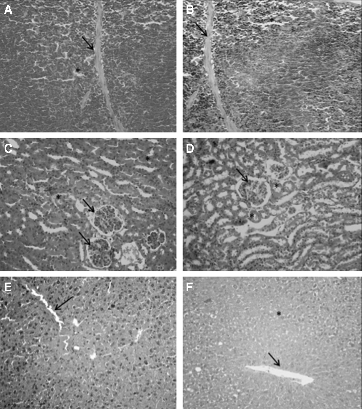
Histopathological evaluation of mice orally treated with vehicle (control group) or 750 mg/kg of HETM for 90 days.
In rats, the spleen connective tissue capsule remained unchanged. Some sinusoidal capillaries of the red pulp were slightly congested, and a reasonable number of megakaryocytes were observed, common in these species. The white pulp shows no change in germinal center (Fig. 2A, B). In the kidney tissue slides, no changes were observed in the renal corpuscles in cortical and proximal tubule and distal and renal pelvis (Fig. 2C, D). Finally, in the liver, no changes were observed in the portal space nor in the radial arrangement of hepatocytes divided into zones 1 and 2 (Fig. 2E, F).
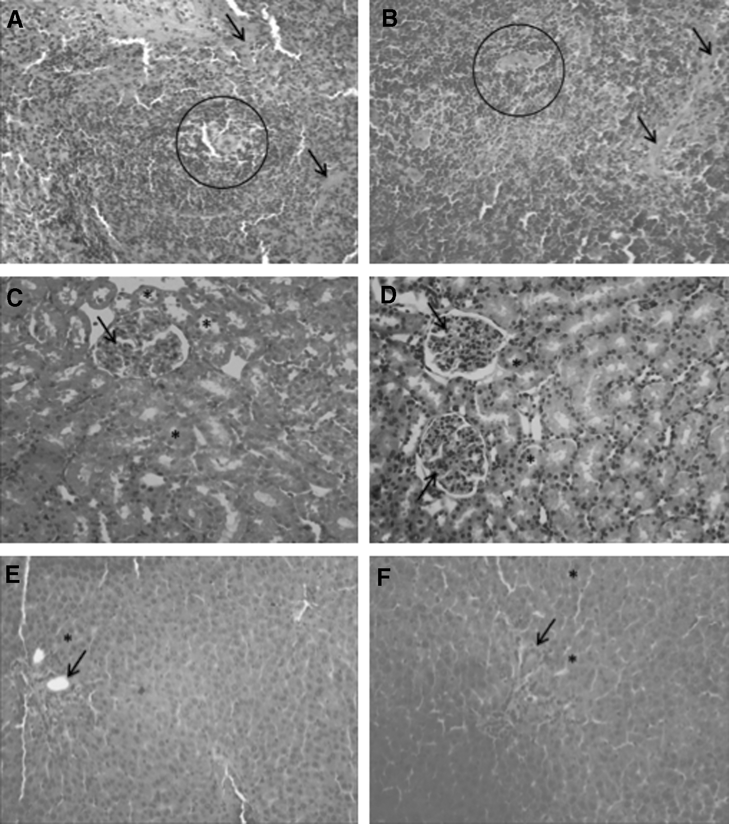
Histopathological evaluation of rats orally treated with vehicle (control group) or 750 mg/kg of HETM for 90 days.
The same histopathological pattern was observed in rabbits. Tissues of all organs collected were morphologically normal and similar between the animals treated with HETM or vehicle. The connective tissue capsule of the spleen presented normal morphology. In addition, the red pulp showed no signs of morphological changes compared to the animals in the control group. The white pulp did not show any changes in the germinal center (Fig. 3A, B). In renal tissue slides, no changes in the renal corpuscles, proximal tubules, and distal and renal pelvis were observed (Fig. 3C, D). In hepatic tissue, portal space and the radial arrangement of hepatocytes (divided into zones 1 and 2) were within the normal range for the species (Fig. 3E, F).
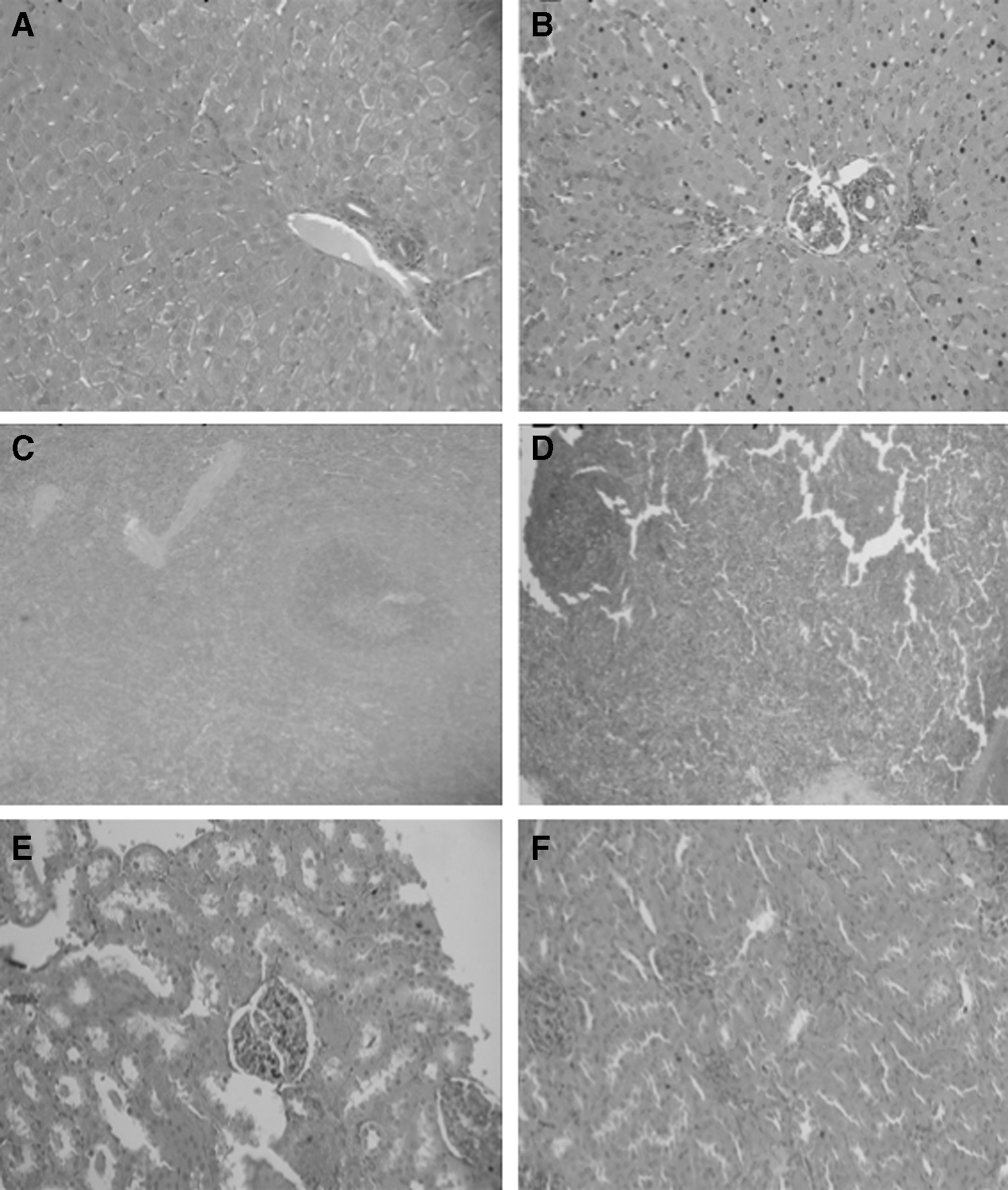
Histopathological evaluation of rabbits orally treated with vehicle (control group) or 300 mg/kg of HETM for 90 days.
Discussion
The use of medicinal plants for pharmaceutical or feeding purposes has always been a common practice for humanity. The species T. majus, for example, is used as infusions for therapeutic uses and as food in the preparation of salads, among others. Thus, it was deemed necessary to assess its toxic effects to demonstrate that the use of this plant is safe. In fact, according to OECD guidelines, 18,19 preclinical safety studies for potential herbal medicines standout, in which toxicity studies after repeated doses used for long periods are recommended.
In a subchronic preclinical research performed by Gomes et al. with HETM for a period of 28 days using the same doses and evaluating the same parameters of this study, the authors did not observe changes in all female or male rat parameters analyzed. However, a 90-day evaluation of T. majus was needed to more accurately assess toxicological effects.
The results of this study demonstrated no signs of toxicity related to the 90-day use of T. majus. No differences were observed with respect to hematopoiesis and blood cell percentage in all tested groups, except in male rats treated with the highest dose of HETM, which have a significant decrease in segmented and neutrophils and a significant increase in the number of lymphocytes compared to vehicle. These changes raised the following question: would it be possible that the extract would have changed in some way the defense system of animals? According to Yusuff et al. 20 and Nishida et al. 21 the prolonged use of some antihypertensive drugs can change the blood parameters, causing hematological toxicity. In fact, T. majus has important diuretic and hypotensive effects, mainly through the inhibition of the angiotensin-converting enzyme 11 –14 ; however, it is still too early to say whether HETM can lead to drug-induced hematological toxicity. So, a potential limitation of our study was the lack of accuracy to identify the role of HETM in the hematological changes evidenced in male rats. Anyway, the changes in the levels of lymphocytes and neutrophils are minimal compared with classes of drugs with the same therapeutic purposes. Moreover, the dose in our study that promoted this change is too high (750 mg/kg) compared to the dose in which the best pharmacological effects in preclinical studies were observed (300 mg/kg). 11 –14
Regardless of serum biochemical parameters used to assess liver, kidney, and pancreas function, no alteration was observed in all groups tested. The absence of HETM nephrotoxicity was also reported by Gasparotto Junior et al. 11 –13 that reported no changes in urea, creatinine, sodium, and plasma potassium levels, even using the semipurified fraction of this extract or the isoquercitrin, a flavonoid present in this species.
Recently, the potential of T. majus to induce genotoxicity was investigated by Traesel et al. 22 on bacterial reverse mutation, genomic lesions, and micronucleus formation in male rats. No revertant colonies were found in any bacterial cultures examined; no significant DNA damage and no significant increases in the frequency of inducing micronuclei were observed in any dose examined. The panoramic parameters evaluated in this research pointed to absence of damage effects in the animals either. No changes were observed in body weight gain and relative and absolute weight of liver, kidneys, and spleen of male and female mice, rats, and rabbits treated with HETM. These safety effects on organs were confirmed by privation of alterations in histopathological evaluation, enhancing therefore the low toxicity of this extract.
In view of the whole exposed points, the results of this research are extremely important since no significant changes were observed after 90-day exposure to T. majus, and the therapeutic use of this plant to treat several diseases requires continuous or long periods of therapy.
Footnotes
Acknowledgments
This work was supported by grants from the Herbarium Pharmaceutical-Grade Herbal Medicine Laboratory, Diretoria Executiva de Gestão da Pesquisa e Pós-Graduação (DEGPP-UNIPAR), Conselho Nacional de Desenvolvimento Científico e Tecnológico (CNPq), and Coordenação de Aperfeiçoamento de Pessoal de Nível Superior (CAPES).
Author Disclosure Statement
No competing financial interests exist.