
Abstract
Hen eggs are a source of bioactive compounds, of which the hen egg white lysozyme (HEWL) protein. HEWL has a demonstrated antibacterial activity. The aim of this study was to evaluate the antimicrobial activity of native and heated HEWL hydrolysates obtained through hydrolysis with pepsin and to identify their peptides using the reversed phase high performance liquid chromatography–electrospray ionization–tandem mass spectrometry (RP-HPLC-ESI-MS-MS) analysis. Native and heat-treated HEWL was hydrolyzed with pepsin at pH 1.2, and their antibacterial activity was tested against Escherichia coli and Staphylococcus carnosus. Two of the hydrolysates obtained presented high antibacterial activity against Gram-positive and Gram-negative bacteria. Native HEWL hydrolysate was a bactericide at 2.0 mg/mL against E. coli. Fifty-one peptide sequences were identified on the two hydrolysates. Peptides identified are cationic peptides. These peptides are rich in Lys and Arg cationic amino acids and have Trp in their sequences.
Introduction
H
Different antimicrobial peptides (AMPs) from HEWL have been reported. Pellegrini et al. 18 reported a pentadecapeptide f(98–112) obtained from hydrolysis using the clostripain enzyme. This peptide has antimicrobial activity, but an absence of enzymatic activity. During et al. 19 identified amphipathic peptide stretches in T4 and HEWL. Two synthetic peptides named A23 f(126–141) and A4 f(143–155) presented antibacterial and antifungal activity. Mine et al. have reported two HEWL isolate peptides with antibacterial activity: f(98–108) and f(15–21). 20 These peptides presented antibacterial activity against Gram-negative bacteria. Ibrahim et al. 21 have reported AMPs from HEWL corresponding with the active site of the enzyme named Helix-Loop-Helix. These peptides were synthetized as Helix-Loop-Helix f(87–114), Helix-1 f(87–100), Loop f(101–106), and Helix-2 f(107–114) and assayed against Gram-positive and negative bacteria. Memarpoor-Yazdi et al. 22 reported the peptide f(46–61) with antioxidant and antibacterial activity identified in a HEWL hydrolysate obtained with papain. You et al. 23 reported multifunctional peptides isolate from lysozyme with antioxidant activity using the 2,2′-azino-bis(3-ethylbenzothiazoline-6-sulphonic acid) and 2,2-diphenyl-1-picrylhydrazyl methods. They identified two fractions with high antioxidant activity and reported 23 antioxidant peptides in fractions 2 and 6. They also identified antioxidant peptides in isolate fractions.
The aim of this study was to evaluate the antimicrobial activity in native and heated HEWL hydrolysate, obtained from hydrolysis with pepsin. It was also studied the identification of peptides from native and heated HEWL hydrolysate using the reversed phase high performance liquid chromatography–electrospray ionization–tandem mass spectrometry (RP-HPLC-ESI-MS-MS) analysis.
Materials and Methods
Materials
HEWL 58000 U/mL Micrococcus lysodeikticus was obtained from Sigma Chemical Co (Saint Louis, MO, USA). Pepsin crystalline 3440 U/mg was obtained from porcine stomach mucus from the company Sigma Chemical Co (Saint Louis). Tryptic soy agar (TSA) and trypticase soy broth (TSB) were from the company Scharlau (Barcelona-Spain). All other reagents were of analytical grade.
Thermal denaturation of lysozyme
Heat-induced inactivation of the used lysozyme (1.0 mg/mL) was performed by incubation at 95°C for 20 min in 10 mM potassium phosphate buffer at a pH 6.0. Insoluble aggregates were removed with centrifugation (3000 g for 15 min). The protein content of the supernatants was determined using the bicinchoninic acid assay (BCA Protein Assay Kit Pierce™, Rockford, USA) according to the protocol described by the manufacturer. The supernatants were dialyzed and lyophilized until their use.
Enzymatic hydrolysis of HEWL
Native and heated–denatured HEWL was initially dissolved at 5 mg/mL in a potassium phosphate buffer 10 mM (pH 1.2). One milliliter of this lysozyme solution was mixed with 50 μL of the pepsin solution (5 mg/mL in solution of 0.35 M NaCl pH 2.0) to obtain an enzyme-to-substrate ratio of 1:20 w/w. This mixture was incubated at 37°C for 1 h. The reaction was stopped by heating at 80°C for 15 min, and the pH was adjusted at 7.0 by addition of 1 M NaOH. 24
Bacteria and grown medium
Escherichia coli ATCC 25922 was from the American Type Culture Collection (ATCC) (Rockville, MD, USA), and Staphylococcus carnosus CECT 4491T was from The Spanish Type Culture Collection (Colección Española de Cultivos Tipo CECT, Valencia, Spain).
Antibacterial assay
To determine antimicrobial activity, 10 mL of TSB was inoculated with a colony of bacteria (E. coli or S. carnosus) and incubated overnight at 37°C. One milliliter of bacterial suspension was then diluted (1:50) in TSB. Bacteria were grown at 37°C until the logarithmic phase was reached as determined by the absorbance at 660 nm. Bacteria were centrifuged at 5000 g for 10 min then washed using a 10 mM sodium phosphate solution with 137 mM sodium chloride buffer pH 7.4 and resuspended (3 × 10⁵ CFU/mL) in the same media. One milliliter of the bacterial suspension was mixed with native and heated HEWL hydrolysates at different concentrations (0.125, 0.25, 0.5, 1.0, and 2.0 mg/mL).
The mixture of sample and bacteria was incubated at 37°C for 15 h, and the absorbance at 660 nm was measured using a Novaspec 2 Pharmacia spectrophotometer (Amersham Pharmacia Biotech). A 100 μL aliquot was placed onto nutrient agar plates. Colony-forming units were obtained after incubation of plates at 37°C for 24 h. The activity was expressed as a logarithmic viability reduction Log (N0/Nf), with Nf and N0 the colony counts after a treatment, in the treated and in the untreated samples, respectively. For all treatments averages ± standard deviations for at least three independent cultures of each strain are shown. 20
Enzymatic activity assay of HEWL
The lytic activity of HEWL was determined by monitoring the decrease in turbidity of a suspension of M. lysodeikticus cells spectrophotometrically at 450 nm at 25°C, according to the Shugar method. 25 One unit of HEWL was defined as a decrease in absorbance at 450 nm of 0.001 min. The enzymatic activity of each sample was assayed in triplicate.
The activity was calculated using the equation:
where ΔA450B min−1 = variation of absorbance at 450 nm of blank; ΔA450S min−1 = variation of absorbance at 450 nm of sample.
Identification of peptides by RP-HPLC-ESI-MS-MS
Native and heated HEWL hydrolysates were analyzed with the RP-HPLC-ESI-MS-MS on a HP Agilent 1100 HPLC System (Agilent Technologies) connected to an Esquire-LC 3000 quadrupole ion trap (Bruker Daltonics GmbH, Bremem, Germany) and equipped with an electrospray ionization source. The variable wavelength detector was set at 214 nm. A C18 precolumn (Nova-Pack® 20 mm × 2826 × 3.9 × 4 μm of particle size; Waters Corp., Milford, MA, USA) was used to protect the analytical column. The column used was a Hi-Pore® RP-318 C18 column (250 × 4.6 mm × 5 μm particle size; Bio-Rad, Richmond, CA, USA).
Chromatographic and mass spectrometry conditions followed the ones described by López-Expósito et al. 26 The sample was eluted at 0.8 mL/min with a linear gradient from 0% to 45% of solvent B (acetonitrile and trifluoroacetic acid [TFA], 1000:0.270 v/v) in solvent A (Milli-Q water and TFA, 1000: 0.370 v/v) in 60 min. The injection volume of sample was of 50 μL, and duplicate of injection was made for each sample. The flow of HPLC was divided ∼1:3 before to ionization source, and the first 6 min of the eluent flow was directed to waste to reduce salt deposit on the transfer capillary of MS instrument and to reduce interferences. For HPLC-MS, spectra were recorded over the mass to charge (m/z) range of 100 to 1500. Helium was used as gas collision with an estimated pressure of 5 × 10⁻³ bar. About 15 spectra were averaged in the MS analysis and the five spectra in the tandem MS analyses. For each fraction, identification of peptides was carried out using the Data Analysis™ (version 3.0; Bruker Daltonics) to process and transform m/z spectral data and BioTools (version 2.1; Bruker Daltonics) to interpret MS/MS spectra and determine the peptide sequences.
Fluorescence microscopy
Cells were observed and photographed with a DM2500 epifluorescence microscope (Leica, Heerbrugg, Switzerland). The LIVE/DEAD BacLight Bacterial Viability Kits L7012 (Invitrogen, Oregon, USA) were used to assess membrane integrity by selective nucleic acid staining. The kit contains two dyes: a first dye, SYTO 9 produces green fluorescence which penetrates and labels all bacteria, and a second dye, Propidium Iodide (PI) produces red fluorescence which penetrates only bacteria with damaged membranes and, in these cells, suppresses SYTO 9 staining. As a result, live cells stain fluorescent green and dead cells stain fluorescent red. The bacteria suspension (106 CFU/mL) was mixed with the antimicrobial agent solution and the mannitol broth and was incubated for 72 h at 30°C. After this time, 1 mL of the mixture was mixed with 3 μL of the stain mixture (SYTO 9-PI 1:1 v/v). After 15 min of incubation in the dark, at room temperature, green and red cells were counted under a fluorescence microscope with a long-pass filter (excitation 420–490 nm; emission 515 nm). Untreated cell suspensions labelled following the same protocol were used as controls. 27
Electron microscopy
Suspensions of E. coli and S. carnosus at mid-log phase (108 cells/mL) made in TSB, pH 7.4, were incubated with different doses of native and heated HEWL hydrolysates at 37°C for 15 h. These bacteria were fixed on the culture plate with 4% paraformaldehyde (Merck, Darmstadt, Germany) and 2% glutaraldehyde (SERVA, Heidelberg, Germany) in a 0.05 M cacodylate buffer pH 7.4 for 120 min at room temperature. Cells were then carefully scraped from the plate, centrifuged at 3000 g for 5 min, and then washed pellet post-fixed with 1% OsO4 and 1% K3Fe(CN)6 (potassium ferricyanide) in water for 60 min. Cells were dehydrated with ethanol and embedded in Epon (TAAB 812 resin; TAAB Laboratories Equipment Limited) according to standard procedures. Ultrathin sections were collected on collodion-carbon coated copper grids, stained with uranyl acetate and lead citrate, and examined at 80 kV in a JEOL 1010 (Tokyo, Japan) electron microscope. Electron micrographs were recorded. 28
Statistical analysis
Results are presented as means ± standard deviations from three replicates of each experiment. Differences between mean values were determined by the analysis of variance. The post hoc analysis was performed by the Tukey test. All tests were considered significant at P < .05. Statistical analysis was performed using the software package Prism 4 for Windows, version 6.0 (GraphPad Software, Inc.,
Results and Discussion
Antibacterial activity of lysozyme hydrolysates
The antimicrobial activity of native and heated HEWL hydrolysates was used to determine the inhibition of bacterial growth of two microorganisms by representing an example of Gram-positive (S. carnosus CECT 4491T) and of Gram-negative bacteria (E. coli ATCC 25922). Different ranges of concentrations were tested for each HEWL hydrolysate according to literature. 29 –34 Figure 1 shows the bacterial count of S. carnosus after 24 h of treatment with native and heated HEWL hydrolysate. The inoculated initial microbial density is shown as a dotted red line in the graphs and allows us to distinguish if the presence of the native and heated HEWL hydrolysate at different concentrations induces or not induces a bacteriostatic or bactericide effect, after 24 h of incubation of the samples.
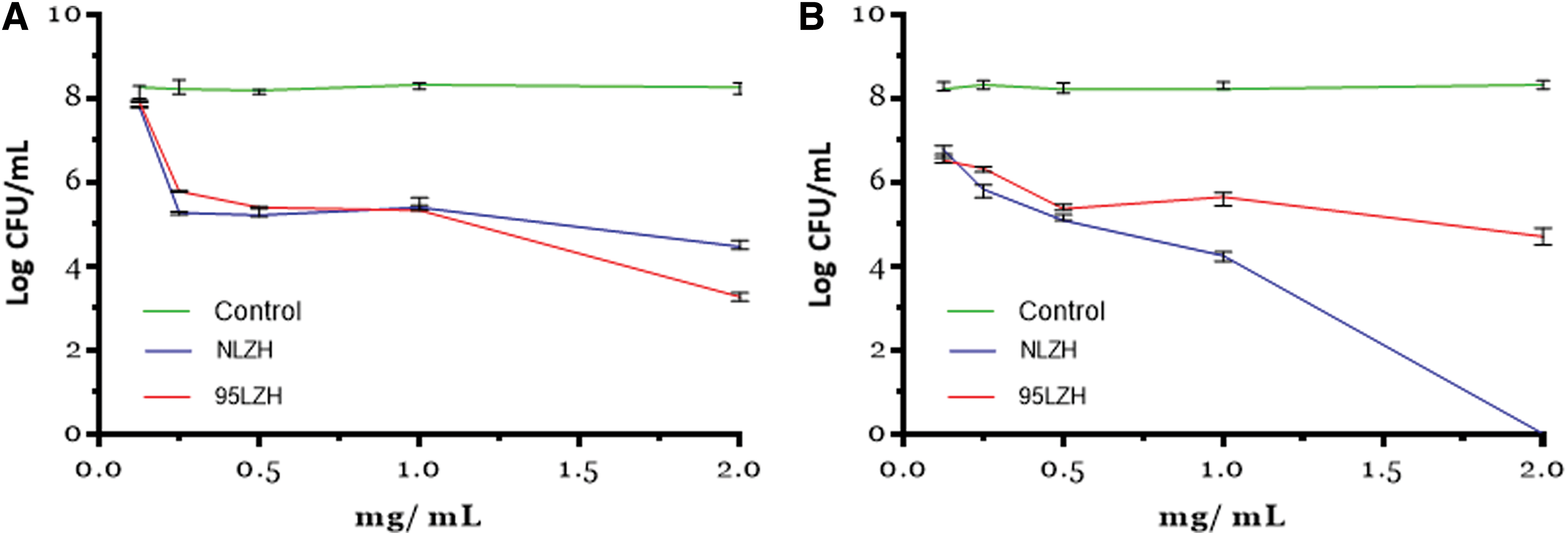
Antibacterial activity of native and heated HEWL hydrolysates at different concentrations.
Native HEWL hydrolysate at 1.0 mg/mL produces a reduction of 3.5 logarithmic cycles of S. carnosus and at 2.0 mg/mL produces a reduction of 4.5 logarithmic cycles of S. carnosus. Heated HEWL hydrolysate against S. carnosus at 1.0 mg/mL produces a reduction of 3.5 logarithmic cycles and at 2.0 mg/mL produces a reduction of 5.5 logarithmic cycles of S. carnosus.
Native HEWL hydrolysate at 1.0 mg/mL induces a reduction of 4.5 logarithmic cycles of E. coli and at 2.0 mg/mL induces a reduction of 8.5 logarithmic cycles. These results show bactericidal activity, as native HEWL was able to reduce all bacterial growth. Heated HEWL hydrolysate at 1.0 mg/mL was able to reduce two logarithmic cycles and at 2.0 mg/mL to reduce three logarithmic cycles of E. coli. These results show that native and heated HEWL hydrolysates present antibacterial activity against Gram-positive and Gram-negative bacteria. Carrillo et al. 24 reported in previous work antibacterial activity of native and heat treated HEWL hydrolysates against oenological lactic acid bacteria and acetic acid bacteria.
Enzymatic residual activity of HEWL
HEWL was treated with heat in a phosphate buffer for 20 min. Native and denatured lysozymes were hydrolyzed with pepsin at pH 1.2. At this pH, native and denatured lysozymes were totally hydrolyzed. Then, enzymatic activity was evaluated using the spectrophotometric method (Fig. 2). Native lysozyme hydrolysate presents 3.7% of residual enzymatic activity and denatured lysozyme hydrolysate presents 1.8% of residual enzymatic activity compared to the native lysozyme without hydrolysis control, which presents 100% of enzymatic activity. The antimicrobial activity of lysozyme hydrolysate is not dependent of its enzymatic activity, because it is residual enzymatic activity.
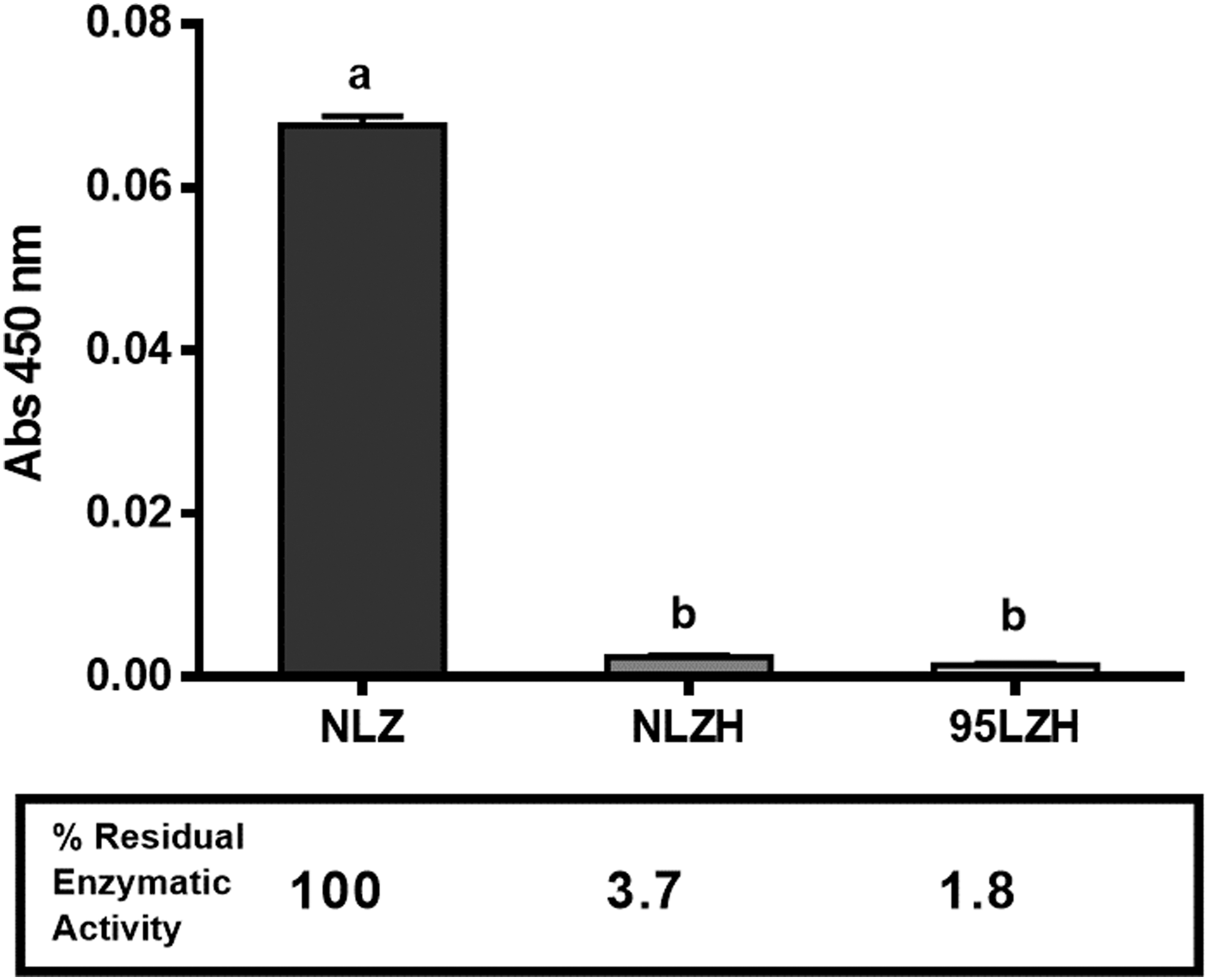
Residual Enzymatic activity of native and heated lysozyme. NLZ = native lysozyme (control), NLZH = native lysozyme hydrolyzed with pepsin at pH 1.2; 95LZH = heat-treated lysozyme at 95°C hydrolyzed with pepsin at pH 1.2. Data are expressed as the means ± standard deviations (n = 3). Values in the same column having different letters differ significantly (P < .05). ANOVA and Tukey's test. ANOVA, analysis of variance.
Different authors have reported hydrolysates obtained from HEWL with antimicrobial activity, but with no enzymatic activity. These same results have been reported with AMPs obtained from HEWL. For example, Mine, Ma & Lauriau, reported two antimicrobial HEWL hydrolysates obtained using pepsin and pepsin/trypsin, respectively. 20 Both hydrolysates presented 0% of enzymatic activity. These hydrolysates were active against Gram-positive bacteria (Staphylococcus aureus 23–394) and Gram-negative bacteria (E. coli K-12). They found two HEWL antibacterial peptides with the sequences f(98–108) I-V-S-D-G-D-G-M-N-A-W and f(15–25) H-G-L-D-N-T-R.
Pellegrini et al. 18 produced a HEWL hydrolysate, using the clostripain enzyme identified with the sequence f(98–112) I-V-S-D-G-D-G-M-N-A-W-V-A-R-W-R. This peptide presented antibacterial activity, but an absence of enzymatic activity. They synthesized a series of modified HEWL peptides presenting no enzymatic activity. Ibrahim et al. 35 described a HEWL hydrolysate obtained using pepsin at pH 4.0 with antibacterial activity. This hydrolysate was fractionated with the help of the size-exclusion chromatography. Three fractions, named S1, S2, and S3, with 0%, 12.3%, and 75.3% of enzymatic residual activity, respectively, were obtained. Our results are in accordance with the results reported in the literature.
In this study, we describe two HEWL hydrolysates with antibacterial activity, but with an absence of enzymatic activity. These results show that the antibacterial activity of native and heated lysosome hydrolysate is independent of its residual enzymatic activity (3.7% and 1.8%, respectively).
Lysozyme is an enzyme/protein that has an active site with a tridimensional structure. When lysozyme is hydrolyzed with pepsin, the active site is modified and produces polypeptides of different sizes. The enzyme needs to have intact its active site to have antibacterial activity against positive bacteria such as M. lysodeikticus. Native and heated hydrolysates from HEWL present antibacterial activity against negative bacteria E. coli. The HEWL derivatives have expanded its antibacterial spectrum against E. coli. This activity is independent of their residual activity (enzymatic activity). Heated HEWL hydrolysate was bactericide against negative bacteria at 2.0 mg/mL. The chromatogram in Figure 7 shows that lysozyme was totally hydrolyzed at the pH used in this study. This confirms that the active site was structurally modified.
Fluorescence microscopy
Fluorescence microscopy was applied to observe changes in cell viability after incubation of E. coli and S. carnosus with native and heated HEWL hydrolysates. Figure 3A, B shows the micrographs of E. coli (control) and S. carnosus (control). Under the fluorescence microscopy, control cells showed green fluorescence, and bacteria have normal movements. Figure 3C, D shows the micrographs of E. coli and Staphylococcus carnosus incubated with native HEWL hydrolysate at 1.0 mg/mL. Under the fluorescence microscopy, cells showed red fluorescence indicating the damage of the bacteria cell membrane, leading to red fluorescence. Cellular aggregation could also be observed (Fig. 3C, D). At all concentrations, red fluorescence, cellular aggregation, and an absence of bacteria movement were observed. Native and heated HEWL hydrolysates produce damages in the membrane of the Gram-positive and Gram-negative bacteria.

Fluorescence micrographs (magnification was of 1.000 × ) of E. coli and S. carnosus nonincubated and incubated with native and heated HEWL hydrolysate for 72 h.
Electron microscopy
Electron microscopy was applied to observe possible damages in the cytoplasm and structure of cells after incubation of S. carnosus and E. coli with native and heated HEWL hydrolysates. Figure 4A shows S. carnosus (control) without an HEWL sample. The morphology is normal in the external membrane and cytoplasm body. The cells can be observed in individual form. Figure 4B, C shows S. carnosus incubated with native HEWL hydrolysate at 1.0 mg/mL. The cells show cellular aggregation, and the morphology is irregular. In addition, broken cells were observed (black arrow). Figure 4C shows E. coli (control) without an HEWL sample. The morphology is normal in the external membrane and cytoplasm body.
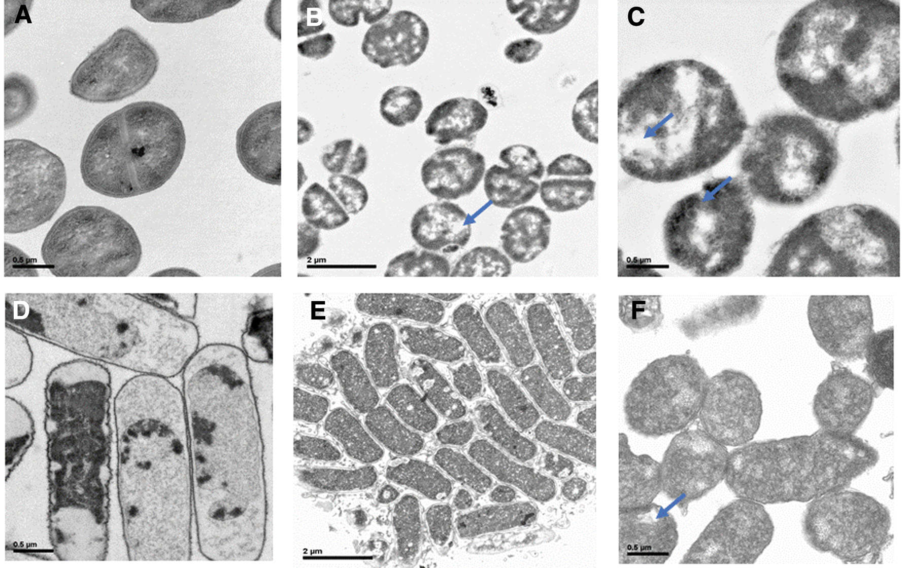
SEM micrographs of bacteria.
Figure 4D, E shows E. coli incubated with native HEWL hydrolysate at 1.0 mg/mL. The cell shows a different content of cytoplasm with respect to the control bacteria. The cytoplasmic content is dark, and the external membrane is irregular. We observed empty spaces in the cytoplasm of bacteria (black arrow). Figure 5 shows E. coli (control) without sample and HEWL sample at 0.5 mg/mL, the cytoplasmic content is dark, and the external membrane is irregular. We observed empty spaces in the cytoplasm of bacteria, and Figure 6 shows S. carnosus (control) without sample and bacteria with HEWL at 0.5 mg/mL; the morphology of bacteria is different at the control.
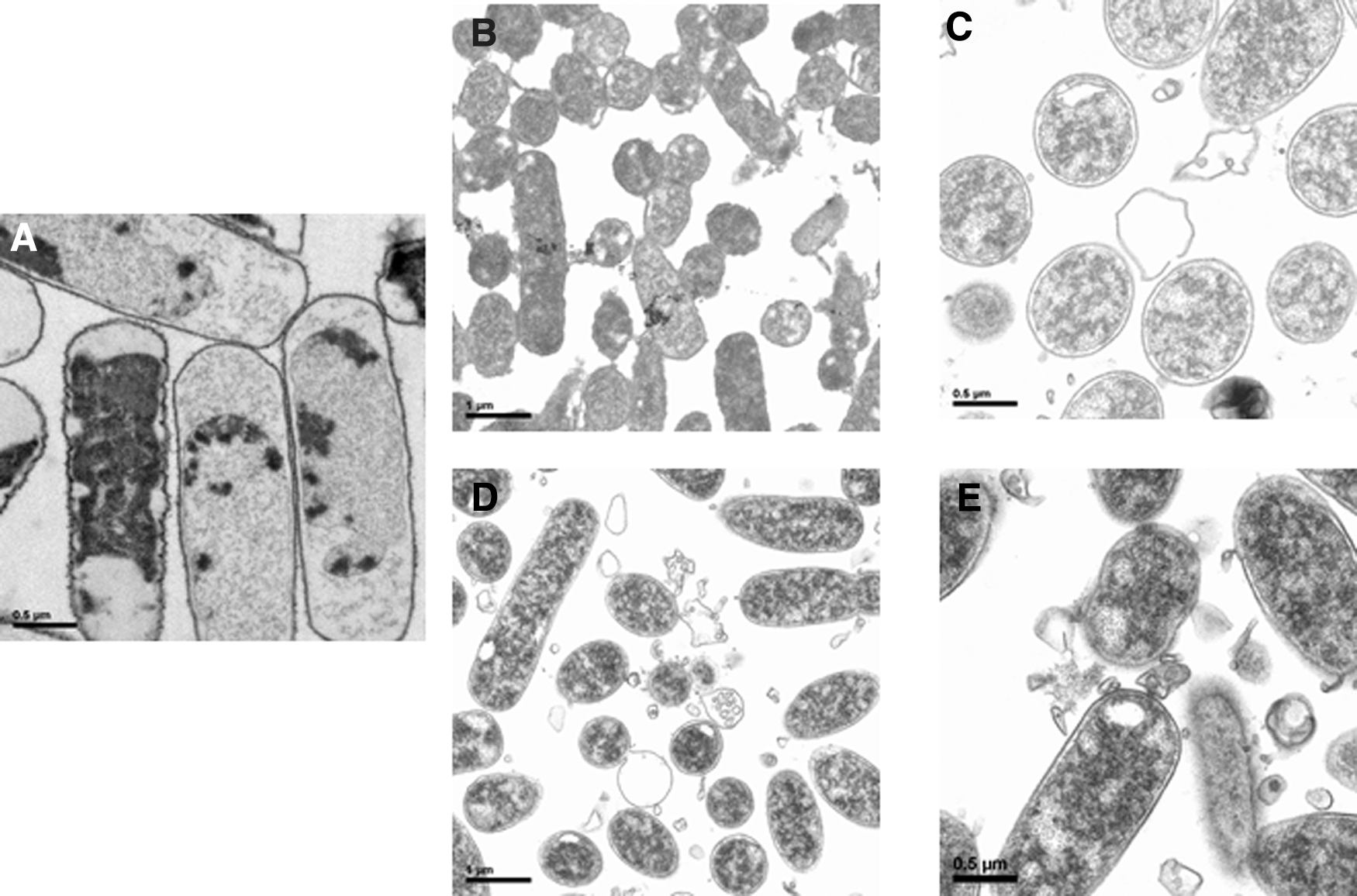
SEM micrographs of bacteria.
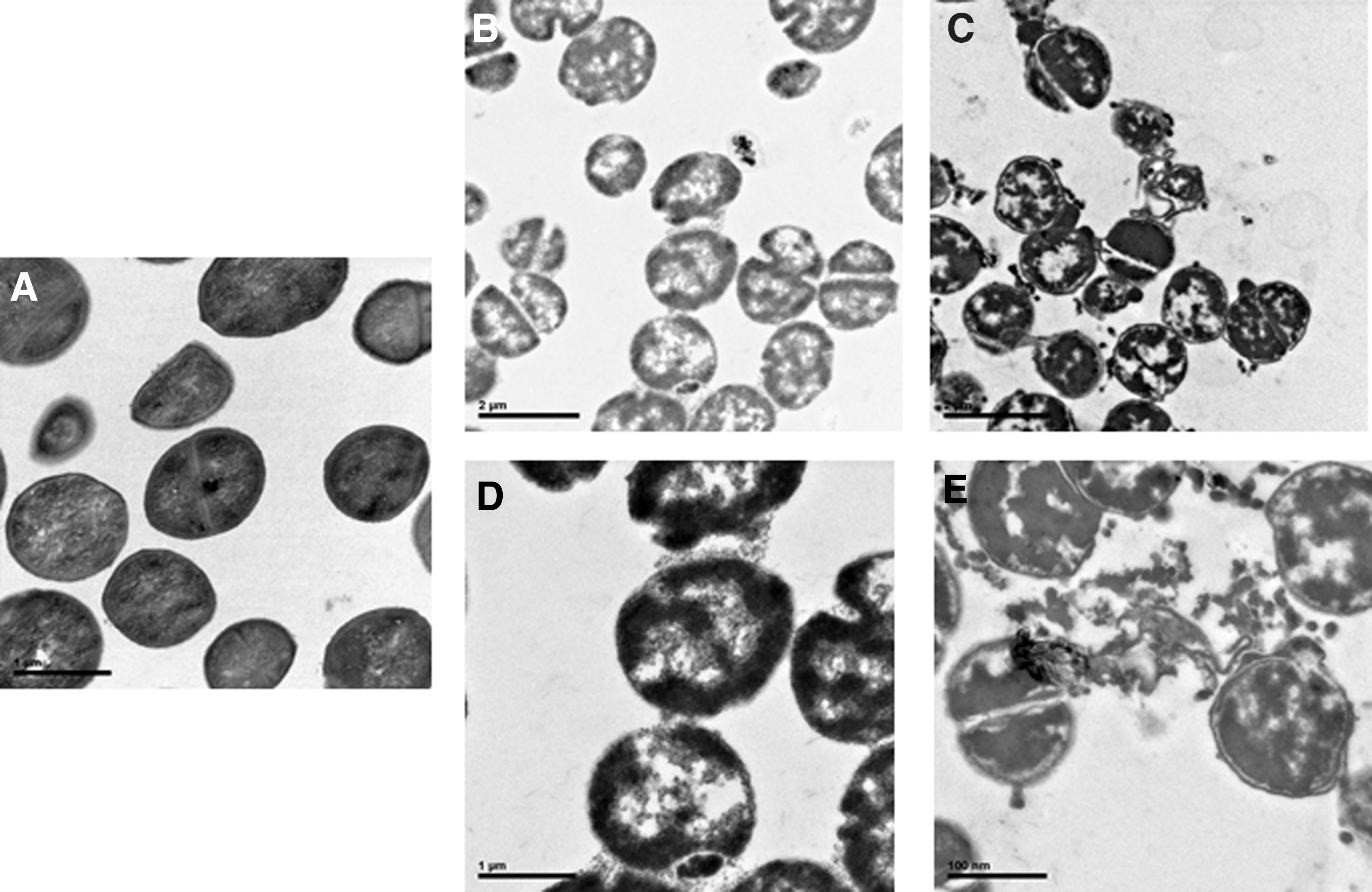
SEM micrographs of bacteria.
Identification of peptides by RP-HPLC-ESI-MS-MS
The peptides present in native and heated HEWL hydrolysates were subject to the RP-HPLC-ESI-MS-MS analysis to characterize their molecular masses and their amino acid sequences (Fig. 7). As an example, the HPLC-chromatogram obtained from the native HEWL hydrolysate is shown in Figure 5A, and one peak eluted around 28 min was used for illustration. The peak was selected by spectrum intensity (over 10000 score) to correct the protein sequence. Figure 5B shows the full MS scan chromatogram relevant to the peak for native and heated HEWL hydrolysates. Figure 5C shows the MS/MS spectrum of a triple charged ion with m/z 947.7 and the amino acid sequence of the identified peptide as f(91–108) SVNCAKKIVSDGNGMNAW. The entire m/z ratio for signal was compared to the Data analysis database and BioTools (chicken lysozyme) and characterized.
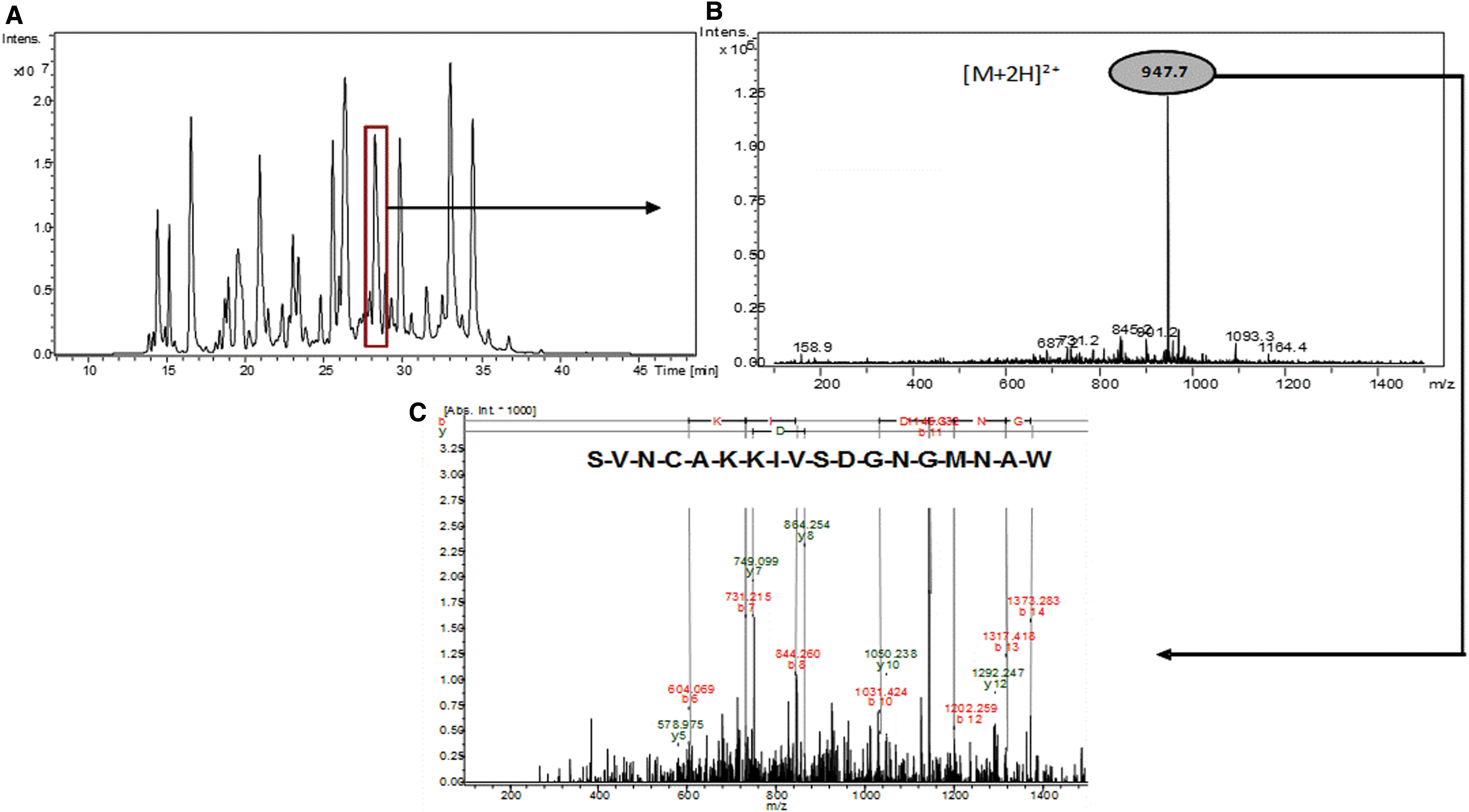
MS-spectra for the native HEWL hydrolysate of HPLC. One example peptide SVNCAKKIVSDGNMNAW was de novo sequencing using their MS/MS spectra by monoisotopic mass of the amino acids:
Native and heated HEWL hydrolysate had 51 identified peptides (Table 1), considering all the m/z ratios proposed by the database for each peptide candidate sequence and the most abundant m/z values in the corresponding MS/MS spectrum. These peptides had range masses of 365.1 to 1494.3.2 Da; these peptides have a small size. We found differences in the presence of peptides. Native HEWL hydrolysate has no presence of seven peptides: f(85–90), f(4–10), f(69–83), f(109–29), f(1–8), f(32–39), and f(97–125), and heated HEWL hydrolysate has no presence of three peptides: f(42–48), f(6–13), and f(60–83). These were differences between the native and heated HEWL hydrolysates with regards to the content of antibacterial peptides.
Peptides f(122–129) AWIRGCRL (974.3 ± 0.5 Da), f(123–129) WIRGCRL (903.3 ± 0.5 Da), and f(124–129) IRGCRL (717.0 ± 0.5 Da) reported in this study were described by Carrillo et al. 17 in native and heated HEWL hydrolysate with the in situ hydrolysis in a cationic column obtained using pepsin. These peptides presented antioxidant activity when using the ORAC and TBARS methods. These peptides presented also antioxidant activity in the zebrafish model. Peptide f(53–56) YGIL (464.1 ± 0.5 Da) described in this study has been described by You et al., as an antioxidant peptide obtained from heated HEWL hydrolysate using the alcalase protease. 23
Ibrahim et al. reported one HEWL hydrolysate with no enzymatic activity. 21 They also described two peptides with strong antibacterial activity identified as f(87–114) D-I-T-A-S-V-N-C-A-K-K-I-V-S-D-G-D-G-M-N-A-W-V-A-R-W-R-N-R and f(107–114) A-W-V-A-W-R-N-R against six strain Gram-positive and Gram-negative bacteria. The sequences identified in this study are the same for some cases and already described in the literature. Other sequences, however, have similitudes with the sequences reported by other groups.
These antibacterial peptides can be used to elaborate new functional ingredients, for the purposes of being used in the food and pharmaceutical industry. AMPs play an important role in the development of many organisms, being an important component of innate immunity response. Antimicrobial function innate immunity is regulated by small peptides with cationic charge (+2 to +9) such as Lys or Arg able to kill Gram-negative and Gram-positive bacteria. The sequences described in this study have in their structure Lys and Arg. These peptides also have a Trp content in their sequences. This amino acid is also suggested as responsible of AMP activity.
Ibrahim et al. reported the peptide f(107–115) RAWVAWRNR from human lysozyme with high antibacterial activity against Gram-negative and Gram-positive bacteria. 21 González et al. 36 used this peptide f(107–115) RAWVAWRNR, and they replaced Ala (108 and 111) for Arg, Asn, His, Ile, Lys, Met, Phe, Pro, Ser,Thr, Trp, Tyr, and Val to evaluate their antibacterial activity. The best amino acids were Lys, Arg, Trp, and Tyr. Trp was chosen as the best sequence in the two positions (108 and 111) for its higher antibacterial activity against E. coli and S. aureus. This same group of González et al. 37 reported the same peptide f(107–115) RAWVAWRNR, and they replaced Ala (108 and 111) for Trp arylated. This peptide presents an increase of its hydrophobicity that produced an increase of its antibacterial activity against S. aureus and Staphylococcus epidermidis. The killing mechanism of the pathogen microorganisms is described as the perturbation of the integrity of the cell membrane. These peptides are characterized to have the presence of positively charged and hydrophobic amino acids and an amphiphilic helical conformation, which interact directly with the bacteria membrane to generate pores that can produce destabilization in the membrane and bacteria death. 38,39
HEWL has a high isoelectric point (pI 10.7) at low pHs, where it is more cationic. Ibrahim et al., reported that HEWL at pH 6.0 has higher hydrophobicity due to the exposition of tryptophan. 33 At this pH, HEWL increases its antibacterial activity against negative bacteria. Different authors have described denatured lysozyme at pH 6.0 with an increase of their antibacterial activity against negative bacteria.
Lfcin is a biofunctional peptide located at the N-terminus of bovine lactoferrin protein. It is obtained from the protein hydrolysis caused by gastric pepsin in vitro digestion.
This peptide has been isolated in various mammals, such as humans (LfcinH: 0GRRRRSVQWCAVSQPEATKCFQWQRNMRKVRGPPVSCIKRDSPIQCI66) and bovines (LfcinB: 17FKCRRWQWRMKKLGAPSITCVRRAF41), among others. LfcinB is a 25 amino acid peptide that has a high proportion of basic residues, with a net charge of +8, and amphipathic properties. 40,41 LfcinB present antibacterial activity against the following (E. coli, B. cereus, K. pneumoniae, B. subtilis, C. perfringens, P. aeruginosa, L. monocytogenes, P. vulgaris, S. aureus, S. bovis, Y. enterocolitica, S. epidermidis, S. haemolyticus, S. Hominis, Y. pseudotuberculosis, S. typhimurium, S. Montevideo, C. albicans, Salmonella enteritidis, P. fluorescens, S. mutans, C. renale, E. Faecalis, B. vulgatus, B. bifidum, B. breve, and B. longum). 42,43
Huertas Méndez et al., reported six synthetic peptides as follows: lineal peptides LfcinB (25 residues), LfcinB 17–31 (15 residues), and LfcinB 20–25 (minimal motif, six residues); a dimer [LfcinB (20–25)2], a tetramer [LfcinB (20–25)4], and a cyclic [LfcinB (20–25) Cyc] peptide. S. enteritidis, E. coli, and S. maltophilia were used to evaluate the antibacterial activity of synthetic peptides. In the susceptibility assays, it was found that the LfcinB (21–25) Pal, LfcinB (20–25)2, and LfcinB (20–25)4 peptides exhibited the greatest inhibition halos against the three tested strains (11 to 14 mm). These synthetic peptides contain Trp and Arg in their sequences. 40
More than 3000 AMPs have been reported on line in different databases. Most of them are cationic peptides, and only a few of them are anionic, which share the ability to fold into amphipathic conformation upon interacting with the cell membranes in the microorganism. 44,45
FDA (Food and Drug Administration) accepted some AMPs to be tested in humans using clinical trials test. Many of these peptides were not accepted, but other peptides are presently in the phase II of assay. This situation opens the possibility of testing these new molecules to be used as antibiotics in the future. 46 –49 Lactoferrin hLF1-11 antimicrobial and antifungal peptides identified from human lactoferrin are in the second phases of experimental trial study due to their strong activity against bacteria and fungi. This sequence is protected with a patent for AM-Pharma, patent number CA2388910 C. This peptide can be used in the development of new natural drugs with antibacterial activity with high spectrum bacteria. 50
Summary
Native and heat treated HEWL hydrolysates were obtained with pepsin with the highest antibacterial activity against Gram-negative bacteria E. coli and Gram-positive bacteria S. carnosus. In both, native and heat treated HEWL hydrolysates, antibacterial peptides were identified. Fifty-one new peptides were identified in the two hydrolysates. These peptides have in their sequences positively charged and hydrophobic amino acids. These amino acids are typically present in antibacterial peptides. LIVE/DEAD method indicates that native and heat HEWL hydrolysates produce damage in the membrane bacteria with a destabilization of the membrane bacteria. The antibacterial HEWL hydrolysate can be used as new molecule to study new natural preservatives in the food and medical industries. These HEWL hydrolysates can be used to elaborate functional foods.
Footnotes
Acknowledgments
The authors thank the Instituto de Investigación en Ciencias de la Alimentación (CIAL-UAM-CSIC), Universidad Estatal de Bolívar (UEB), and Universidad Técnica de Ambato (UTA).
Author Disclosure Statement
No competing financial interests exist.