
Abstract
Native and heated hen egg white lysozyme (HEWL) hydrolysates were isolated by hydrolysis with pepsin at pH 2.0 in situ in a cation exchange membrane to isolate and identify antibacterial peptides of the HEWL hydrolysates. Native and heated HEWL was partially hydrolyzed with pepsin at pH 2.0. The fractions were eluted with 5 M ammonia to identify 23 antibacterial peptides using a tandem mass spectrometry. Then, these fractions were eluted with a solution of NaCl 1 M, and seven positively charged peptides f(23–28) YSLGNW, f(122–129) AWIRGCRL, f(123–129) WIRGCRL, f(124–129) IRGCRL, f(82–96) ALLSSDITASVNCAK, f(103–129) VAWRNRCKGTDVQAWIRGCRL, and f(97–123) KIVSDGNGMNAWVAWRNRCKGT were identified using tandem mass spectrometry. Native HEWL hydrolysate presented an enzymatic activity of 23.0%, heated HEWL hydrolysate at pH 6.0 presented a residual enzymatic activity of 22.0%, and heated HEWL hydrolysate at pH 7.0 presented an enzymatic activity of 21.33%. Native and heated HEWL hydrolysate presented antibacterial activity against Escherichia coli and Staphylococcus carnosus. Native HEWL hydrolysate presented a higher enzymatic activity than heated HEWL hydrolysates.
Introduction
H
Hen egg white lysozyme (HEWL) is a globular basic protein consisting of 129 amino acid residues with a molecular weight of 14.3 kDa. The sequences of amino acid residues are cross-linked by four disulfide bridges and have a high isoelectric point of pI (10.7). HEWL has a positive charge at physiologic pH (pH 7.0) with a high content of positive amino acids such as Lys, Hys, and Arg. The rest of the proteins of the egg white are negatively charged. Many cationic proteins such as lactoferrin, lactoperoxidase, and lysozyme may be purified using ion exchange chromatography. 2 Lysozyme has also been purified using cation-exchange membranes and technical resins. 3,4
HEWL belongs to a type of enzymes that lyses the cell wall of certain Gram-positive bacteria by splitting β (1–4) linkages between N-acetylmuramic acid and N-acetylglucosamine of the peptidoglycan. It is known that HEWL has antibacterial activity against Staphylococcus carnosus and Staphylococcus epidermis. 5 –7 Different groups of research have reported antibacterial activity of native lysozyme against Gram-negative bacteria such as Escherichia coli and Salmonella typhimurium. 8,9 Native and heated HEWL hydrolysates obtained with pepsin at pH 1.2 have been used to control lactic bacteria and acetic bacteria in wine. 10 HEWL has also been used in the conservation of meat products such as sausages, salami, pork, beef, or turkey. HEWL has also been used to prevent growth of Clostridium tyrobutyricum in cheese production. HEWL can also be used in other pharmaceutical (Lizipaina, antibacterial drug) and cosmetic applications. 11 –16 Other biological activities from HEWL have been reported such as antiviral, 17 –19 immune modulatory, 20 anti-inflammatory and antinociceptive, 21 antitumor, 22 antihypertensive, 23 and antioxidant activities. 24 HEWL is considered a potent allergen (named allergen Gald4) for their resistance to enzymatic hydrolysis. 25 Some proteolytic enzymes such as trypsin, chymotrypsin, and papain can hydrolyze HEWL. It is known that HEWL is resistant to hydrolysis with pepsin A at pHs higher than 2.0. 26 These authors have reported total hydrolysis of HEWL at pH 1.2 in SGF (Fluid Gastric Simulated, NaCl 0.35 M). However, these authors have also reported hydrolysis of HEWL with pepsin at lower pHs. Carrillo et al. reported hydrolysis in situ of native and heated HEWL with pepsin at pH 2.0 in a cationic exchange column and identified five potent antioxidant peptides evaluated in the zebrafish model larvae. 27
The aim of this work was to produce hydrolysis in situ in a cationic exchange membrane and evaluate the antibacterial activity of native and heated hydrolysates from HEWL and identify antibacterial peptides using high-performance liquid chromatography–electrospray ionization–tandem mass spectrometry in a reverse phase (RP-HPLC-ESI-MS-MS).
Materials and Methods
Materials
HEWL 58000 U/mL of activity, Micrococcus lysodeikticus, and pepsin A crystalline 3440 U/mg of activity were obtained from the company Sigma Chemical Co. (St. Louis, MO, USA). Tryptic soy agar and trypticase soy broth (TSB) were obtained from the company Scharlau (Barcelona, Spain). All other reagents were of analytical grade. Cationic exchange membrane S100 was obtained from Sartorius (Rotterdam, The Netherlands).
Thermal denaturation of HEWL
Heat-induced inactivation of the HEWL (1 mg/mL) used was performed by incubation at 95°C for 20 min in a 10 mM potassium phosphate buffer at pH 6.0 and pH 7.0. Insoluble aggregates were removed with centrifugation (3000 g for 15 min). The protein content of the supernatants was determined using the bicinchoninic acid assay (BCA Protein Assay Kit; Pierce™, Rockford, USA) according to the protocol described by the manufacturer. The supernatants were dialyzed and lyophilized until they were used.
Sodium dodecyl sulfate–polyacrylamide gel electrophoresis
Sodium dodecyl sulfate–polyacrylamide gel electrophoresis (SDS-PAGE) of HEWL digests was carried out in a Mini-PROTEAN electrophoresis system (Bio-Rad, Hercules, CA, USA) using 20% polyacrylamide gels (Bio-Rad). Polypeptide bands were stained with Coomassie Brilliant Blue G-250 for 12 h. Relative molecular masses of protein were determined by comparing them with molecular weight markers of 14.4–94 kDa, (GE Healthcare, Uppsala, Sweden).
Hydrolysis of HEWL in situ on a cation-exchange membrane and isolation of peptides
Denaturation of HEWL was performed as previously described by Carrillo et al. 10 HEWL was denatured using a heating treatment. HEWL at 5 mg/mL was resuspended in buffer phosphate, and pH 6.0 and pH 7.0 were heated at 95°C for 20 min. Then, HEWL was lyophilized and stored at −20°C. To carry out the hydrolysis of membrane-bound native and denatured HEWL, 1000 mL of a 0.1 mg/mL solution of protein in 10 mM NaCl, adjusted to pH 2.0 with HCl, was pumped through a cation-exchange membrane S100 (Sartorius). The binding of native and denatured HEWL was carried out at room temperature (25°C) with a flow rate of 20 mL/min. This process was generated using a peristaltic pump (Verder-Vleuten, Vleuten, The Netherlands). The process was monitored by an UV detector with a 2-mm light path flow cuvette (Model EM-1 Econo UV Monitor; Bio-Rad) at 280 nm. Before being used, the cation-exchange membrane was pre-equilibrated with water acidified with HCl (pH 2.0). The native and denatured HEWL bound to the membrane was hydrolyzed at 37°C by recycling with 100 mL of an aqueous solution (pH 2.0) of porcine pepsin A (25 mg/mL) at 20 mL/min for 16 h. The membrane was washed sequentially with acidified water at pH 2.0. Solvent A was 10 mM ammonium hydrogen carbonate acidified to pH 7.0 with formic acid, and solvent B was 5 M of ammonia solution. Finally, the membrane was treated with 1 M NaCl to remove more cationic peptides. The effluent was monitored at 280 nm of absorbance. All fractions were collected (10–20 mL) with the equipment fast protein liquid chromatography of GE Pharmacia, freeze-dried, and analyzed with a RP-HPLC-ESI-MS-MS.
Enzymatic activity assay
The HEWL lytic activity was determined by monitoring the decrease in turbidity of a suspension of M. lysodeikticus cells spectrophotometrically at 450 nm at 25°C, according to the Shugar method. 28 One unit of HEWL was defined as a decrease in absorbance at 450 nm of 0.001 min. The enzymatic activity of each sample was assayed in triplicate. The enzymatic activity was expressed as a percentage related to the activity of the untreated HEWL. The enzymatic activity was expressed as a percentage relative to the activity of the untreated HEWL.
Bacteria and growth medium
E. coli ATCC 25922 was from the American Type Culture Collection (ATTC, Rockville, MD, USA), and S. carnosus CECT 4491T was from The Spanish Type Culture Collection (Colección Española de Cultivos Tipo CECT; Valencia, Spain).
Antibacterial assay
To determine antimicrobial activity, 10 mL of TSB was inoculated with a colony of bacteria (E. coli or S. carnosus) and incubated overnight at 37°C. One milliliter of bacterial suspension was then diluted (1:50) in TSB. Bacteria were grown at 37°C until the logarithmic phase was reached as determined by the absorbance at 660 nm. Bacteria were centrifuged at 5000 g for 10 min then washed with a 10 mM sodium phosphate solution and 137 mM NaCl buffer pH 7.4 and resuspended (3 × 10⁵ CFU/mL) in the same media. One milliliter of the bacterial suspension was mixed with an equal volume of various concentrations of native and heated lysozyme hydrolysates (0.5, 1.0, and 2.0 mg/mL). The mixture of sample and bacteria was incubated at 37°C for 15 h, and the absorbance at 660 nm was measured using a Novaspec 2 Pharmacia spectrophotometer (Amersham Pharmacia Biotech). A 100 μL aliquot was placed onto nutrient agar plates. Colony-forming units (CFUs) were obtained after incubation of plates at 37°C for 24 h. The activity was expressed as a logarithmic viability reduction Log (N0/Nf), with Nf and N0 the colony counts after a treatment, in the treated and in the untreated sample, respectively. For all treatments, averages ± standard deviations (SDs) for at least three independent cultures of each strain are shown. 29
Identification of peptides by RP-HPLC-ESI-MS-MS
The fractions eluted with 5 M and 1 M NaCl of native and heated HEWL hydrolysates were analyzed with the RP-HPLC-ESI-MS-MS on a HP Agilent 1100 HPLC System (Agilent Technologies) connected to an Esquire-LC 3000 quadrupole ion trap (Bruker Daltonics GmbH, Bremem, Germany) and equipped with an electrospray ionization source. The variable wavelength used was set at 214 nm. A C18 precolumn (Nova-Pack®, 20 mm × 2826 × 3.9 × 4 μm of particle size; Waters Corp., Milford, MA, USA) was used to protect the analytical column. The column used was a Hi-Pore® RP-318 C18 column (250 mm × 4.6 mm × 5 μm particle size; Bio-Rad, Richmond, CA, USA). Chromatographic and mass spectrometry conditions followed the ones described by López-Expósito et al. 30 The sample was eluted at 0.8 mL/min with a linear gradient from 0% to 45% of solvent B (acetonitrile and TFA, 1000:0.270 v/v) in solvent A (Milli-Q water and TFA, 1000:0.370 v/v) in 60 min. The injection volume of sample was of 50 μL, and duplicate of injection was made for each sample. The flow of HPLC was divided ∼1:3 before to ionization source, and the first 6 min of the eluent flow was directed to waste to reduce salt deposit on the transfer capillary of MS instrument and to reduce interferences. For HPLC-MS, spectra were recorded over the mass to charge (m/z) range of 100–1500. Helium was used as gas collision with estimated pressure of 5 × 10⁻³ bar. About 15 spectra were averaged in the MS analysis and the five spectra in the tandem MS analyses. For each fraction, identification of peptides was carried out using the Data Analysis™ (version 3.0; Bruker Daltonics) to process and transform m/z spectral data and BioTools (version 2.1; Bruker Daltonics) to interpret MS/MS spectra and determine the peptide sequences.
Fluorescence microscopy
Cells were observed and photographed with a DM2500 epifluorescence microscope (Leica, Heerbrugg, Switzerland). The LIVE/DEAD BacLight bacterial Viability Kits L7012 (Invitrogen, Oregon, USA) were used to assess membrane integrity by selective nucleic acid staining. The kit contains two dyes: a first dye, SYTO 9 produces green fluorescence which penetrates and labels all bacteria and a second dye, Propidium Iodide (PI) produces red fluorescence which penetrates only bacteria with damaged membranes and, in these cells, suppresses SYTO 9 staining. As a result, live cells stain fluorescent green, and dead cells stain fluorescent red. The bacteria suspension (106 CFU/mL) was mixed with the antimicrobial agent solution and the mannitol broth and was incubated for 72 h at 30°C. After this time 1 mL of the mixture was mixed with 3 μL of the stain mixture (SYTO 9-PI 1:1 v/v). After 15 min of incubation in the dark at room temperature, green and red cells were counted under a fluorescence microscope with a long-pass filter (excitation 420–490 nm; emission 515 nm). Untreated cell suspensions were labeled following the same protocol, as they were used as controls. 31
Electron microscopy
Suspensions of E. coli and S. carnosus at mid-log phase (108 cells/mL) made in TSB, pH 7.4 were incubated with different doses of native and heated HEWL hydrolysates at 37°C for 15 h. These bacteria were fixed on the culture plate with 4% paraformaldehyde (Merck, Darmstadt, Germany) and 2% glutaraldehyde (SERVA, Heidelberg, Germany) in a 0.05 M cacodylate buffer pH 7.4 for 120 min at room temperature. Cells were then carefully scraped from the plate, centrifuged at 3000 g for 5 min, and then washed pellet post-fixed with 1% OsO4 and 1% K3Fe(CN)6 (potassium ferricyanide) in water for 60 min. Cells were dehydrated with ethanol and embedded in Epon (TAAB 812 resin; TAAB Laboratories Equipment Ltd.) according to standard procedures. Ultrathin sections were collected on collodion carbon-coated copper grids, stained with uranyl acetate and lead citrate, and examined at 80 kV in a JEOL 1010 (Tokyo, Japan) electron microscope. Electron micrographs were recorded. 32
Peptide synthesis
The small peptides from NaCl fraction of lysozyme were chosen to be synthetized f(111–119) AWIRGCRL, f(122–129), WIRGCRL, f(123–129) and IRGCRL, f(124–129) using a conventional FMOC solid phase synthesis method with a 431A peptide synthesizer (Applied Biosystems, Inc., Überlingen, Germany). The purity of the synthetic peptides was above of 90%.
Statistical analysis
Results are presented as means ± SDs from three replicates of each experiment. Differences between mean values were determined using the analysis of variance (ANOVA). The post hoc analysis was performed by the Tukey test. All tests were considered significant at P < .05. Statistical analysis was performed using the software package Prism 4 for Windows, version 6.0 (GraphPad Software, Inc.,
Results and Discussion
Hydrolysis in situ of HEWL with pepsin in a cation-exchange membrane S100
Native and heated (95°C, 20 min at pH 6.0 and pH 7.0) HEWL was subject to hydrolysis in situ with a solution pepsin A in a cation-exchange membrane S100 at 5 mg/mL of concentration. The aim of this work was to obtain, in one step, rich peptides with positively charged amino acids derived from the hydrolysis of native and heated HEWL and to assess whether denatured HEWL could generate peptides other than native lysozyme. It is known that HEWL has resistance to the in vitro hydrolysis with pepsin. It has been recently described that HEWL at pH 1.2 has total susceptibility to the hydrolysis with pepsin A. In this work, native and heated HEWL was hydrolyzed at pH 2.0 with an excess of pepsin for long time. Native and heated HEWL hydrolysates were eluted with a solution of ammonia 5 M. Native and heated HEWL hydrolysates were analyzed with the SDS-PAGE and RP-HPLC methods to determine the grade of hydrolysis of HEWL. SDS-PAGE showed that bands of HEWL are less intensive compared to the band of HEWL control without hydrolysis (Fig. 1). RP-HPLC showed the same situation in the behavior of HEWL hydrolysis. The hydrolysis at pH 2.0 of native and heated HEWL was partial. This is according to the RP-HPLC analysis. In the chromatogram, we observed residual HEWL (Fig. 2).

SDS-PAGE analysis of native and heat hydrolysates of HEWL. Lane 1: molecular weight; lane 2: HEWL control without hydrolysis; lane 3: native HEWL hydrolysate; lane 4: heat HEWL hydrolysate at pH 6.0; and lane 5: heat HEWL hydrolysate at pH 7.0. HEWL, hen egg white lysozyme; SDS-PAGE, sodium dodecyl sulfate–polyacrylamide gel electrophoresis. Color images available online at

MS-spectra for the native HEWL hydrolysate of RP-HPLC. One example peptide f(1–8) KVFGRCEL was de novo sequencing using their MS/MS spectra by monoisotopic mass of the amino acids:
Enzymatic activity from native and heated HEWL hydrolysates
Figure 3 shows the percentage of residual enzymatic activity of native and heated HEWL hydrolysates obtained with pepsin. HEWL without hydrolysis was used as positive control. The activity of HEWL control was expressed as 100% of enzymatic activity. Native HEWL hydrolysate presented a value of 23.0% of residual enzymatic activity, heated HEWL hydrolysate at pH 6.0 presented a value of 22.0% of residual enzymatic activity, and the heated HEWL hydrolysate at pH 7.0 presented a value of 21.33% of residual enzymatic activity. This residual enzymatic activity is produced by the presence of HEWL intact. Native and heated HEWL hydrolysates presented an absence of statistical differences between them. Only the hydrolysates presented statistical differences compared to the enzymatic activity of HEWL control. The presence of enzymatic activity of native and heated HEWL hydrolysates is shown according to the hydrolysis profiles observed using the SDS-PAGE and RP-HPLC methods. The antibacterial activity of native and heated HEWL hydrolysates can be caused for the presence of HEWL without hydrolysis.
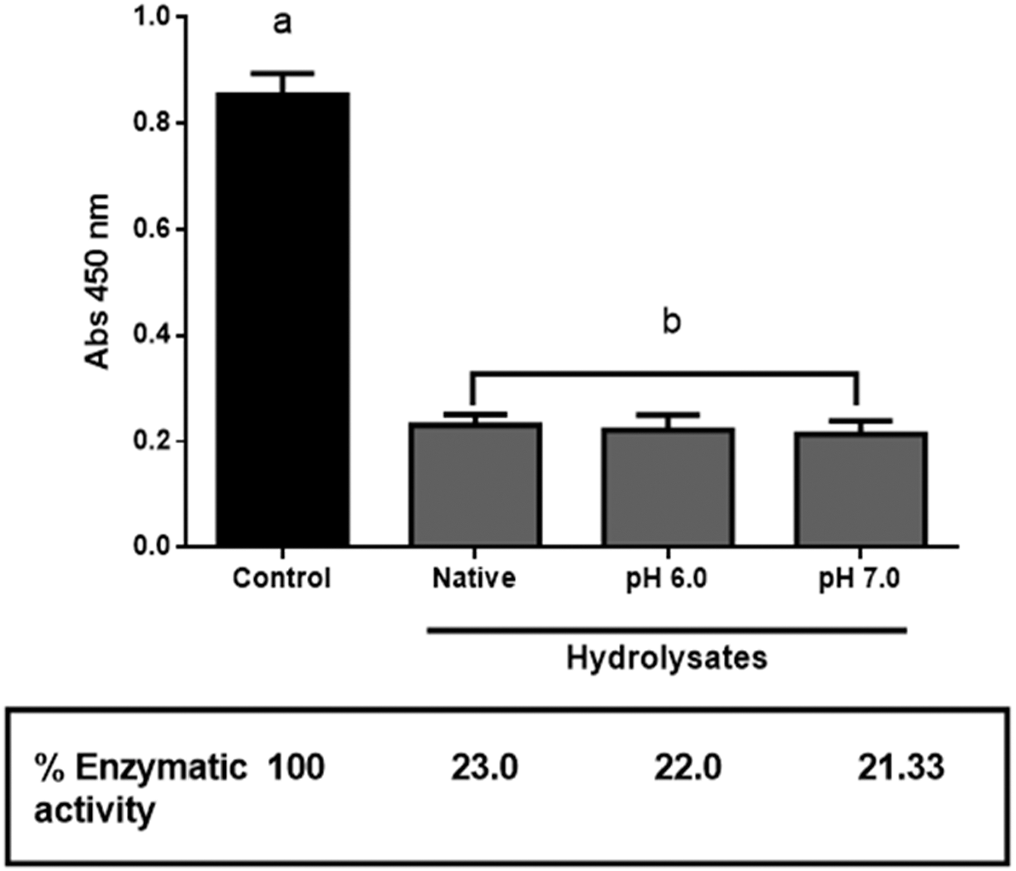
Enzymatic activity residual of HEWL. Control = HEWL without hydrolysis; Native = native hydrolysate of HEWL, pH 6.0 = heat hydrolysate HEWL at pH 6.0 and heat hydrolysate HEWL at pH 7.0. Data are expressed as means ± SDs (n = 3). Values in the same column having different letters differ significantly (P < .05). Was applied at an ANOVA one way and Tukey's test. ANOVA, analysis of variance; SD, standard deviation.
Carrillo et al. reported native HEWL hydrolysate and heated HEWL hydrolysate obtained with pepsin at pH 1.2 with percentage of residual enzymatic activity of 4.1% and 2.6%, respectively. 10 These hydrolysates presented antibacterial activity against bacteria of wine, and this antibacterial activity was not dependent of their residual enzymatic activity. Mine et al. reported HEWL hydrolysates with antibacterial activity against Gram-negative and positive bacteria, but without enzymatic activity. 29
Ibrahim et al. reported HEWL hydrolysates obtained with pepsin at pH 4.0 for 2 h with antibacterial activity with 75% of residual enzymatic activity. 33 This hydrolysate was fractionated, and the fractions S0 presented 0% of enzymatic activity, fraction S1 presented 12.3% of enzymatic activity, and S3 presented 75.3% of residual enzymatic activity. During et al. reported modified lysozyme treated with a chemical reduction agent without enzymatic activity. 34
Identification of peptides from HEWL
The peptides present in native and heated HEWL hydrolysates were subject to the RP-HPLC-ESI-MS-MS analysis to characterize their molecular mass and their amino acid sequence (Fig. 2). As an example of analysis, the RP-HPLC-chromatogram obtained from the native HEWL hydrolysate is shown in Figure 4A, and one peak eluted around 16 min was used for illustration. The peak was selected by spectrum intensity (over 10,000 score) to correct the protein sequence. Figure 4B shows the ion extracted (951.4 m/z) chromatogram relevant to the peak for native and heated HEWL hydrolysates. Figure 4C shows the MS/MS spectrum of a charged ion with m/z 951.4 [H+1 ] and the amino acid sequence of the identified peptide as the sequence f(1–8) KVFGRCEL. The entire m/z ratio for good signal was compared to the Data analysis database and BioTools (HEWL) and characterized. This [f(1–8) KVFGRCEL] sequence was described by Ibrahim et al. HEWL was hydrolyzed with pepsin at pH 4.0, and the peptides were identified using MALDI-TOF. 35
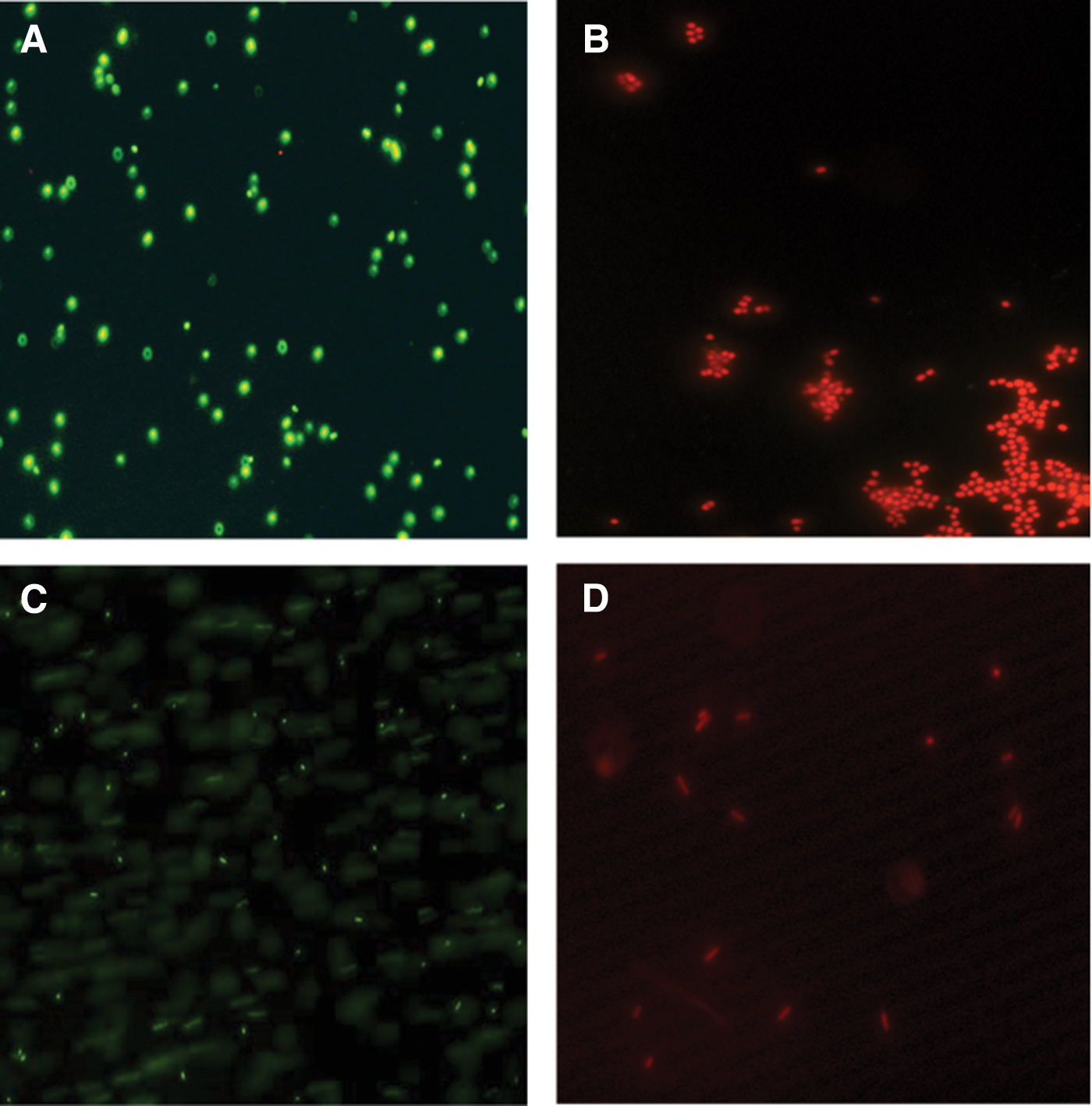
Fluorescence micrographs (1000 × ) of Escherichia coli and Staphylococcus carnosus nonincubated and incubated with native and heated HEWL hydrolysate for 72 h.
Native and heated hydrolysates from HEWL were eluted with a solution of ammonia 5 M (Fig. 3), this fraction was analyzed with RP-HPLC-ESI-MS-MS, and 23 peptides (Table 1) were identified. A number of peptides were identified such as f(11–22) AMKRHGLDNYRG with an observed mass of 1415.8 Da; this peptide was only found in the heated hydrolyzed at pH 6.0 HEWL. In the fraction of NaCl 1 M seven peptides f(23–28) YSLGNW, f(122–129) AWIRGCRL, f(123–129) WIRGCRL, f(124–129) IRGCRL, f(82–96) ALLSSDITASVNCAK, and f(97–123) KIVSDGNGMNAWVAWRNRCKGT (Table 2) were eluted. Seven peptides were identified in the native, heated at pH 6.0 and pH 7.0 HEWLs. This fact indicates that native and denatured HEWL hydrolysates present the same point of bound as the functional groups of cationic exchange membrane. For this reason, during the hydrolysis the same sequences of peptides were produced. These peptides were rich in positive amino acids Lys, His, and Arg; they also contain Trp in their sequences.
Monoisotopic mass for the neutral molecule, calculated from amino acid sequence.
Molecular ion (m/z) selected for MS/MS charge.
Protein fragment.
Nat-LZ, native lysozyme hydrolysate; LZ pH 6.0, hydrolysate of lysozyme heated at pH 6.0; LZ pH 7.0, hydrolysate of lysozyme heated at pH 7.0.
Monoisotopic mass for the neutral molecule, calculated from amino acid sequence.
Molecular ion (m/z) selected for MS/MS charge.
Protein fragment.
Nat-LZ, native lysozyme hydrolysate; LZ pH 6.0, hydrolysate of lysozyme heated at pH 6.0; LZ pH 7.0, hydrolysate of lysozyme heated at pH 7.0.
Carrillo et al. have reported hydrolysis in situ of native and heated HEWL with pepsin A in a cationic exchange column. 27 They described five positively charged peptides obtained with the NaCl 1 M fraction; the sequences identified were f(109–119) VAWRNRCKGTD, f(111–119) WRNRCKGTD, f(122–129) AWIRGCRL, f(123–129) WIRGCRL, and f(124–129) IRGCRL. 27 These positive peptides presented antioxidant activity using the in vitro (ORAC method) and the in vivo antioxidant model (TBARS in zebrafish larvae). Three identical peptides were found in the two methods of hydrolysis in situ of native and heated HEWL with the sequences f(122–129) AWIRGCRL, f(123–129) WIRGCRL, and f(124–129) IRGCRL.
Antibacterial activity of native and heated hydrolysates from HEWL
Antibacterial activity of native and heated hydrolysates from HEWL was assayed against Gram-negative (E. coli ATCC 25922) used as model of this type of bacteria and Gram-positive bacteria (S. carnosus CECT 4491T) used as model of this type of bacteria. Native and heated hydrolysates from HEWL were assayed at different concentrations (0.5, 1.0, and 2.0 mg/mL). All samples of native and heated HEWL hydrolysates presented antibacterial activity against Gram-negative and Gram-positive bacteria (Table 3). Native HEWL hydrolysates present higher antibacterial activity against S. carnosus. At 0.5 mg/mL, native HEWL hydrolysates produce a reduction of 6.8 logarithmic cycles of S. carnosus, at 1.0 mg/mL produces a reduction of 7.0 logarithmic cycles of S. carnosus, and finally at 2.0 mg/mL produces a reduction of 7.2 logarithmic cycles of S. carnosus.
Data are expressed as the means ± SDs (n = 3). a,bIndicate significant differences between groups of bacteria (Nat-LZ, LZ pH 6.0 and LZ pH 7.0) (P < .05).
Nat-LZ, native lysozyme hydrolysate; LZ pH 6.0, hydrolysate of lysozyme heated at pH 6.0; LZ pH 7.0, hydrolysate of lysozyme heated at pH 7.0.
HEWL, hen egg white lysozyme; SD, standard deviation.
Native HEWL hydrolysate also was more active against E. coli with values of 5.3, 6.1, and 5.7 cycles of logarithmic reduction at 0.5, 1.0, and 2.0 mg/mL, respectively. Heated HEWL hydrolysate at pH 6.0 and pH 7.0 only presented high antibacterial activity against S. carnosus at concentrations higher than 2.0 mg/mL with values ranging from 4.7 to 5.2 reduction of logarithmic cycles. Native HEWL hydrolysate when is compared statistically against heat HEWL hydrolysate at pH 6.0 and pH 7.0 has difference significantly at P < .05 to both bacteria at all concentrations assayed. No differences exist significantly between heat HEWL hydrolysates at pH 6.0 and pH 7.0 in both bacteria at all concentrations assayed.
Antibacterial activity of peptides from HEWL
Peptides obtained from HEWL with small sequences were synthetized. We evaluated their antibacterial activity against E. coli and S. carnosus at 50 μg/mL (Table 4). All peptides from native and heated HEWL hydrolysates presented antibacterial activity against both bacteria. Three peptides contain Trp amino acid in their sequences f(23–28) YSLGNW, f(123–129) WIRGCRL, and f(122–129) AWIRGCRL. The sequences described in this study contain in their structure Lys and Arg. These peptides also have a Trp content in their sequences. This amino acid is also suggested as responsible of antimicrobial peptide activity. 36 González et al. used this peptide f f(107–115) RAWVAWRNR, and they replaced Ala (108 and 111) for Arg, Asn, His, Ile, Lys, Met, Phe, Pro, Ser,Thr, Trp, Tyr, and Val to evaluate their antibacterial activity. The best amino acids were Lys, Arg, Trp, and Tyr. Trp was chosen as the best sequence in the two positions (108 and 111) for their higher antibacterial activity against E. coli and Staphylococcus aureus. 37
Data are expressed as the means ± SDs (n = 3). a,b,c,dIndicate significant differences between bacteria (P < .05). ANOVA and Tukey's test.
ANOVA, analysis of variance.
Huertas Méndez et al. reported six synthetic lineal peptides LfcinB (25 residues), LfcinB 17–31 (15 residues), and LfcinB 20–25 (minimal motif, six residues); a dimer [LfcinB (20–25)2], a tetramer [LfcinB (20–25)4], and a cyclic [LfcinB (20–25) Cyc] peptide in Salmonella enteritidis, E. coli, and Stenotrophomonas maltophilia. In the susceptibility assays, it was found that the LfcinB (21–25) Pal, LfcinB (20–25)2, and LfcinB (20–25)4 peptides presented the greatest inhibition halos against the three tested strains (11–14 mm). These synthetic peptides have Trp and Arg in their sequences. 38
Memarpoor-Yazdi et al. reported that HEWL hydrolyzed with papain and trypsin showed antioxidant and antibacterial activity. The fraction two (F2) was more active. 39 In this fraction, it was identified that the peptide with sequence f(46–61) NTDGSTDYGILQINSR inhibited the growth of Gram-negative and Gram-positive bacteria.
More than 3000 AMPs have been reported on line in different databases. Most of them are cationic peptides, and only a few of them are anionic, which share the ability to fold into amphipathic conformation upon interacting with the cell membranes in the microorganism. 40,41
The mechanism of action of antimicrobial peptides has been extensively investigated, and it has been shown that an amphiphilic form, mostly α-helical formation, and an overall net positive charge are proposed to initiate the interaction with the bacterial surface to enter the membrane. 42 Cationic peptides are thought to inhibit Gram-negative bacteria through a variety of mechanisms, including interacting with the lipopolysaccharides and electrostatic interactions with the negatively charged lipid groups in the membrane that produces interruption of the membrane integrity. 43 –48 Peptides reported in this study have positive charge and have Trp in their sequence. These results are in accordance with the sequences of antibacterial peptides reported in the literature.
Fluorescence microscopy (LIVE/DEAD)
LIVE/DEAD is a kit of fluorescence that observes damage in the cell membrane. Red fluorescence indicates damage in the bacteria membrane, and green fluorescence indicates no damage in the bacteria membrane. The controls of E. coli and S. carnosus without HEWL hydrolysate sample were stained with green fluorescence (Fig. 4A, C). The cells in these controls presented normal morphology and movement. E. coli and S. carnosus in presence of native HEWL hydrolysate under fluorescence microscopy presented red fluorescence (Fig. 4C, D). Cellular aggregation was observed in the S. carnosus. The cells presented an absence of movement, and the cellular morphology was different to the controls. The same behavior was observed with heated HEWL hydrolysates (no data shown).
Carrillo et al. reported damage of membrane of bacteria of wine in the presence of native and heated HEWL hydrolysates, cellular aggregation, and a loss of movement in bacteria. 10 Masschalck et al. reported membrane damage produced by the modified lysozyme; the action mechanism is not dependent of the enzymatic activity of lysozyme. In this study, the native and heated HEWL hydrolysates produced damage in the integrity of cellular membrane. 31 We suggest that this can be the action mechanism of this sample with the help of residual enzymatic activity.
Electronic microscopy
Electron microscopy was applied to observe possible damages in the cytoplasm and structure of cells after incubation of S. carnosus and E. coli with native and heated HEWL hydrolysates. Figure 5A shows S. carnosus (control) without a HEWL sample. Figure 5C shows E. coli (control) without a HEWL sample. The morphology of both bacteria is normal in the external membrane and cytoplasm. The cells can be observed in an individual form and have normal movement. It was found that the majority of bacterial cells were cylindrical when grown in a media without a HEWL sample. Figure 5B shows S. carnosus incubated with heated HEWL hydrolysate at pH 6.0 at a concentration of 1.0 mg/mL. The cells show cellular aggregation, and the morphology is irregular. White spaces in the cellular cytoplasm are observed in all cells. The cellular wall is irregular and broken around the cell.
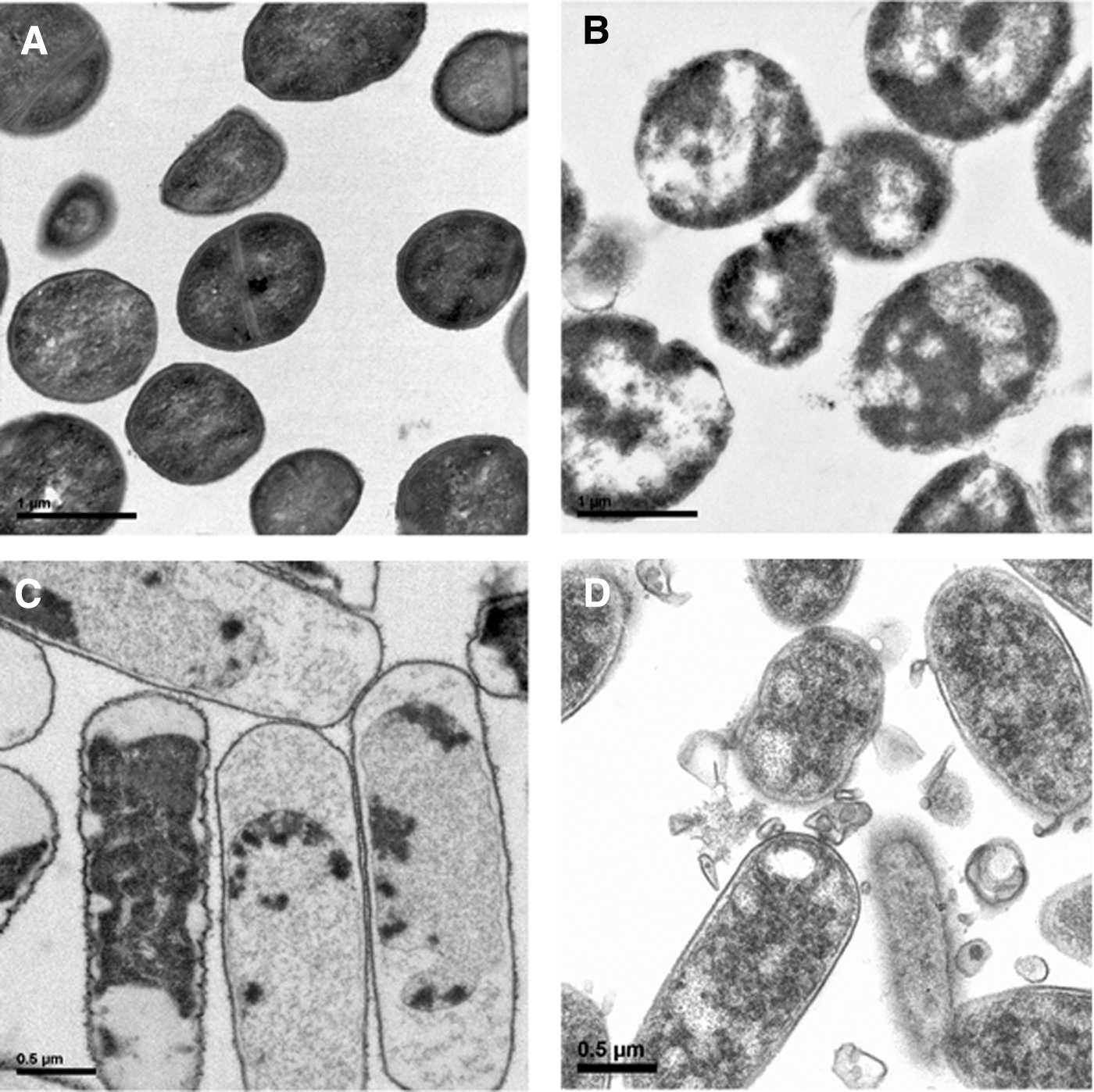
SEM micrographs of bacteria.
Figure 5D shows E. coli incubated with native HEWL hydrolysate at pH 6.0 at a concentration of 1.0 mg/mL. The cell shows different contents of cytoplasm with respect to the control bacteria. White spaces in the cytoplasm are visible, condensed cytoplasm with breaks of the outer membrane and cellular aggregation. In general, cells presented a different morphology than the control bacteria.
Ulvatne et al. reported morphological changes in E. coli ATCC 25922 produced for incubation of lactoferricin B (30 μg/mL). The bacterial cells were intact, and the content of the cell was still inside. 49 The bacterial cells did not appear swollen, but there was a separation of the cell envelope. Other observations revealed blebs containing bilayer structures on the bacterial surface, extruding from the cytoplasmic membrane, and released to the surroundings of the bacteria. Pellegrini et al. reported morphological changes in E. coli incubated with lysozyme. 50 They observed condensed cytoplasm and many bacteria ghosts. Our results on morphological changes in the presence of HEWL are in accordance to the ones reported with lysozyme, hydrolysates, and peptides from HEWL.
Summary
HEWL was used to obtain native and heated hydrolysates with pepsin at pH 2.0 with antibacterial activity against Gram-negative and positive bacteria. Residual enzymatic activity in HEWL can be related to the antibacterial activity presented in the hydrolysates. New peptide sequences from HEWL were identified in native and heated HEWL hydrolysates. The peptides identified in native and heated HEWL hydrolysates contained tryptophan, lysine, and histidine amino acids. These amino acids are responsible for antibacterial activity in the peptides. LIVE/DEAD fluorescence method indicates that native and heated HEWL hydrolysates produce damage in the Gram-negative and positive membrane bacteria with destabilization of the bacteria membrane. Positive charged antibacterial peptides in HEWL can be an alternative to be used in bacteria control with interesting applications in the medical and food industries.
Footnotes
Author Disclosure Statement
No competing financial interests exist.
Acknowledgments
This work has received financial support from the project 1373-CPU-UTA-2014 and project UEB.