
Abstract
“Quelites” are edible plants that are part of the traditional agro-ecosystems in Mexico. These plants, despite their already known nutritional properties, are now considered neglected and underutilized species. With the objective of promoting their reinsertion in the markets and mainly, in daily diets, efforts have been made to study them from multidisciplinary approaches to demonstrate their beneficial properties. To generate evidence of an added health-promoting value that would encourage quelites consumption, in the present work, the anti-Helicobacter pylori activity of three representative quelite species, Anoda cristata (Alache), Cnidoscolus aconitifolius (Chaya), and Crotalaria pumila (Chepil), was tested. H. pylori is considered the etiological agent of gastritis, ulcer, and gastric cancer, and represents a public health problem in Mexico and worldwide. Aqueous (AQ) and dichloromethane–methanol (DM) extracts were obtained from the three species of quelites to investigate their effect on H. pylori growth and on two of its colonization factors (adherence and urease activity). DM extracts from Chaya, Chepil, and Alache exert the best inhibitory effect on bacterial growth, with minimum inhibitory concentrations of 62.5, 125, and 250 μg/mL, respectively. AQ and DM extracts inhibit bacterial adhesion by 30% to 50%. None of them has an effect on urease activity. The two flavonoids present in A. cristata, acacetin and diosmetin, inhibit H. pylori growth by ∼90% with 3.9 μg/mL. These results provide new information about the anti-H. pylori potential of three edible quelites, and give an added value, since their routine consumption may impact on the prevention and/or control of H. pylori-associated diseases.
Introduction
T
The traditional agro-ecosystem in Mexico, known as “milpa,” houses besides the Mesoamerican triad (beans, corn, and squash) several edible plants considered Neglected and Underutilized Species (NUS), 2 among which quelites are included. 3
Currently in Mexico, quelites are used by different ethnic groups in all regions of the country. The form of consumption is varied and, due to their importance in local traditions and to their nutritional value, these plants are promising species with the potential to enrich nutrition in areas of the country with greater food vulnerability and to empower marginalized communities by strengthening their local identity. 4 Unfortunately, in terms of production and market value, the quelites, like other NUS, have a minor significance and have been ignored by agricultural researchers, plant breeders, and policymakers.
With the objective of rescuing the consumption of quelites and their reintegration in the Mexican markets and in the daily diet, a multidisciplinary project was initiated with the objective of studying the following three representative native species of quelites: Anoda cristata (L.) Schltdl., Cnidoscolus aconitifolius (Mill.) I.M. Johnst., and Crotalaria pumila Ortega, commonly known as Alache, Chaya, and Chepil, respectively. In addition to their well-known use as food, two of these species have been traditionally recognized for having beneficial health properties. In some areas, A. cristata is used to treat fever, cough, skin wounds, alopecia, renal disorders, and digestive ailments such as stomachache. 5 –8 Moreover, some pharmacological studies using different animal models have reported the hypoglycemic and antihyperglycemic effects of this plant. 9 In the case of C. aconitifolius (Chaya), its traditional usage in muscular discomforts, rheumatism, and arthritis has been documented. The consumption of Chaya has also been reported for the treatment of other diseases such as diabetes, hypercholesterolemia, hypertension, renal and urinary maladies, and digestive disorders. 5,6,8,10 Some pharmacological approaches have assessed Chaya's anti-inflammatory and cardioprotective activities. 11 In this context, in vivo experiments in diabetic rats have demonstrated that a 4-week Chaya aqueous (AQ) extract administration produced hypoglycemic effects and was also helpful in reducing hyperlipidemia. 12 More recently, antiprotozoal, antibacterial, and anti-inflammatory activities of a Chaya CHCl3-MeOH extract have been reported. 13 Regarding C. pumila (Chepil), in the XVI century, this plant was traditionally used for skin conditions and cough; however, nowadays these uses seem to be discontinued. 5
In the present work, the anti-Helicobacter pylori potential of the three mentioned quelites was explored.
H. pylori is a Gram-negative bacteria that colonizes the stomach of more than half of the world's population and is the main etiological factor of gastritis, peptic ulcer, and gastric cancer. 14 –16 H. pylori uses unique strategies (colonization, virulence, and survival factors) to overcome the protective elements of the gastric mucosa and to evade the host immune response. 17
For the present study, two extracts were obtained from each one of the three species of quelites to test, in vitro, their anti-H. pylori activity and the effect over two colonization factors (urease and adhesion to gastric epithelial cells). In addition, the anti-H. pylori activity of two flavonoids present in A. cristata was investigated. The purpose of this work is to generate evidence to give an added health-promoting value to these plants, besides their already known nutritional properties, which would encourage their consumption.
Materials and Methods
Chemicals
Gallic acid, quercetin, and acacetin were purchased from Sigma-Aldrich. Diosmetin was from Santa Cruz Biotechnology. Analytical-grade solvents were from Merck.
Plant material
A. cristata (L.) Schtdl (Alache) was collected and identified by MS Edelmira Linares and Dr. Robert Bye (Instituto de Biología, UNAM) at Tepetlixpa, Estado de México (18° 58′ 57.5″ N, 98° 49′ 40.1″ W), on June 2, 2015. A voucher specimen is deposited in the Herbario Nacional (MEXU 1445645) located at the Universidad Nacional Autónoma de México. C. aconitifolius (Mill.) I.M. Johnst (Chaya) was collected at Timucuy, Mérida, Yucatán (20° 48′ 43.5″ N, 89° 30′ 56.8″ W) by Dr. Amanda Gálvez and identified by MS Clarisa Jiménez (Unidad de Recursos Naturales del Centro de Investigación Científica de Yucatán) on August 28, 2015. A voucher specimen is deposited in the Herbario CICY (068784). C. pumila Ortega (Chepil) was collected at Ocotlán de Morelos, Oaxaca (16° 48′ 30.7″ N, 96° 40′ 21.3″ W), on September 2, 2015, and identified by MS Magali Martínez. A voucher specimen is deposited in the Herbario Nacional (MEXU 1457315).
Extract preparation
The leaves were separated and cleaned. After air drying, the material was pulverized. The dichloromethane–methanol (DM) (1:1) extracts were prepared by maceration of 50 g of plant material in 500 mL of the solvent mixture for 72 h (this procedure was repeated three more times). The solvents were filtered and evaporated to dryness under reduced pressure to obtain the DM extracts. The AQ extracts were obtained by preparing an infusion with 10 g of dried plant material in 500 mL of boiling distilled water and left to stand for 30 min. After filtration the extract was lyophilized. A. cristata and C. pumila contain mucilage, which was removed from the filtered infusion by precipitation with ethanol (1:1, v/v). The precipitated mucilage was collected by centrifugation and then oven dried (70°C, 5 h).
Quantification of total phenolic and flavonoid contents
For each extract, a 1 mg/mL stock solution was prepared (methanol or bidistilled water was used as solvent for DM or AQ extracts, respectively).
Total phenolic concentration was determined according to García-Rodríguez et al. 11 with modifications. Briefly, 0.05 mL of each extract solution was mixed with 2.5 mL of 10% (v/v) Folin–Ciocalteu reagent and 2 mL of 7.5% (w/v) Na2CO3. The absorbance was measured at 765 nm after a 15-min incubation at 45°C in a water bath. Gallic acid was used for the standard calibration curve. Results were expressed as μg of gallic acid equivalent (GAE) per mg of extract.
Total flavonoid quantification was performed according to Formagio et al. 18 with modifications. To 0.5 mL of each extract solution, 1.5 mL of methanol was added and then mixed with 0.01 mL of 10% AlCl3 (w/v) and 0.1 mL of 1 M CH3COONa ·3H2O. The final volume was adjusted with water to 5 mL. The absorbance was measured at 415 nm after a 30-min incubation in the dark. Quercetin was used for the standard calibration curve. Results were expressed as μg of quercetin equivalent per mg of extract.
Anti-H. pylori activity
H. pylori strain ATCC 43504 was cultured on Casman agar base as previously reported. 19
The antibacterial activity of the extracts, acacetin, diosmetin, and the reference antibiotics (amoxicillin and metronidazole), was assessed by the broth dilution method using Mueller-Hinton broth (DIFCO), supplemented with 0.2% β-cyclodextrin, 10 mg/L vancomycin, 5 mg/L trimethoprim, 2 mg/L amphotericin B, and 2.5 mg/L polymyxin B. 19 A volume of 0.01 mL of different concentrations of the extracts, dissolved in bidistilled water or DMSO, was added to 1.5 mL of H. pylori broth culture at the beginning of the exponential growth phase (∼108 CFU/mL). ΔA600 was measured after 24 h of incubation at 37°C under microaerophilic conditions and with shaking (150 rpm). The value obtained was used to calculate the percentage of growth inhibition with respect to a control that grew only in the presence of water or DMSO (0.66% final concentration, without any effect on bacterial growth). The minimum inhibitory concentration (MIC) was defined as the lowest concentration of the extract that inhibited 100% of bacterial growth.
Inhibition of bacterial adhesion to AGS cells
Human gastric adenocarcinoma cells (AGS) ATTCC 1739 were cultured in Dulbecco's modified Eagle's medium (DMEM) supplemented with 5% (v/v) heat-inactivated FBS, 2 mM
H. pylori was collected from a liquid culture at the beginning of the exponential growth phase, suspended in PBS (1 × 10⁹ UFC/mL), and incubated with 10 μL of 0.1% fluorescein isothiocyanate (FITC) diluted in DMSO for 1 h at 37°C in the dark. The excess FITC was removed with two washing steps in PBS. Formerly, 1 × 10⁵ AGS cells/well were plated in 96-well plates with supplemented DMEM and incubated for 24 h in a 5% CO2 atmosphere at 37°C. Subsequently, AGS cells were washed and infected with 1 × 10⁸ FITC-labeled H. pylori in a final volume of 110 μL of DMEM. After 1 h of incubation under 5% CO2 and 37°C in the presence or absence of the dissolved extracts (0.025–1 mg/mL), AGS cells were washed with PBS for eliminating nonadherent bacteria and the extract remnants. The adherence was quantified fluorometrically by measuring the FITC-labeled H. pylori bound to AGS cells (FITC excited at 460 nm and detected at 544 nm).
Obtention of urease
H. pylori cells were collected from a liquid culture at the exponential growth phase and washed twice with PBS pH 7.2. Bacteria suspended in PBS-protease inhibitor cocktail (cOmplete Roche™) were lysed by sonication (Branson Sonifier 250 at 40 W for 90 sec at 4°C). The lysate was centrifuged at 17,000 g at 4°C and the supernatant was collected and ultracentrifuged at 130,000 g at 4°C. The supernatant was used as the urease source and stored at 4°C until its utilization. Protein content was determined by Lowry's method. 20
Urease inhibition assay
The urease activity was determined by measuring the amount of ammonia released in the reaction, which was detected by the Berthelot's method with some modifications. 21 The reaction mixture consisted of 2 μg protein of H. pylori urease, 10 μL of the DM or AQ extracts dissolved in DMSO or in bidistilled water, respectively (1.95–250.0 μg/mL), and PBS in a final volume of 130 μL. The reaction was initiated with the addition of 20 μL 37.5 mM urea (5 mM final concentration). After a 10-min incubation at 37°C, the reaction was stopped by adding 50 μL of solution A (0.714 M phenol). To start the colorimetric reaction, 100 μL of solution B (0.714 M NaOH/0.357 M NaOCl) was added. After a 5-min incubation at room temperature, the absorbance was read at 600 nm in a Bio-Rad 2550 EIA Reader. (NH4)2SO4 was used as standard for the calibration curve.
The activity of noninhibited urease was considered 100% enzyme activity (negative control). Acetohydroxamic acid (AHA) was used as positive control. The percentage of activity inhibition was calculated with respect to the negative control.
Statistical analysis
All experiments were performed in triplicate and at least three independent experiments were recorded. Data are presented as mean ± SEM. A one-way analysis of variance followed by the Bonferroni's multiple comparison test was performed. A P value <.05 was considered to be statistically significant.
Results
Yields and total phenolic and flavonoid contents of DM and AQ extracts
Table 1 shows the extraction yields obtained from A. cristata, C. aconitifolius, and C. pumila, and the abbreviations assigned to each one of the extracts. The total phenolic (TPC) and flavonoid contents (TFC) of each extract are also presented. The TPC of the three analyzed quelites ranges from 0.4% to 1.2% (per 100 g of dried plant), independently of the solvent used for extraction. It can be observed that in the AQ extracts, the TPC is higher in A. cristata and in C. pumila, compared with the corresponding DM extracts, whereas in the case of C. aconitifolius, the TPC is practically the same with either solvent used. Regarding the TFC, the range varies from 0.1% to 1%. However, a higher content of flavonoids was quantified in the three species of quelites when DM was used for extraction.
Abbreviation, Yield, and Total Phenolic and Flavonoid Contents of the Quelite Extracts
Data are presented as mean ± SEM of at least three independent experiments in triplicate.
Yields are expressed with respect to 100 g of dry plant material.
Mucilage-free yield.
AQ, aqueous extract; DM, dichloromethane–methanol extract; GAE, gallic acid equivalent; QE, quercetin equivalent.
Anti-H. pylori activity
The effect of DM extracts against H. pylori growth is shown in Figure 1. The results demonstrate that DM extracts obtained from the three species of quelites inhibited the growth of H. pylori in a concentration-dependent manner but with different effectiveness. The Cha-DM extract exerted the highest inhibitory effect on H. pylori with an MIC = 62.5 μg/mL, followed by the DM-Che and DM-Ala extracts, with MIC values of 125 and 250 μg/mL, respectively (Table 2). The anti-H. pylori activity of DM extracts obtained from C. aconitifolius and C. pumila was even more effective than the reference antibiotic metronidazole (Fig. 1 and Table 2), but less effective than amoxicillin (Table 2).

Effect of DM extracts on H. pylori growth. The antibacterial activity was assessed by broth dilution method. The percentage of growth inhibition was calculated with respect to a control that grew only in the presence of DMSO. Data are presented as mean ± SEM of at least three independent experiments in triplicate. Ala, A. cristata (Alache); Cha, C. aconitifolius (Chaya); Che, C. pumila (Chepil); DM, dichloromethane–methanol extract; Met, metronidazole (positive control).
Summary of the Effect of Three Species of Quelites (Extracts and Mucilages) Against H. pylori and Two Colonization Factors (Adhesion to Cells and Urease Activity)
Data are presented as mean ± SEM of at least three independent experiments in triplicate.
The MIC was defined as the lowest concentration of the extract that inhibited 100% of bacterial growth. MIC values are within the range reported by the Clinical and Laboratory Standards Institute (CLSI) and were determined by broth dilution method.
AHA, acetohydroxamic acid; MIC, minimum inhibitory concentration; MU, mucilage obtained from the AQ extract.
With respect to AQ extracts, none of them had activity against H. pylori. The same lack of antibacterial activity was observed in the case of mucilages obtained from the AQ extracts of Alache (Ala-MU) and Chepil (Che-MU). The MIC values obtained for all the quelite extracts are summarized in Table 2.
Antiadherent activity of H. pylori to AGS cells
All quelite extracts exhibited an incomplete inhibition. At the highest tested concentration (1000 μg/mL), the three DM extracts inhibited the adhesion around 50%, (49.8%, 50.2%, and 47.6% for Alache, Chaya, and Chepil, respectively) (data not shown). This percentage was almost achieved with 100 μg/mL of DM-Ala and DM-Che extracts (Table 2).
The AQ extracts, in general, were less efficient than DM extracts at all the tested concentrations. Nevertheless, with 100 μg/mL, the Cha-AQ extract was more effective (33% of adherence inhibition) than the corresponding DM extract (Table 2). The mucilages studied also presented a low but significant antiadherent activity.
Urease inhibition
In the last column of Table 2, it is shown that, compared to the positive control, none of the quelite extracts provoked a significant inhibitory effect on H. pylori urease activity, even at the highest concentration tested (250 μg/mL, data not shown).
Anti-H. pylori activity of acacetin and diosmetin
Figure 2 shows the effect of diosmetin and acacetin on H. pylori growth. Acacetin shows an MIC value of 62.5 μg/mL, meanwhile diosmetin inhibited a maximum of about 90% at this concentration. Interestingly, the inhibitory effect is practically achieved with a very low concentration. In the case of acacetin, it inhibited the bacterial growth by 86.70% with only 3.9 μg/mL, while diosmetin by 91.94%.
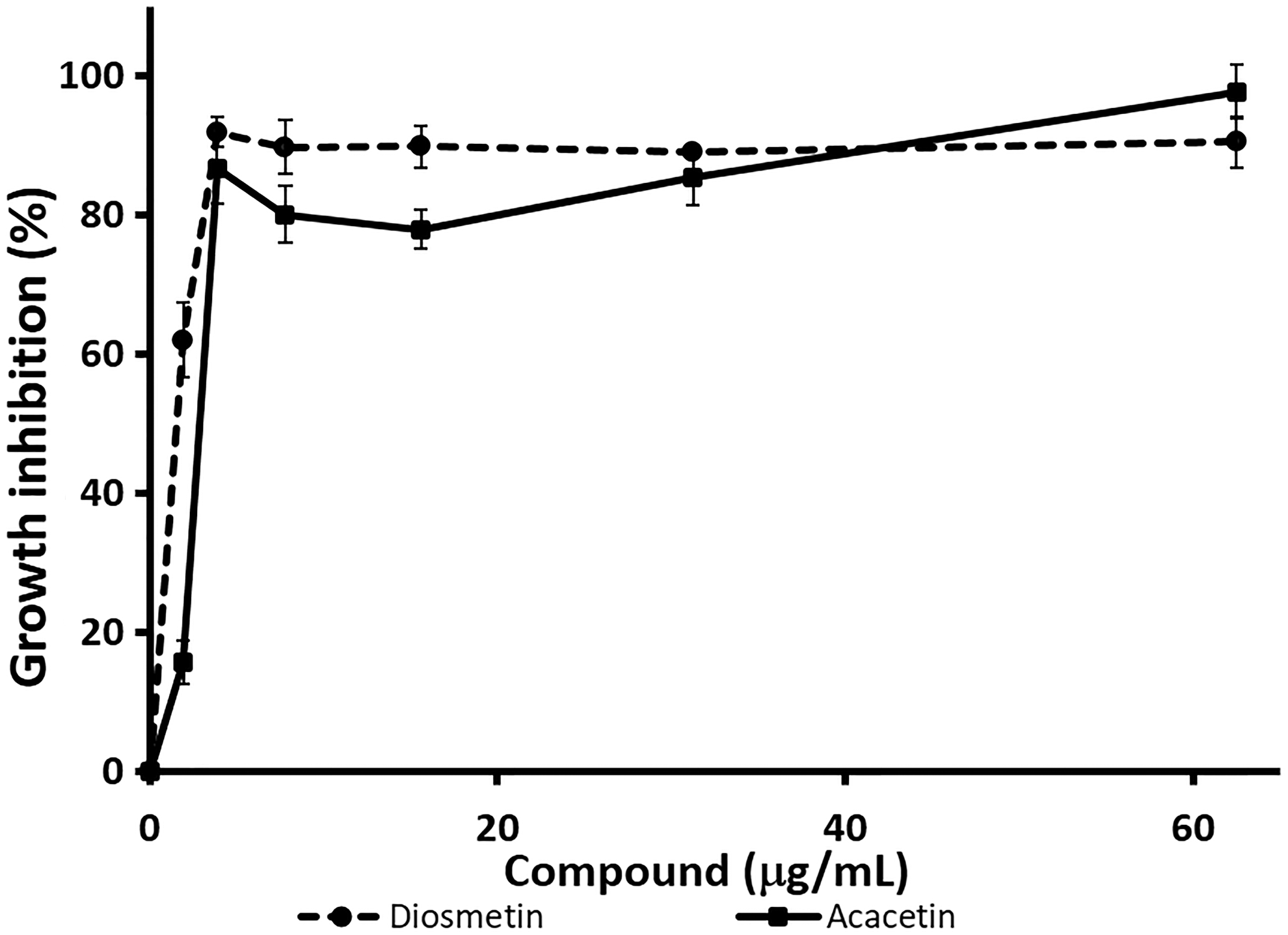
Effect of the flavonoids acacetin and diosmetin on H. pylori growth. The antibacterial activity was assessed by broth dilution method. The percentage of growth inhibition was calculated with respect to a control that grew only in the presence of DMSO. Data are presented as mean ± SEM of at least three independent experiments in triplicate.
Discussion
Antibiotic phytocomponents can offer an alternative for the treatment of infectious diseases. 22 In the present work, the effect on H. pylori growth of some edible Mexican quelites was investigated for determining whether they could have, besides their nutritional value, some other health benefits.
Several previous reports, working with different plant extracts, have classified their anti-H. pylori activity from strong to null according to their MIC values. 23,24 Taking into account these classifications, we considered five categories of antibacterial activity depending on the MIC: (1) strong (MIC ≤15.6 μg/mL); (2) good (MIC 15.6–125 μg/mL); (3) moderate (MIC 125–300 μg/mL); (4) low (MIC >300–500 μg/mL); and (5) null (MIC >500 μg/mL). From this perspective, all quelite DM extracts exerted inhibition on H. pylori growth. The anti-H. pylori activity produced by the Ala-DM extract can be considered moderate, while both Cha-DM and Che-DM extracts had a good activity. On the contrary, AQ extracts did not have antibacterial activity (Table 2).
Some concentrations of DM extracts produced more than 100% of bacterial growth inhibition (Fig. 1), an effect that is due to a decrease in the culture absorbance at the end of the experiment with respect to the initial one. This absorption decrement indicates that the extracts are not only promoting inhibition of bacterial growth but also bacterial lysis.
When comparing the obtained MICs of DM extracts with the MIC of amoxicillin (0.005 μg/mL), it is evident that much higher concentrations of DM extracts are required to inhibit bacterial growth; however, the antibacterial activity displayed by each one of the DM extracts was equal or even better than the other positive control, metronidazole (250 μg/mL) (Table 2). The latter antibiotic is widely used in the H. pylori eradication therapy, despite its high resistance rates. 25
Adherence of H. pylori to the gastric epithelium is a primary step for a successful infection. Many studies have shown that the binding of bacteria is mediated by multiple adhesins; among the most studied are BabA and SabA, which are known to interact through fucosylated or sialylated blood group antigens, 26 respectively. It is also known that there are other bacterial proteins involved in H. pylori adhesion, however, for most of them, their corresponding host receptors remain unknown. Thus far, the adherence receptor network related to H. pylori infection is not completely elucidated. 27,28
In this context, antiadhesive compounds capable of inhibiting the docking process of H. pylori to gastric cells have drawn great attention in recent years as a new strategy to prevent and combat bacterial infection.
In the present work, the evaluation of antiadhesive potential of quelite extracts showed that, at the highest concentration tested (1000 μg/mL), DM extracts inhibited around 50% the adhesion of bacteria to AGS cells, while the AQ extracts and mucilages blocked the interaction between bacteria and cells in a lesser (30–40%), but statistically significant, extent. It is possible that the compounds responsible for the antiadherent activity are different in each one of the extracts (DM, AQ, or mucilage); hence, their combination could enhance the antiadherent activity over the individual value obtained for each extract.
Other research works have demonstrated a partial inhibition of H. pylori adherence to cells with different kinds of extracts 29 –32 ; and only a few works have reported a total inhibition, just as in the case of a chloroform extract obtained from Phyllanthus urinaria, which inhibited the adhesion to AGS cells by 100% with 0.5 mg/mL. 33
As stated before, bacterial binding to epithelial cells is mediated by multiple adhesins, which explains the difficulty to reach a complete inhibition of H. pylori attachment to cells with only one compound. Considering this, the antiadherent activity exhibited by the quelite extracts could be part of a prophylactic or eradication therapy by participating in the inhibition of the bacterial binding to host cells.
For a successful infection, H. pylori also requires the participation of urease. 34 The importance of this enzyme can be appreciated considering that urease-negative mutant strains are unable to colonize animal models. 35 Regarding our results, we can conclude that the anti-H. pylori effect observed with the quelite extracts does not rely on the abolishment of the urease activity.
Collectively, the results obtained for DM extracts indicate that they could be a good source for the isolation of compounds with antibiotic or antiadhesive activity.
Dietary plant phenolic compounds exert a variety of biological functions (including the antimicrobial activity) 36 –38 and have received particular attention because of their beneficial health properties. In this work, we determined that all the assayed extracts contain both kinds of compounds (Table 1).
Only a few studies have assessed the phytochemical composition of the quelite species utilized in this work. With respect to phenolic compounds, in C. aconitifolius has been reported the presence of some flavonoids, including amentoflavona, quercetin, kaempferol, catechin, hesperidin, kaempferol-3,7-dimethyl ether, epigallocatechin gallate, naringenin, and some flavonoid glycosides of quercetin and kaempferol. In addition, phenolic acids such as protocatechuic, chlorogenic, 4 hydroxybenzoic, caffeic, p-coumaric, ferulic, and ellagic acids, among others, have been detected in the species. 12,13,39 –41 In the case of A. cristata, only two flavones have been reported: acacetin and diosmetin. 9 For C. pumila, none compound of this group has been identified.
Several studies have determined the anti-H. pylori activity of phenolic acids and flavonoids, 23 including the majority of those reported for C. aconitifolius 42 –47 ; nevertheless, concerning the two flavonoids of A. cristata, only one work describes some effect of diosmetin on H. pylori, 48 but there is no report regarding acacetin. With this background, we decided to study the effect of both flavonoids on bacterial growth.
In our experiments, the MIC obtained for diosmetin (Fig. 2) was 62.5 μg/mL. With the same H. pylori strain that we used in this work, Bae et al. 48 reported an MIC >100 μg/mL. The discrepancy could be due to the source of the compound or to the method they used in the inhibition assay (agar dilution method). However, for both flavones, we obtained a very good inhibitory effect (∼90%) with only 3.9 μg/mL. The results indicate that it is possible that these flavonoids may be, in part, responsible for the anti-H. pylori activity achieved with the Ala-DM extract.
The anti-H. pylori activity and the inhibitory effect of the bacterial adhesion to AGS cells obtained with the quelites are probably due not only to a particular compound but to the combined effect of several molecules as well. Furthermore, the two main flavones of A. cristata, acacetin and diosmetin, wield a strong activity against the bacteria, and could represent new anti-H. pylori agents of natural origin.
H. pylori has lived with us for thousands of years and it is estimated that around 50% of the world's population is infected. It is associated with a wide group of human gastrointestinal diseases, but only some of the infected people develop any pathology. The main factors that influence the transition from H. pylori infection to an overt clinical disease are bacterial polymorphism, host susceptibility, and diet.
Since the beginning of mankind, plants have fulfilled basic functions for humans, including food or/and medicine. The proposed mechanism by which plants and plant-derived drugs exert their action against H. pylori is, on the one hand, by preventing, mitigating, or eradicating the infection; and on the other hand, by protecting against the damage provoked by the bacteria to the host. Even though plant alternative therapies do not achieve permanent eradication of H. pylori, the routine consumption of some edible and/or medicinal plants may help to maintain a delicate balance between health and illness, where bacteria remain in the gastric mucosa without causing the most severe clinical outcomes such as peptic ulcer or gastric cancer.
In the case of the three species of quelites studied in this work, the results suggest that, considering their antibacterial and antiadhesive properties, these plants not only represent a promising source for a complementary therapy but also their regular consumption may play an important key role in reducing bacterial colonization.
This work provides new information about the anti-H. pylori potential of three edible quelites and provides the basis for further studies to establish whether their consumption in a daily diet could have an impact on the prevention and/or control of H. pylori-associated diseases.
Footnotes
Acknowledgments
This study was supported by CONACYT 214286, “Rescate de especies subvaloradas tradicionales de la dieta mexicana y su contribución para el mejoramiento de la nutrición en México,” and by DGAPA-UNAM (PAPIIT IN214317).
Author Disclosure Statement
No competing financial interests exist.