




Abstract
The aim of this study is to examine the therapeutic effects of Olea europaea L. leaf extract on carbon tetrachloride (CCl4)-induced liver damage in rats. In the experiments, 3- to 4-month-old 28 male Sprague–Dawley rats were divided into four groups: control, O. europaea leaf extract, CCl4, and curative. The CCl4 and curative groups received CCl4 (0.2 mL/kg) intraperitoneally for 10 days to form hepatic injury. O. europaea (80 mg/kg) leaf extract was given orally to the curative group dissolved in distilled water the following 14 days. Hepatic and antioxidant enzyme levels, p53, caspase 3, lipid peroxidation marker malondialdehyde (MDA), and also DNA fragmentation levels were determined to establish oxidative stress in hepatic cell damage and its consequences. After formation of liver damage, oral administration of the O. europaea significantly reduced CCl4-induced elevations of serum alkaline phosphatase, aspartate aminotransferase and alanine aminotransferase levels (P < .001), MDA levels of both blood (P < .001) and liver tissues (P < .001), DNA fragmentation (P < .001), p53 (P < .001), and caspase 3 (P < .001) levels of liver tissues. Also this administration in curative group significantly increased CCl4-induced reductions of superoxide dismutase (SOD) (P < .001) and catalase (CAT) (P < .001) activity of blood samples and decreased SOD (P < .001) and CAT (P < .05) activity observed in liver tissue curative groups compared with CCl4 curative group. In CCl4 group, liver tissue samples exhibited remarkable damage because of CCl4 and reduction of these damages were observed in the curative group. Our results showed that O. europaea leaf extract was effective in reducing hepatic damage caused by CCl4 by reducing lipid peroxidation, regulating antioxidant enzymes, and minimizing DNA damage.
Introduction
T
Liver injury has been shown to be associated with many diseases, such as cirrhosis, fibrosis, fatty liver (steatosis), steatohepatitis, and malignant transformation (hepatocellular carcinoma). 3
CCl4 is widely used in rat models for the generation of liver damage 4 and is a biologically inactive and stable substance. The cytochrome P-450-dependent monooxygenase system metabolizes CCl4 and these processes cause the free radical production. These radicals initiate lipid peroxidation in membranes. 1,5 As a result of imbalance between free radical production and antioxidant systems, oxidative stress is observed in tissues. 6 –8 Large amounts of ROS and oxidative stress will cause cell death through necrotic or apoptotic mechanisms leading to cellular and tissue damage. 9
Natural antioxidants found in edible or medicinal plants often have strong antioxidant and free radical-scavenging properties that will form the basis of other biological activities and health benefits. 9 O. europaea is a plant that spreads widely in the Mediterranean countries and has been among medicinal plants since ancient times. O. europaea, an important part of the daily diet, has been shown to have blood pressure-lowering effects, to accelerate blood flow, and to lower blood glucose. Besides these, O. europaea is an antimicrobial, anti-inflammatory, and antioxidant agent, thanks to its phenolic profile containing caffeic acid, verbascoside, oleuropein, luteolin 7-O-glucoside, routine, apigenin 7-O-glucoside, and luteolin 4′-O-glucoside. 10 –12
In our study, effects of O. europaea leaf extract on CCl4-induced liver damage and antioxidant system were investigated.
Materials and Methods
Plant materials
Twenty percent oleuropein-containing O. europaea leaf extract powder was provided by Kale Naturel Herbal Products Company, Ltd., Balikesir, Turkey.
Animals
Twenty-eight Sprague–Dawley male rats were used for the study. All rats were housed in the laboratory under the same conditions and fed with standard pellet diet and tap water. The study was carried out with approval from the Eskisehir Osmangazi University Local Ethics Committee of Animal Experiments (26.04.2011/36_210).
Experimental protocol
After a week of acclimatization, the rats were divided into four groups (n = 7) as control, CCl4 control, Olea leaf extract, and curative group.
Control group
Around 0.2 mL/kg saline solution (0.9% NaCl) was injected intraperitoneally twice daily to rats for 10 days. The following 2 weeks, tap water was administered to rats by gavage method.
CCl4 control
To induce liver injury during the first 10 days of the study, 0.2 mL/kg CCl4 (Merck, Darmstadt, Germany) was administered intraperitoneally to the rats twice daily. The following 2 weeks, tap water was administered to rats by gavage method.
Olea leaf extract group
Around 0.2 mL/kg saline solution (0.9% NaCl) was injected intraperitoneally twice daily to rats for 10 days. The O. europaea leaf extract (80 mg/kg) was dissolved in tap water and administered orally to rats.
Curative group
To induce liver injury during the first 10 days of the study, 0.2 mL/kg CCl4 was administered intraperitoneally to the rats twice daily. The following 2 weeks, O. europaea leaf extract (80 mg/kg) was dissolved in tap water and administered by gavage method.
Tissue collection and sample preparations
Intracardiac blood samples were withdrawn to the proper tubes and standardized hemoglobin pipettes under anesthesia 24 h after the last treatment. Hemoglobin concentration was determined spectrophotometrically at 540 nm by oxyhemoglobin method. 13 To separate serum, blood samples were centrifuged at 3500 g for 10 min and then stored at −80°C until analysis of liver function tests. Blood samples collected into 2-mL ethylenediaminetetraacetic acid (EDTA) tubes were centrifuged for 10 min at 3000 g to separate plasma. The supernatant was removed and erythrocyte pellets were washed three times with physiological sodium chloride solution (0.9% NaCl) and the supernatant was removed each time. Then, prepared erythrocyte packets were hemolyzed by adding an equal volume of double-distilled water and centrifuged at 3000 g for 10 min. The pellet was discarded and supernatant was collected as hemolysate and stored at −80°C until the measurements of malondialdehyde (MDA), superoxide dismutase (SOD), and catalase (CAT). 14,15
Livers from sacrificed rats were transferred to ice-cold isotonic NaCl solution and the residues were removed. Of the liver samples divided in two, the specimens were taken in 10% formalin (Sigma-Aldrich, Taufkirchen, Germany) and made stable for histopathological examination. The other part was stored at −80°C until the homogenate was prepared. The liver tissue samples were weighed (1 g) and homogenized in cold potassium chloride (1%, pH 7.4) using an ultrasonic homogenizer at 4°C. The homogenates were centrifuged at 3000 g at 4°C. The supernatants were collected and stored at −80°C until being assayed.
Liver tissue protein quantities were determined spectrophotometrically according to the kit (Total Protein LiquiColor 10 570; Wiesbaden, Germany).
Assessment of liver function test
Liver alkaline phosphatase (ALP), aspartate aminotransferase (AST), and alanine aminotransferase (ALT) enzymes levels were analyzed by a Roche Modular P chemistry analyzer (Roche Diagnostics, IN). Product test kits were supplied by Roche (Roche Diagnostics).
Lipid peroxidation determination
Lipid peroxidation was estimated by MDA, one of the lipid peroxidation end products. MDA concentration was determined according to the method based on color reaction of MDA with thiobarbituric acid (Sigma-Aldrich) as previously described. 16 MDA levels are expressed as nmol/g protein for liver homogenate and kU/g Hb for hemolysate.
Assay of SOD activity
The SOD enzyme activity was measured in both liver homogenate and hemolysate using the SOD Determination Kit (Cat. No: 19160; FLUKA, St. Louis, MO) based on water-soluble tetrazolium salt reaction. SOD activity was expressed as% inhibition.
Assay of CAT activity
The CAT activity in both liver homogenate and hemolysate samples was measured by Goth's method 17 as described previously. 18 Results were expressed as kU/g protein for liver homogenate and kU/mL for hemolysate.
DNA fragmentation of liver tissue
DNA fragmentation as previously described was determined by a spectrophotometric method using the diphenylamine (Sigma-Aldrich) reaction. 19 The percentage of DNA fragmentation was calculated using the formula fragmented DNA/(fragmented DNA + intact DNA) and expressed as a percentage of total DNA appearing in the supernatant fraction.
p53 Levels of liver tissues
The concentrations of p53 protein in liver tissue were assessed using the ELISA Kit (Cat. no. 11828789001; Roche Molecular Biochemicals, Mannheim, Germany), 18 and p53 concentrations were determined as pg/mL.
Caspase 3 levels of liver tissues
Caspase 3 concentrations in the liver tissues were measured by the USCN Life Science, Inc. Caspase 3 ELISA Kit (Cat. No: SEA626Ra) according to the manufacturer's instructions as previously described. 18 Caspase 3 concentrations were determined as ng/mL.
Histopathological evaluation
After the chemical fixation (into 10% formalin) was completed, the first parts of the tissues were converted into paraffin blocks by the routine procedure in the ethyl alcohol series. Four to six micrometer sections were taken from these blocks for standard Hematoxylin and Eosin staining. Histopathological examination was performed under a light microscope (NIKON, Japan) on all these preparations to be prepared. Samples were determined as (+) less damaged, (++) moderately damaged, (+++), or severely damaged.
Statistical analyses
SPSS version 21 was used for data analysis. The normal distribution of continuous variables was determined by the Kolmogorov–Smirnov test. The groups were compared using normal distribution of variance, one-way analysis of variance and Tukey's test. The results were expressed as mean ± standard deviation. The P values <.05 were considered statistically significant.
Results
Administration of CCl4 increased the levels of the liver marker enzymes (ALP, AST, and ALT) significantly (P < .001) in CCl4 group, and O. europaea leaf extract treatment after CCl4 administration in the curative group decreased liver marker enzymes significantly (P < .001) (Table 1). Serum concentrations of ALP, AST, and ALT were not altered by the daily oral administration of O. europaea leaf extract in olive leaf extract group without any CCl4 application significantly (P > .05).
Data are presented as mean ± SD (n = 7).
Significant differences were found: *** P < .001 compared with control group; +++ P < .001 compared with CCl4 group.
ALP, alkaline phosphatase; AST, aspartate aminotransferase; ALT, alanine aminotransferase; SD, standard deviation.
The hepatocurative effects of O. europaea leaf extract on MDA levels, SOD, and CAT activity of the liver tissue are shown in Table 2. As a result of CCl4 administration, MDA levels were increased significantly (P < .001) in the CCl4 group compared with control group. The treatment with O. europaea leaf extract in the curative group after CCl4 administration reduced the MDA levels significantly (P < .001) by reducing lipid peroxidation. O. europaea extract application had not changed the MDA levels in olive leaf extract group. Administration of CCl4 decreased SOD activity significantly (P < .001) in CCl4 group compared with control and O. europaea treatment reversed the depletion toward the normal range (P < .01 compared with the control group, P < .001 compared with CCl4 group) into the curative group. Same results were also obtained for CAT activity. While CCl4 application in CCl4 group decreased the CAT activity (P < .001), after treatment with O. europaea extract, it was increased significantly (P < .05).
Data are presented as mean ± SD (n = 7).
Significant differences were found: ** P < .01, *** P < .001 compared with control group; + P < .05, +++ P < .001 compared with CCl4 group.
MDA, malondialdehyde; SOD, superoxide dismutase; CAT, catalase.
The hepatocurative effects of O. europaea leaf extract on MDA levels, SOD, and CAT activity of the blood are summarized in Table 3. CCl4 administration has caused an increase in levels of MDA significantly (P < .001) in the CCl4 group with respect to control. After 10 day CCl4 administration, 2 weeks treatment with O. europaea leaf extract decreased the MDA levels (P < .001, compared with CCl4 group) in curative group. SOD and CAT activities were significantly decreased in CCl4 group (P < .001). Treatment with O. europaea leaf extract in curative group has caused an increase in both SOD and CAT activities (P < .001) with respect to the CCl4 group.
Data are presented as mean ± SD (n = 7).
Significant differences were found: *** P < .001 compared with control group; +++ P < .001 compared with CCl4 group.
The hepatocurative effects of O. europaea leaf extract on DNA% fragmentation, p53, and caspase 3 levels of the liver tissue are shown in Table 4. After 10 days of CCl4 administration, DNA% fragmentation, p53, and caspase 3 levels of the liver tissue were significantly increased in CCl4 group with respect to control. Treatment with O. europaea leaf extract in curative group has caused a decrease in DNA% fragmentation, p53, and caspase 3 levels of the liver tissue (P < .001) with respect to CCl4 group.
Data are presented as mean ± SD (n = 7).
Significant differences were found: *** P < .001 compared with control group; +++ P < .001 compared with CCl4 group.
Histological evaluation of the study was demonstrated in Figure 1. Liver tissue samples of control and Olea leaf extract group exhibited normal tissue structure, whereas extensive/intensive cellular damage with necrotic cell structures, asymmetric nuclear structures, sinusoidal dilation, cellular infiltration, and vascular congestion were determined in CCl4 treated group.
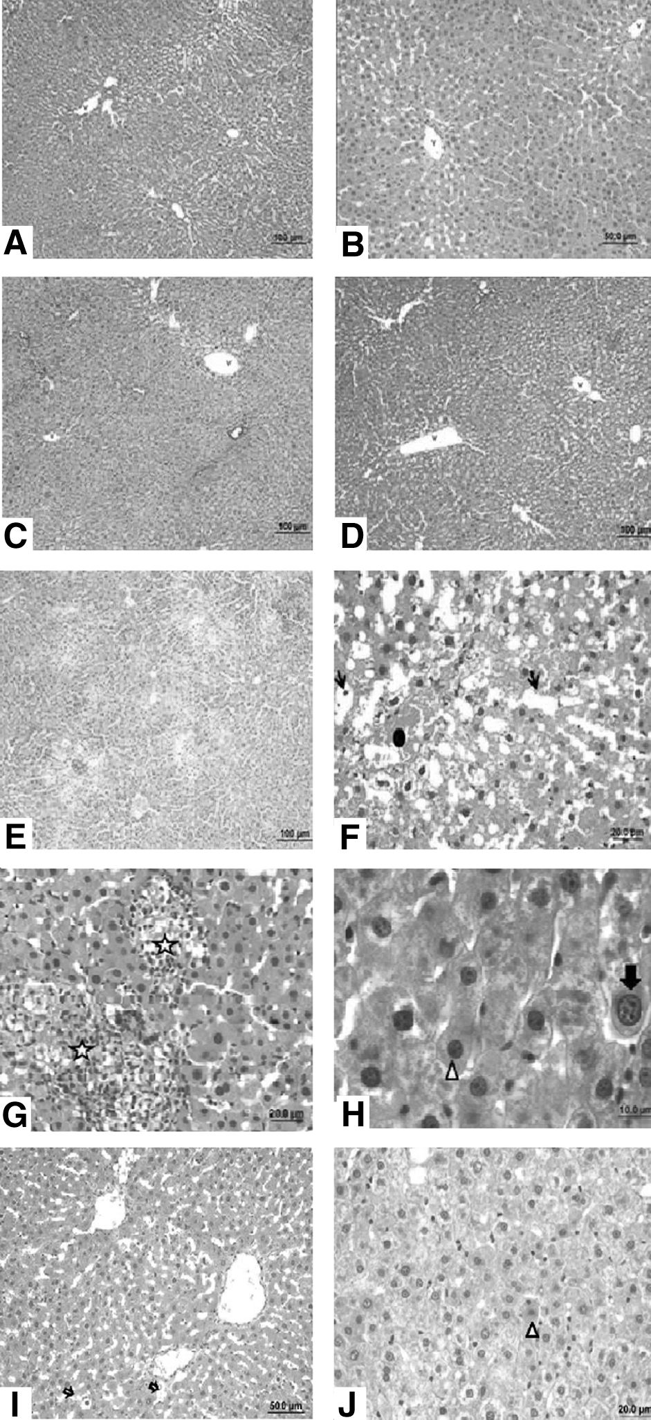
Histological examinations of liver tissues. Normal histological liver structure of Control group
Discussion
The generated free radicals initiate the lipid peroxidation that cause degradation of the hepatocellular membrane. 20 This is followed by secretion of liver-specific enzymes, such as ALP, AST, and ALT into the bloodstream. 4,7,21 In our study, the elevation of the ALP, AST, and ALT in serum of CCl4 group demonstrated the hepatocellular membrane damage. O. europaea leaf extract treatment after induction of hepatocellular injury by CCl4 significantly decreased all liver-specific enzyme levels. These results suggested that O. europaea leaf extract has curative effects on cell membrane damage because of having in vivo antioxidant properties. This result was endorsed by both lipid peroxidation product MDA levels and histological evaluation results. Similar to our results, it is shown that CCl4 administration caused several structural changes in liver tissue. 18
Nonenzymatic and enzymatic antioxidant systems have been developed in organisms to overcome the harmful effects of ROS, which is the natural end result of various metabolic activities. The basic elements of enzymatic systems are SOD, CAT, and GPx. 22 Several studies have reported that, CCl4 leads to a significant reduction in the activity of SOD and CAT, the endogenous antioxidants found naturally in our bodies. 23,24 We demonstrated that application of CCl4 triggered ROS production and adversely affected the endogenous antioxidant system. According to our findings, it was observed that O. europaea treatment significantly increased SOD and CAT activities in blood and liver tissues, whereas CCl4 induced reduction. These data suggested that O. europaea was protective against the oxidative stress caused by CCl4 by stimulating the antioxidant defense mechanism.
ROS triggered by CCl4 causes oxidative damage in DNA as well as cellular injury. The products of CCl4 chemical decomposition are covalently bound to DNA leading to mutations and chromosomal abnormalities. 25 Oxidation of DNA induces cell proliferation and apoptosis pathways and disrupts cellular stability. 5 The tumor suppressor, p53 protein, blocks the cell cycle, repairing DNA damage. When DNA damage is persistent, apoptotic signals are induced, and mitochondrial cytochrome c releases so that cell death begins by stimulating caspase 3. 26 Caspase 3 cleaves and activates the DNA fragmentation factor, resulting in DNA fragmentation. 27 In support of our findings, Domitrović et al. found that oleuropein increased SOD activity and decreased caspase 3 levels compared with the CCl4-treated group. 28 In the present study, CCl4 triggered apoptosis and caused a significant increase in p53, caspase 3 protein levels, and DNA fragmentation as previously described. 18 Hence, the increased levels of DNA fragmentation, caspase 3, and p53 were considered as important molecular markers for apoptosis. Several CCl4-induced rat models have reported that p53-mediated hepatic cell death occurs through the mitochondrial pathway. 18,25,26 Besides, decreased p53, caspase 3 levels and low levels of DNA fragmentation were found in O. europaea treatment group. These data indicate that O. europaea significantly blocked the toxic effect of CCl4 on liver. The regulatory effect of O. europaea on apoptotic parameters may be due to its being a potent antioxidant and ROS scavenger. Thus, inhibition of p53-associated hepatocyte apoptosis may be an effective therapeutic strategy in the treatment and prevention of CCl4-induced hepatic injury.
In the current study, our outcomes supported that oxidative damage is one of the basic mechanisms of CCl4-induced hepatotoxicity. However, ROS-triggered apoptosis plays an important role in the development of liver damage in rats. Liver diseases are seen as an important problem in the world. Despite new developments in hepatology, methods used to treat liver diseases remain inadequate to overcome problems caused by chemical toxins. Detoxification of liver, which is the most important organ affected by toxic substances at the cellular level, will be beneficial. Taking advantage of natural components to protect liver cells is crucial for the development of new drugs. In addition, consuming rich foods with these natural ingredients in our daily diet will mitigate the deleterious effects of hepatotoxicity.
In conclusion, this study indicated the curative effects of O. europaea leaf extract on the oxidative stress and liver damage induced by CCl4. This therapeutic effect was revealed through reducing lipid peroxidation, regulating antioxidant enzymes, and providing the improvement of DNA damage.
Footnotes
Acknowledgment
The authors would like to thank the Kale Naturel Herbal Products Company, Ltd., Balikesir, Turkey for providing the Olea europaea leaf extract.
Author Disclosure Statement
No competing financial interests exist.