
Abstract
After cultivation of ginseng, ginsenosides, which are the major active ingredients of gingeng, were approved for use by the food industry, and began to be used as added functional ingredients to try to improve the quality and price of functional foods. However, the interaction between different types of ginsenosides and nutrients needs further study. We investigated the effect of B-complex vitamins (which are essential nutrients) on the pharmacokinetics of the ginsenosides protopanaxatriol-type saponin Rg1, protopanaxadiol-type saponin Rb1, and oleanolic acid-type saponin Ro after oral administration. Ginsenosides Rg1, Rb1, and Ro, with or without B-complex vitamins, respectively, were administered orally to rats to evaluate their pharmacokinetics. The concentration of ginsenosides in plasma was determined by liquid chromatography/tandem mass spectrometry. Pharmacokinetic parameters were fitted using WinNonlin v6.2. After oral coadministration with B-complex vitamins, the area under the concentration–time curve from zero to infinity (AUC0–∞) of ginsenoside Rg1 was reduced by 70%, that of ginsenoside Rb1 was reduced by 43%, and that of ginsenoside Ro was reduced by 34%. The AUC0–∞ of ginsenosides Rg1 and Rb1 showed significant differences between different treatments, but the AUC0–∞ of ginsenoside Ro did not. These results suggest significant ginsenoside-nutrient interactions between ginsenosides Rg1, Rb1, and B-complex vitamins.
Introduction
G
Ginsenosides are the main active ingredients in ginseng and are classified into protopanaxatriol-type saponin, protopanaxadiol-type saponin, and oleanolic acid-type saponin. 7 Ginsenoside Rg1 is the principal protopanaxatriol-type saponin, ginsenoside Rb1 is the main kind of protopanaxadiol-type saponin, and ginsenoside Ro is a representative oleanolic acid-type saponin (Fig. 1).
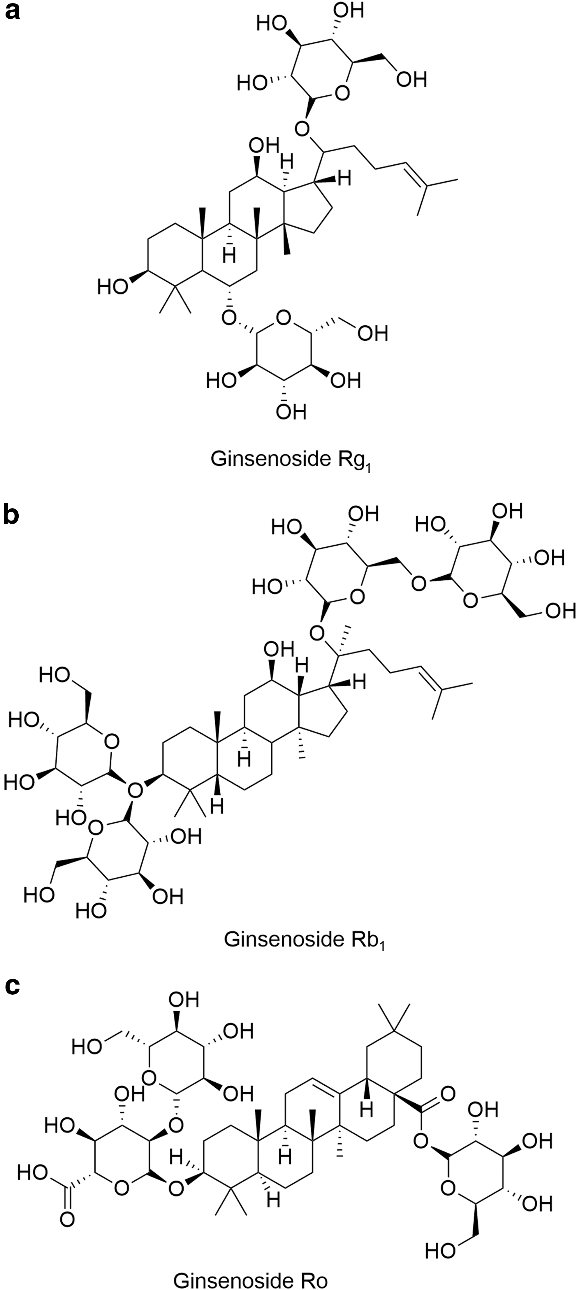
The chemical structures of ginsenosides Rg1
The main members of the B-vitamin family are B1, B2, B3, B5, B6, B7, B9, and B12, and these are essential nutritional compounds. 8 B vitamins are required as cofactors for certain enzymes essential for cell function and energy production. 9 As a family, B vitamins usually coexist in nature. The synergistic effect is an important feature of B vitamins. To achieve the best results, all types of B vitamins should be consumed together. 10,11 Therefore, B-complex vitamins are often recommended as nutritional supplements in the market.
People often focus on the pharmacologic efficacy of a product and consider that the product mixed with certain components will have wider and better efficacy while ignoring the potential interactions between components. Previously, we found an interaction between ginsenoside Re and B-complex vitamins. 12 However, a comparative study on the effect of B-complex vitamins on the different types of ginsenosides is lacking. We chose the representative ginsenosides Rg1, Rb1, and Ro and B-complex vitamins to ascertain if there are interactions between different types of ginsenosides and B-complex vitamins. Our results could provide valuable theoretical guidance for the combined use of ginsenosides Rg1, Rb1, and Ro and B-complex vitamins or for their use in foods fortified with B-complex vitamins.
Materials and Methods
Chemicals and reagents
Ginsenoside standards for Rg1, Rb1, and Ro with a purity of ≥98.0% were purchased from the School of Chemistry within Jilin University (Changchun, China). The internal standard (IS) digoxin (purity ≥98.0%) was obtained from Pure Chemical Standard (Chengdu, China). Vitamin B1 (purity ≥99%), B2 (≥98%), B3 (≥99%), B5 (≥99%), B6 (≥99%), B7 (≥97%), B9 (≥97%), and B12 (≥98%) were purchased from Shanghai Yuanye Bio-Technology (Shanghai, China). Ammonium hydroxide (high-performance liquid chromatography [HPLC] grade) was purchased from Beijing Chemical Works (Beijing, China).
Solid-phase extraction columns (Oasis® HLB 3 cm3/60 mg) were purchased from Waters (Milford, MA, USA). Methanol and acetonitrile of HPLC grade were obtained from Fisher Scientific (Springfield, NJ, USA). Milli-Q (Millipore, Milford, MA, USA) water was used throughout the study. All other chemicals were of HPLC or analytical grade.
Animals
Male Sprague–Dawley rats used for pharmacokinetic experiments (230–260 g) were purchased from Changsheng Biotechnology (Dalian, China). The animals were kept at standard conditions in an animal house with free access to food and water under a 12-h light/12-h dark cycle at a constant temperature (22°C ± 2°C) and humidity (50% ± 10%). Experiments were designed in accordance with the Guide for the Care and Use of Laboratory Animals (US National Institutes of Health, Bethesda, MD, USA) and approved by the Committee of the Institute of Special Economic Animals and Plants of the Chinese Academy of Agricultural Science (Changchun, China).
Experimental protocols and blood sampling
After a 7-day acclimatization period to laboratory conditions, rats were fasted for 12 h but had free access to water. Then, they were divided randomly into six groups of five: Rg1; Rg1 and B-complex vitamin mixture; Rb1; Rb1 and B-complex vitamin mixture; Ro; Ro and B-complex vitamin mixture. Ginsenoside monomer groups were administered as ginsenoside monomer (200 mg/kg body weight, p.o.). Mixture groups were administered ginsenoside monomer (200 mg/kg, p.o.) mixed with B-complex vitamins (B1, 1.5 mg/kg; B2, 1.5 mg/kg; B3, 5 mg/kg; B5, 2.3 mg/kg; B6, 1 mg/kg; B7, 15 μg/kg; B9, 40 μg/kg; B12, 1 μg/kg). 13 –16
Blood samples (0.2 mL) were collected at predetermined time points (0, 0.083, 0.25, 0.5, 1.0, 1.5, 2.0, 3.0, 5.0, 8.0, 10.0, 12.0, 24.0, 48.0, and 72.0 h) via the caudal vein after dosing and placed in tubes containing heparin. Subsequently, plasma samples were prepared by centrifugation at 1400 g for 10 min at room temperature and then stored at −80°C for further analyses.
Preparation of blood samples
Plasma samples (50 μL) were removed from storage at −80°C and thawed under ambient temperature. Preconditioning of the solid-phase extraction column was done by washing it with 3.0 mL of methanol and 3.0 mL of deionized water in succession. Plasma samples were spiked with 10 μL of an IS solution (8 μg/mL digoxin dissolved in water) and then loaded onto a preconditioned solid-phase extraction column after diluting 10-fold with 4% phosphoric acid solution. Then, the column was washed with 1.0 mL of water and 1.0 mL of methanol in succession. Finally, the methanol eluant was dried under a flow of nitrogen at 37°C and dissolved in 100 μL of water/methanol (50:50, v/v) for liquid chromatography/tandem mass spectrometry (LC-MS/MS) analyses.
LC-MS/MS analyses
A 5-μL aliquot of prepared sample solution was used for LC-MS/MS analyses. LC-MS/MS analyses were done on an UPLC/XEVO TQ system with an electrospray ionization source (Waters). Separation was achieved using a BEH C18 column (2.1 × 50 mm, 1.7 μm; Waters) with a mobile phase consisting of 0.1g/L ammonium hydroxide/acetonitrile solution as solvent A and 0.1g/L ammonium hydroxide solution as solvent B.
The gradient elution system for ginsenoside Rg1 was as follows: 0–3.3 min, 20–35% A; 3.3–4.3 min, 35–95% A; 4.3–6.3 min, 95% A; 6.3–6.5 min, 95–20% A; 6.5–8.5 min, 20% A. The gradient elution system for ginsenoside Rb1 was as follows: 0–3 min, 25–42% A; 3–5.5 min, 42–95% A; 5.5–7.5 min, 95% A; 7.5–8.5 min, 95–25% A; 8.5–10.5 min, 25% A. The gradient elution system for ginsenoside Ro was as follows: 0–2 min, 25–40% A; 2–4 min, 40–90% A; 4–4.5 min, 90–95% A; 4.5–6.5 min, 95% A; 6.5–7 min, 95–25% A; 7–9 min, 25% A. The flow rate was 0.5 mL/min, and the column temperature was maintained at 35°C.
Mass spectrum analyses were carried out using negative multiple reaction monitoring (MRM) mode. The detection parameters were optimized as follows: nebulizer pressure, 2.8 kV; source temperature, 150°C; drying gas temperature, 450°C; drying gas flow, 1000 L/h; nebulizer gas flow, 50 L/h; collision gas flow, 0.16 mL/min. The precursor/product ion pairs, fragmentor voltage (Frag. V), and collision energy (CE. V) for the analytes were as follows: m/z 799.484 > 637.432 for the quantitative ion pair of ginsenoside Rg1 (Frag. 60 V, CE. 34 V) and m/z 799.484 > 475.378 for the qualitative ion pair of ginsenoside Rg1 (Frag. 60 V, CE. 34 V); m/z 1107.595 > 945.542 for the quantitative ion pair of ginsenoside Rb1 (Frag. 94 V, CE. 39 V) and m/z 1107.595 > 783.490 for the qualitative ion pair of ginsenoside Rb1 (Frag. 94 V, CE. 43 V); m/z 955.490 > 793.437 for the quantitative ion pair of ginsenoside Ro (Frag. 91 V, CE. 41 V) and m/z 955.490 > 569.384 for the qualitative ion pair of ginsenoside Ro (Frag. 91 V, CE. 56 V); m/z 779.653 > 649.556 for the quantitative ion pair of the IS (Frag. 66 V, CE. 32 V) and m/z 779.653 > 475.451for the qualitative ion pair of the IS (Frag. 66 V, CE. 44 V).
Statistical analyses
Values are the mean ± standard deviation (SD). Pharmacokinetic parameters were estimated using a noncompartment model with WinNonlin v6.2 (Pharsight, Mountain View, CA, USA). The parameters were area under the concentration–time curve from zero to the final sampling time or infinity (AUC0–t, AUC0–∞), maximum observed concentration (Cmax), and peak-reaching time (Tmax). The AUC0–t was calculated using the linear trapezoidal with linear interpolation rule. The AUC0–∞ was extrapolated by the AUC0–t. Cmax and Tmax were read directly from individual plasma concentration–time data.
All statistical procedures were undertaken using SAS v9.2 (SAS, Cary, NC, USA). P < .05 was considered significant and P < .01 was considered highly significant. Figures were drawn using SigmaPlot (Systat, San Jose, CA, USA) and ChemDraw (CambridgeSoft, Cambridge, UK).
Results
Effect of B-complex vitamins on the pharmacokinetics of ginsenoside Rg1
We undertook oral administration of ginsenoside Rg1 (200 mg/kg) or ginsenoside Rg1 (200 mg/kg) mixed with B-complex vitamins to rats. Pharmacokinetic results showed that B-complex vitamins reduced the bioavailability of ginsenoside Rg1 after oral administration highly significantly. Compared with the ginsenoside Rg1 group, the AUC0–t and AUC0–∞ of ginsenoside Rg1 were 3.3-fold higher than that of the coadministration mixture-treated group, and the AUC0–t and AUC0–∞ of ginsenoside Rg1 were significantly different between these two treatments (P < .01; P < .01). The Cmax1 of the mixture group was reduced significantly (P < .01), but there were no significant differences in Tmax1 (P > .05), Tmax2 (P > .05), or Cmax2 (P > .05) between these two treatments (Table 1; Fig. 2).
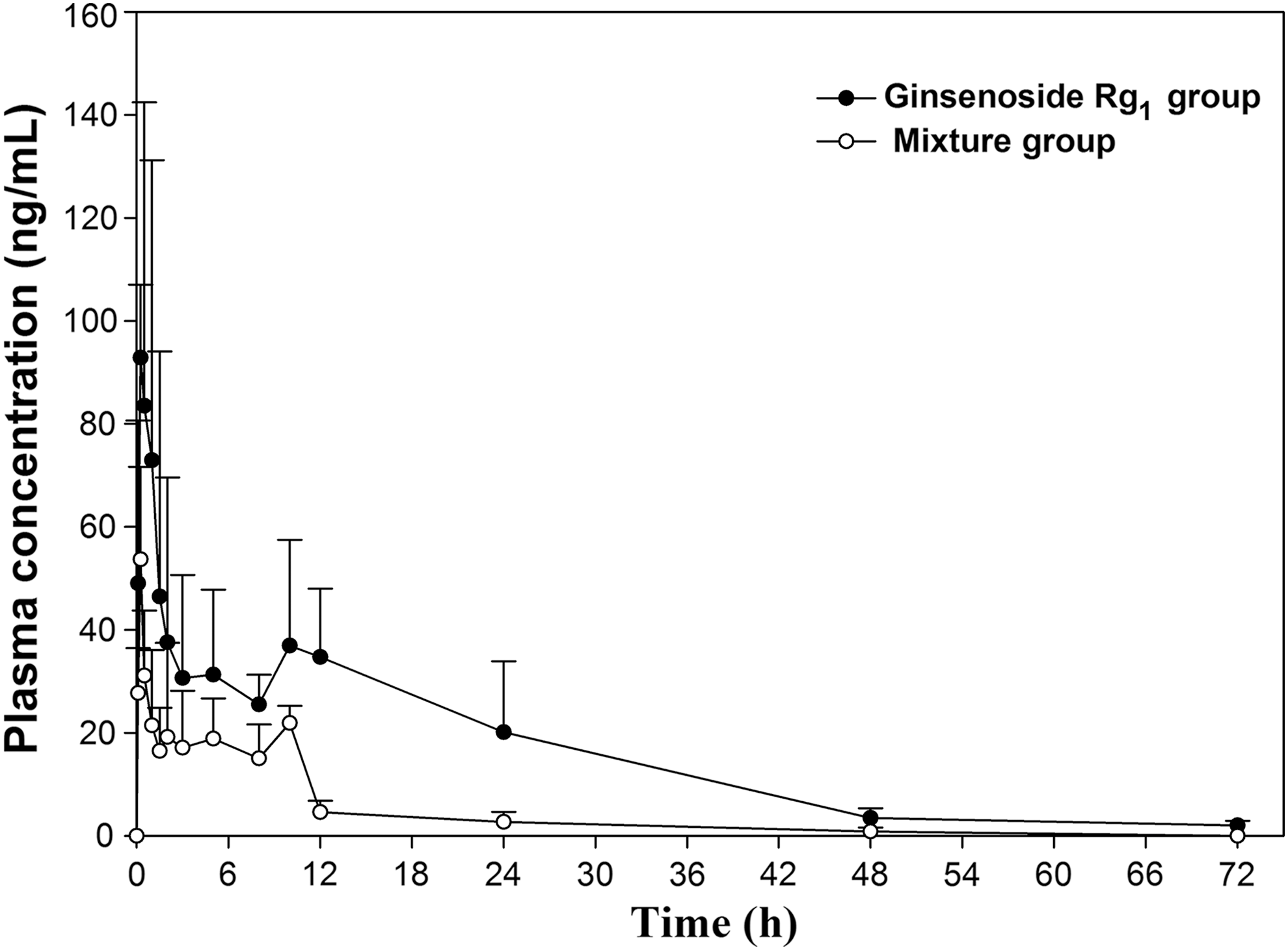
Mean plasma concentration–time profiles of ginsenoside Rg1 in rats. In the ginsenoside Rg1 group, rats were administered ginsenoside Rg1 (200 mg/kg, p.o.). In the mixture group, rats were administered ginsenoside Rg1 (200 mg/kg, p.o.) in combination with B-complex vitamins (p.o.).
Data are mean ± SD, compared with ginsenoside Rg1 group.
SD, standard deviation
P < .05, ** P < .01.
Effect of B-complex vitamins on the pharmacokinetics of ginsenoside Rb1
We also undertook oral administration of ginsenoside Rb1 (200 mg/kg) or ginsenoside Rb1 (200 mg/kg) mixed with B-complex vitamins to rats. Pharmacokinetic results showed that B-complex vitamins reduced the bioavailability of ginsenoside Rb1 after oral administration highly significantly. Compared with the ginsenoside Rb1 group, the AUC0–t and AUC0–∞ of ginsenoside Rb1 were 1.9-fold and 1.8-fold higher in ginsenoside Rg1-treated rats than in coadministration mixture-treated rats, respectively, and the AUC0–t and AUC0–∞ of ginsenoside Rb1 were significantly different between these two treatments (P < .01; P < .01). The mixture group exhibited a significantly lower Cmax1 (P < .01) and Cmax2 (P < .01), but there were no significant differences in Tmax1 (P > .05), Tmax2 (P > .05), mean residence time from zero to the final sampling time (MRT0–t) (P > .05), or mean residence time from zero to infinity (MRT0–∞) (P > .05) between these two treatments (Table 2; Fig. 3).

Mean plasma concentration–time profiles of ginsenoside Rb1 in rats. In the ginsenoside Rb1 group, rats were administered ginsenoside Rb1 (200 mg/kg, p.o.). In the mixture group, rats were administered ginsenoside Rb1 (200 mg/kg, p.o.) in combination with B-complex vitamins (p.o.).
Data are mean ± SD, compared with ginsenoside Rb1 group.
P < .01.
Effect of B-complex vitamins on the pharmacokinetics of ginsenoside Ro
We next undertook oral administration of ginsenoside Ro (200 mg/kg) or ginsenoside Ro (200 mg/kg) mixed with B-complex vitamins to rats. Compared with the ginsenoside Ro group, the AUC0–t and AUC0–∞ of ginsenoside Ro were 1.6-fold and 1.5-fold higher in ginsenoside Ro-treated rats than with coadministration of the mixture to the rats, respectively. The AUC0–t of ginsenoside Ro was significantly different between these two treatments (P < .05), but the AUC0–∞ of ginsenoside Ro was not significantly different between these two treatments (P > .05). The mixture group had a reduced Cmax2 (P < .05), but there were no significant differences in Cmax1 (P > .05), Tmax1 (P > .05), Tmax2 (P > .05), MRT0–t (P > .05), or MRT0–∞ (P > .05) between these two treatments (Table 3; Fig. 4).
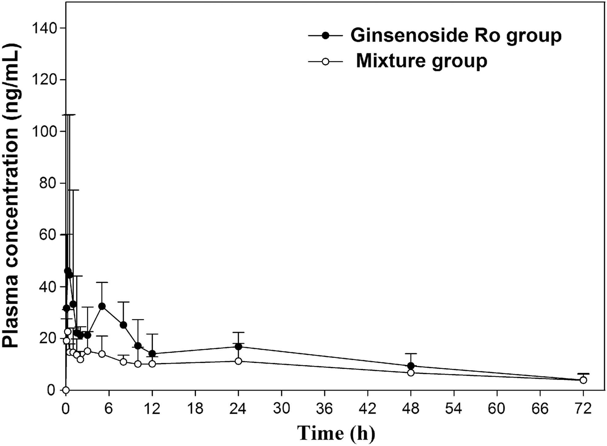
Mean plasma concentration–time profiles of ginsenoside Ro in rats. In the ginsenoside Ro group, rats were administered ginsenoside Ro (200 mg/kg, p.o.). In the mixture group, rats were administered ginsenoside Ro (200 mg/kg, p.o.) in combination with B-complex vitamins (p.o.).
Data are mean ± SD, compared with ginsenoside Ro group.
P < .05.
Validation of LC-MS/MS analyses
The UPLC/XEVO TQ system operating in MRM mode was suitable for quantitative analyses of ginsenoside Rg1, Rb1, and Ro in rat plasma collected at different time points. Calibration standards were prepared by spiking working solutions into blank rat plasma. Ginsenosides Rg1, Rb1, and Ro presented good linearity with a correlation coefficient (R 2 ) of 0.995, 0.998, and 0.996 over ranges (in ng/mL) of 1.0–500.0, 100.0–3000.0, and 0.5–75, respectively.
The lower limit of quantitation (LLOQ signal-to-noise [S/N] ratio = 10) of three ginsenosides was (in ng/mL) 0.6, 50, and 0.5, respectively. The lower limit of detection (LLODS/N ratio = 3) was (in ng/mL) 0.2, 20, and 0.2, respectively. The LLOQ and LLOD were acquired by spiking decreasing concentrations of working solutions into blank plasma to yield the expected S/N ratio. The specificity of the method was confirmed by comparing MRM chromatograms of ginsenosides and the IS for a blank sample of rat plasma with a spiked sample of rat plasma. The analytes could be detected without any significant interference. The recoveries of ginsenoside Rg1 ranged from 89.0% to 93.0%, which were estimated by six replicates using spiked plasma at high, middle, and low concentrations. The recoveries of ginsenoside Rb1 ranged from 95.5% to 99.2%. The recoveries of ginsenoside Ro ranged from 88.2% to 91.9%. The precision of the method was determined by derivation of the peak areas of quality control (QC) plasma samples at six consecutive sampling times. The relative SD was 1.8–2.6% for Rg1, 2.3–4.1% for Rb1, and 3.2–4.7% for Ro. The intraday precision was determined by six replicates using derivation of the peak areas of QC plasma samples at different sampling times on the same day. The interday precision was determined by six replicates using derivation of the peak areas of QC plasma samples on five consecutive days. The intraday precision was 4.5–6.8% for Rg1, 4.8–7.8% for Rb1, and 6.2–8.4% for Ro. The interday precision was 5.8–7.1% for Rg1, 4.9–8.7% for Rb1, and 6.7–10.6% for Ro.
Discussion
Rg1, Rb1, and Ro are three representative ginsenosides with different structures. B-complex vitamins are important nutrients and crucial for some metabolic processes. However, the present study showed that there were ginsenoside-nutrient interactions between ginsenosides Rg1, Rb1, and B-complex vitamins. The B-complex vitamins decreased the absorption of ginsenosides Rg1 and Rb1 significantly if administered simultaneously via the oral route, but B-complex vitamins had no effect on the absorption of ginsenoside Ro by oral administration. These results suggest that the absorption of different types of ginsenosides are differentially affected by the coadministration with B-complex vitamins. These different results may be due to the different structures of ginsenosides. The aglycone of ginsenosides Rg1 and Rb1 is a tetracyclic triterpenoid, but the aglycone of ginsenoside Ro is a pentacyclic triterpenoid. The specific reasons for these differences merit further study.
Through observation of the concentration–time curves of ginsenosides in pharmacokinetic experiments, we found that the concentration of ginsenosides Rg1, Rb1, and Ro in plasma decreased immediately after combined administration with B-complex vitamins, and that the half-life (which reflects the clearance rate of a drug in vivo) of ginsenosides was unchanged by different drug treatments. Hence, the differences should occur mainly in the absorption phase rather than in the metabolic phase. 17
P-glycoprotein (P-gp) is expressed at high levels on the apical surfaces of epithelial cells in the small intestine. 18 P-gp as an efflux transporter has an important role in many drug–drug interactions. 19,20 In 2014, Liang et al., using LLC-PK1 and L-MDRE in Caco-2 cells, confirmed that ginsenoside Rb1 was a substrate of P-gp. 21 In 2007, Meng et al. indicated that the efflux of ginsenoside Rg1 was energy dependent, and that P-gp is likely to be involved its efflux. 22 In 2007, Braga et al. found that ginsenoside Ro had no inhibitory effect on P-gp activity, 23 but whether ginsenoside Ro is a substrate of P-gp was not shown. Hence, one of the reasons for the interaction may be that B-complex vitamins enhance the expression and activity of P-gp in the small intestine, resulting in a reduction of the absorption of ginsenosides Rg1 and Rb1.
Poor membrane permeability is a major factor limiting the intestinal absorption of ginsenosides. This can be attributed mainly to their high molecular weight, low solubility in lipids, and numerous sugar moieties. 24 Ginsenosides can combine easily with other compounds. 25,26 B vitamins are water soluble and highly polar. Hence, interaction between ginsenosides and B-complex vitamins may be due to the formation of new compounds that have even higher molecular weight and even greater water solubility. This would result in the extinction effect of B-complex vitamins on the absorption of ginsenosides after oral administration. However, this hypothetical mechanism of action needs further research.
In conclusion, the results presented in this work showed that when ginsenosides Rg1, Rb1, and Ro were administered via the oral route in combination with B-complex vitamins, respectively, there were significant reductions in the absorption of ginsenosides Rg1 and Rb1. These results suggest pharmacokinetic interactions between ginsenosides Rg1 and Rb1 and B-complex vitamins. Thus, it may be better not to administer ginsenosides Rg1 and Rb1 and B-complex vitamins, or food fortified with B-complex vitamins, simultaneously. Our work may provide some guidance for designing products that include ginsenosides in the food industry.
Footnotes
Acknowledgments
This work was financially supported by the Public Welfare Industry (Agriculture) Scientific Research Projects (No. 20130311102) and the National International Science and Technology Cooperation Projects (No. 2015DFA31290).
Author Disclosure Statement
No competing financial interests exist.