
Abstract
During roasting, major changes occur in the composition and physiological effects of coffee beans. In this study, in vitro antioxidant effects and anti-inflammatory effects of Coffea arabica green coffee extracts were investigated at different roasting levels corresponding to Light, Medium, City, and French roast. Total caffeine did not show huge difference according to roasting level, but total chlorogenic acid contents were higher in light roasted coffee extract than other roasted groups. In addition, light roasted coffee extract had the highest antioxidant activity in the 2,2-diphenyl-1-picrylhydrazyl (DPPH) assay. To determine the in vitro antioxidant property, coffee extracts were used to treat AML-12 cells. Intracellular glutathione (GSH) concentration and mRNA expression levels of genes related to GSH synthesis were negatively related to roasting levels. The anti-inflammatory effects of coffee extracts were investigated in lipopolysaccharide-treated RAW 264.7 macrophage cells. The cellular antioxidant activity of coffee extracts exhibited similar patterns as the AML-12 cells. The expression of mRNA for tumor necrosis factor-alpha and interleukin-6 was decreased in cells treated with the coffee extracts and the expression decreased with increasing roasting levels. These data suggest that coffee has physiological antioxidant and anti-inflammatory activities and these effects are negatively correlated with roasting levels in the cell models.
Introduction
C
The quality of coffee is significantly related to the roasting process. Although roasting is not a fully standardized process and many variations are used, the simplest description of the coffee roasting level is based on evaluation of the color of the prepared beans. In this approach, four main roast levels are defined: Light, Medium, City, and French. 6
During roasting, there are numerous changes in coffee bean compound profiles and the aroma is increased. Major changes in coffee bean composition occur during roasting as a result of the Maillard reaction. The resulting melanoidins are nitrogen-containing end products of the Maillard reaction, and are formed through the reaction of reducing sugars with the lysine residue of proteins and peptides. 7,8 Roasting markedly affects chlorogenic acid, leading to hydrolysis of chlorogenic acid. 9 New compounds are formed during the roasting process; one of these is melanoidin. Its formation might alter the overall antioxidant capacity of coffee beans after roasting. 10,11
Global health concerns about chronic diseases resulting from oxidative stress, such as cancer, diabetes, cardiovascular disease, and inflammation are incressing. 12 Oxidative stress is caused by reactive oxygen species (ROS), including hydrogen peroxide (H2O2), superoxide anion radical (O2 −), and hydroxyl radical (OH−), which are likely to damage several cellular components (lipids, proteins, nucleic acids, and DNA) through oxidation. 13,14
Coffee is a rich source of antioxidants that may contribute to prevention of oxidative stress-related diseases. 15,16 The antioxidant properties of coffee may reflect the presence of both phenolic and nonphenolic bioactive compounds, such as caffeine and chlorogenic acids. 17 –19 Previous studies have shown that coffee has protective effects against oxidation and DNA damage in human cell models 20,21 and has been shown to possess an in vitro antioxidant activity that lessens lipid peroxidation and neoplastic activity. 22 –24 However, little data exist on the effects of roasting levels on coffee composition or on the differences in antioxidant activity.
The chemical-based assays that provide information on the antioxidant properties of coffee do not reflect the cellular physiological conditions. Therefore, there is a need for actual biological models to support the previous antioxidant research. In this study, we examined the effects of coffee beans prepared with various roasting levels on oxidative stress and inflammation using mouse hepatocyte and macrophage models.
Materials and Methods
Coffee extracts
Green coffee beans (200 g, Coffea arabica L, Brazil Ipanema Euro Natural, Coiners International Ltd., Bucheon-si, Korea) were put into a model Kn-8828-2 coffee roaster (Hottop USA, Cranston, RI, USA). Four different levels of roasting were applied for the experiment. Initial roasting was followed by the actual roasting. The roasting conditions of initial and final temperature, roasting time, roasted weight, percentage loss, and roasting level are presented in Table 1.
The roasted coffee beans were ground with a commercial coffee grinder (900N, Yang-Chia Machine Works, Co. Ltd., Taichung, Taiwan) for espresso coffee extraction. The ground coffee (18.0 g) was placed in a porter filter and tamped. Espresso coffee was extracted using a commercial espresso machine (Faema E98; Faema, Milan, Italy) with a two-shot option, which extracted ∼60 mL of coffee. Extraction was conducted at 9 atm (atmospheres) with 98°C steam, which was a preset condition by the manufacturer. The extraction was performed thrice and the extracts were combined together for freeze-drying (FD8505; IlshinBioBase Co., Ltd., Dongducheon-si, Korea).
Determination of caffeine and chlorogenic acid contents
Total phenolic compounds and caffeine contents were determined using high-performance liquid chromatography (HPLC; Agilent, Santa Clara, CA, USA). The separation of the constituents of the extracted coffee beans was performed using an SP column C18 (5 μm, 4.6 × 250 mm). The mobile phase consisted of two components: 0.1% (v/v) acetic acid (mobile phase, A) and acetonitrile (mobile phase B). Gradient elution was performed using the following gradient profile (% v/v): initial (5% B), 20 min (25% B), 21–23 min (80% B), and 24–30 min (5% B). The mobile phase flow rate was 1.0 mL/min and the temperature of the column was set at 40°C.
2,2-Diphenyl-1-picrylhydrazyl (DPPH) free radical scavenging activity
The free radical scavenging activities of the coffee extracts were evaluated by the 2,2-diphenyl-1-picrylhydrazyl (DPPH) assay. Briefly, 0.5 mL of 0.1 mM DPPH (Sigma-Aldrich, St. Louis, MO, USA) in methanol was added to a test tube containing 0.5 mL of the sample. The mixture was then shaken vigorously for 1 min and kept at room temperature for 30 min in the dark. The absorbance of each sample solution was measured at 515 nm.
2,2′-Azino-bis(3-ethylbenzthiazoline-6-sulfonic acid) (ABTS) radical scavenging activity
The 2,2′-azino-bis(3-ethylbenzthiazoline-6-sulfonic acid) (ABTS) (Sigma-Aldrich) was dissolved in water to a concentration of 7 mmol/L. ABTS (Sigma-Aldrich) was produced by reacting the ABTS stock solution with 2.45 mM potassium persulfate (Sigma-Aldrich) (final concentration) and allowing the mixture to stand in the dark at room temperature for 12 h before use. The ABTS stock solution was diluted with phosphate-buffered saline (5 mM/L, pH 7.4) to an absorbance of 0.70 at 734 nm. After the addition of 980 μL of diluted ABTS to 20 μL of sample, the absorbance reading was taken 5 min after the initial mixing and the percent ABTS scavenging activity was calculated.
Cell culture
AML-12 cells derived from mouse liver were obtained from the American Type Culture Collection (ATCC, Manassas, VA, USA). Cells were grown in a 1:1 mixture of Dulbecco's modified Eagle's medium (DMEM; GE Healthcare Life Science, South Logan, UT, USA) and Ham's F12 medium supplemented with 10% fetal bovine serum (FBS; HyClone Laboratories, Inc., South Logan, UT, USA), 0.005 mg/mL insulin, 0.005 mg/mL transferrin, 5 ng/mL selenium, and 40 ng/mL dexamethasone at 37°C in a humidified incubator with 5% CO2.
RAW 264.7 mouse macrophage cells (ATCC) were maintained in DMEM, supplemented with 10% FBS at 37°C in a humidified incubator with 5% CO2.
Determination of cell viability
3-(4,5-Dimethylthiazol-2-yl-2,5-diphenyl-tetrazolium-bromide) (MTT) reduction assay was used to determine the in vitro cell viability. RAW 264.7 cells were plated at a density of 0.2 × 10⁶ cells/well in 96-well plates. The cells were preincubated with coffee extracts for 18 h. The amount of MTT formazan product formed was determined by measuring absorbance using a microplate reader at a wavelength of 570 nm.
Antioxidant in vitro assay
AML-12 cells were seeded in six-well plates (0.2 × 10⁶ cells/well) 6 h before treatment. To determine the cellular antioxidant properties of coffee extracts, coffee extracts with different roasting levels (0.5 and 1.0 mg/mL) were diluted into the medium (5%, v/v) for 12 h.
Anti-inflammatory in vitro assay
RAW 264.7 cells were seeded in six-well plates (0.2 × 10⁶ cells/well) 6 h before treatment. To determine the anti-inflammatory effect of coffee extracts, coffee extracts with different roasting levels (0.5 and 1.0 mg/mL) were added to the medium (5%, v/v). After pretreatment, they were stimulated with lipopolysaccharide (LPS) for 4 h.
Intracellular glutathione concentration
At the end of the treatment, cells were scrapped and homogenized in phosphate buffered saline. Then, they were centrifuged at 10,000 g for 30 min at 4°C and aliquots (0.1 mL) of the supernatants were added to 0.1 mL of 0.6 M perchloric aicd to remove protein portions of the cells. The intracellular glutathione (GSH) concentration was measured by a spectrophotometric cycling assay. 25
mRNA expression levels of GSH synthesizing enzymes and anti-inflammatory factors
Total RNA was isolated with Trizol (Invitrogen, Carlsbad, CA, USA). Complementary DNA (cDNA) was reverse transcribed using a RevertAid First Strand cDNA Synthesis Kit (Thermo Fisher Scientific, Waltham, MA, USA) and used as a template for quantitative polymerase chain reaction (qPCR). qPCR was performed using Maxima SYBR Green qPCR Master Mixes (Thermo Fisher Scientific). The primer sequences were as follows: glutathione synthetase (GS): 5′-CCAGGAAGTTGCTGTGGTGTAC-3′ (forward), 5′-GCTGTATGGCAATGTCTGGACAC-3′ (reverse), glutatmate cysteine ligase catalytic subunit (GCLC): 5′-ACACCTGGATGATGCCAACGAG-3′ (forward), 5′-CCTCCATTGGTCGGAACTCTAC-3′ (reverse), glutamate-cysteine ligase modifier subunit (GCLM): 5′-TCCTGCTGTGTGATGCCACCAG-3′ (forward), 5′-GCTTCCTGGAAACTTGCCTCAG-3′ (reverse), tumor necrosis factor-α (TNF-α): 5′-GGTGCCTATGTCTCAGCCTCTT-3′ (forward), 5′-GCCATAGAACTGATGAGAGGGAG-3′ (reverse), and inducible nitric oxide synthase (iNOS): 5′-GAGACAGGGAAGTCTGAAGCAC-3′ (forward), 5′-CCAGCAGTAGTTGCTCCTCTTC-3′ (reverse).
Statistical analysis
Data are presented as the mean ± SEM. These data were analyzed by one-way analysis of variance using SAS ver. 9.3 (SAS Institute, Inc., Cary, NC, USA). The differences among the treatments were tested with Duncan's new multiple range test. P value under .05 is considered significant.
Results
Caffeine and chlorogenic acid contents in coffee extracts with different roasting levels
The antioxidant effects of coffee extracts are derived from the constituent components. So, we determined the concentration of caffeine and chlorogenic acid in coffee extracts prepared with different roasting levels. Quantitative results demonstrated that the caffeine concentration was slightly decreased in the City and French roast samples compared with Light and Medium roast (Table 2). The chlorogenic acid concentration was significantly decreased with increased roasting levels (P < .05) (Table 2).
Means with different superscript letters (a,b) are significantly different (P < .05) (n = 3).
AVG, average; SE, standard error.
Antioxidant properties of coffee extracts with different roasting degrees
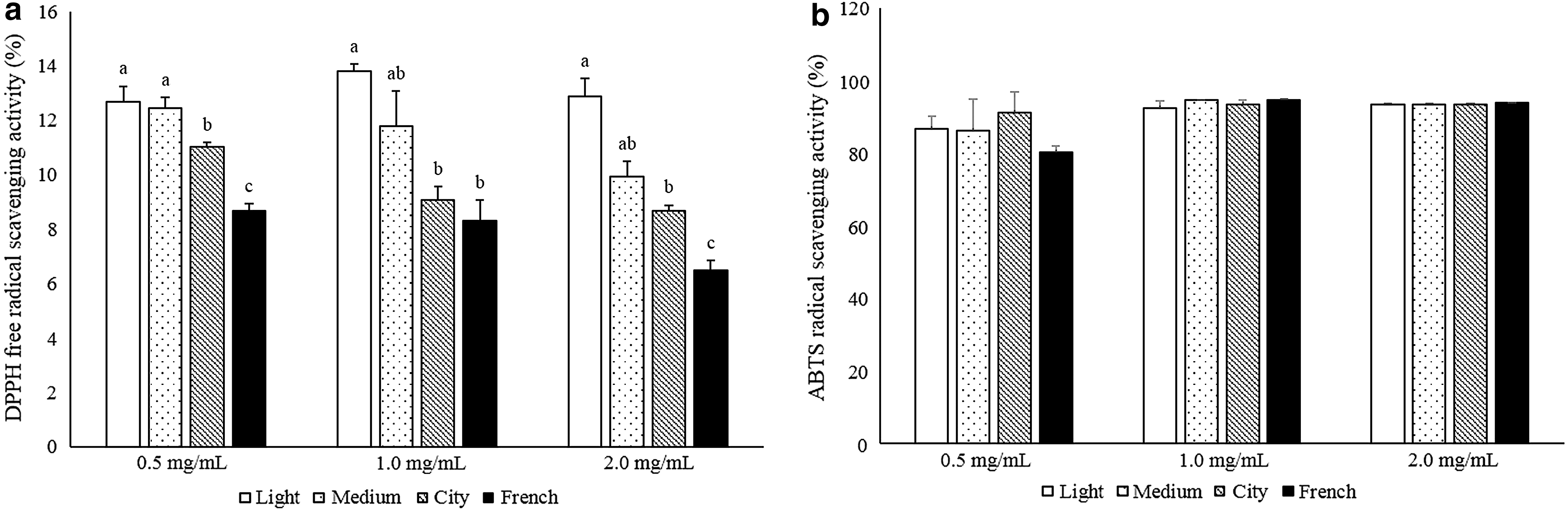
The DPPH test was used to measure cation radical scavenging effects. The extracts exhibited a DPPH cation radical scavenging activity with increasing concentrations in the range of 0.5 to 2 mg/mL. Each concentration of extract (0.5, 1.0, and 2.0 mg/mL) exhibited the highest DPPH scavenging effect in Light roasted coffee extract and lowest in French roasted coffee extract (Fig. 1a). The reduction capability of ABTS induced by antioxidants suggested that the extracts were free radical scavengers. The scavenging effects of coffee extracts from different roasting levels on ABTS radicals were compared. The ABTS test revealed no differences among coffee extracts with different roasting levels (Fig. 1b).
Cytotoxicity of coffee extracts in AML-12 cells and RAW 264.7 cells
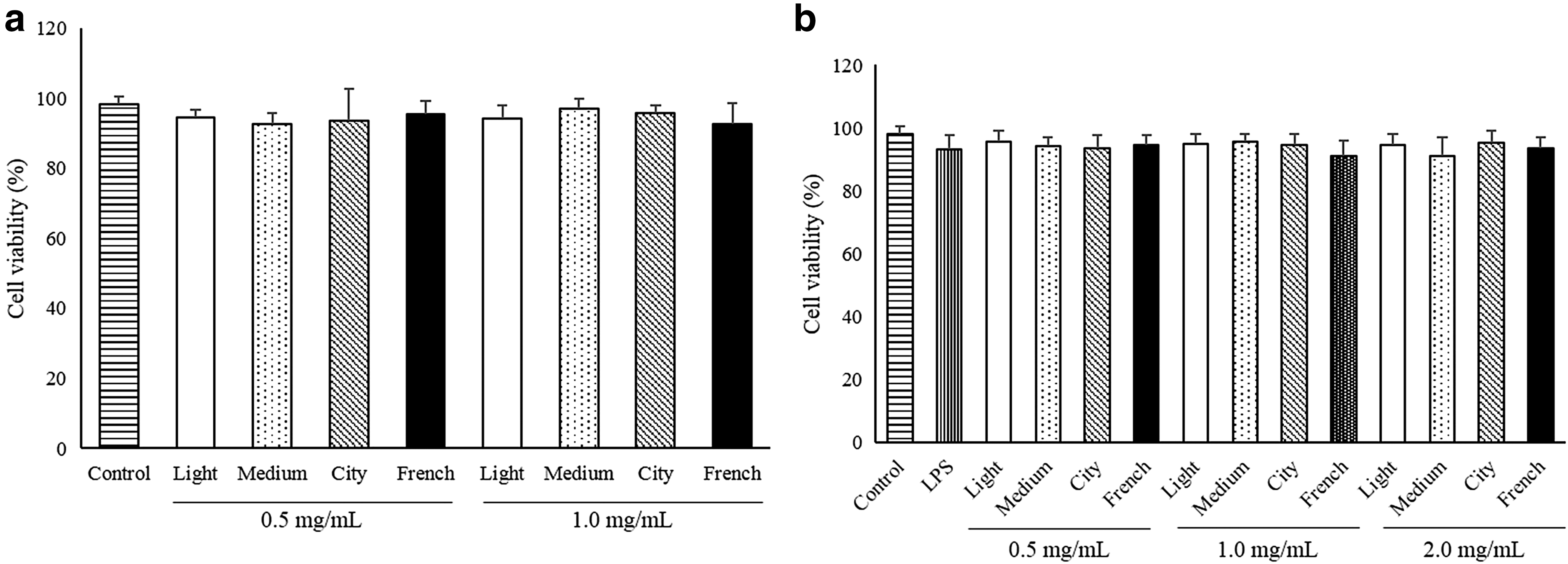
The MTT assay was conducted to evaluate the effects of coffee extracts on the viability of AML-12 and RAW 264.7 cells. Our data indicated that concentrations of 0.5, 1.0, and 2.0 mg/mL of coffee extracts were not cytotoxic to AML-12 cells and RAW 264.7 cells (Fig. 2). Cell viability was >80% in all groups and there was no significant difference among groups.
Effects of coffee extracts on the viability of AML-12
Intracellular GSH concentration in AML-12 cells treated with coffee extracts prepared with different roasting levels
Intracellular GSH is a physiological antioxidant defense system against oxidative stresses. Cells treated with coffee extracts with different roasting levels had increased intracellular GSH concentrations compared with the control group. Light, Medium, City, and French roast extracts in the 0.5 mg/mL treatment exhibited decreased intracellular GSH concentration with increased roasting degree (Fig. 3). However, at the extract concentration of 1.0 mg/mL, no difference between groups was evident with different roasting levels, and all were significantly higher than control.
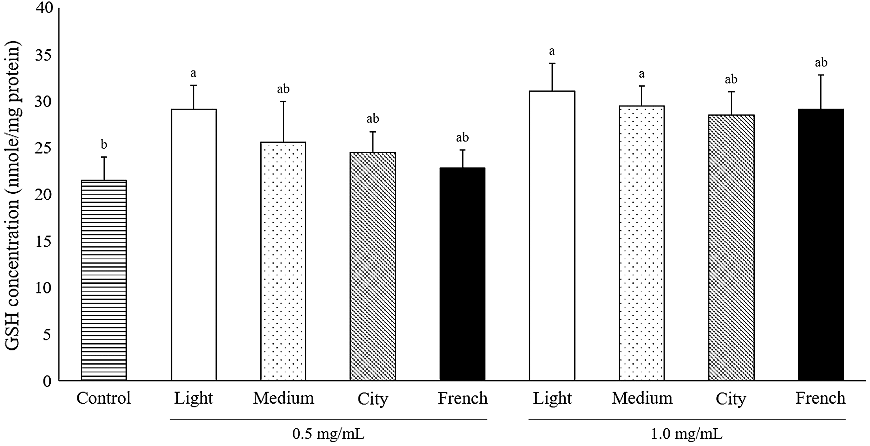
Effect of coffee extracts obtained from various roasting levels on intracellular GSH concentration in AML-12 cells. Different superscripts are significantly different (P < .05). Each bar represents the mean ± standard error (n = 3). GSH, glutathione.
Effects of coffee extracts with different roasting levels on intracellular GSH concentration in LPS-treated RAW 264.7 cells
To investigate the antioxidant property of coffee extracts with various roasting levels in LPS-challenged RAW 264.7 cells, intracellular GSH concentrations were measured. When RAW 264.7 cells were treated with LPS, intracellular GSH concentration was significantly decreased compared to the control (Fig. 4a). When the coffee extracts were treated, the lower GSH concentration was increased and these effects were negatively correlated with the roasting level. However, when 1 mg/mL of coffee extracts were used to treat LPS-challenged RAW 264.7 cells, all treatments showed increased intracellular GSH concentration without differences among different roasting levels.
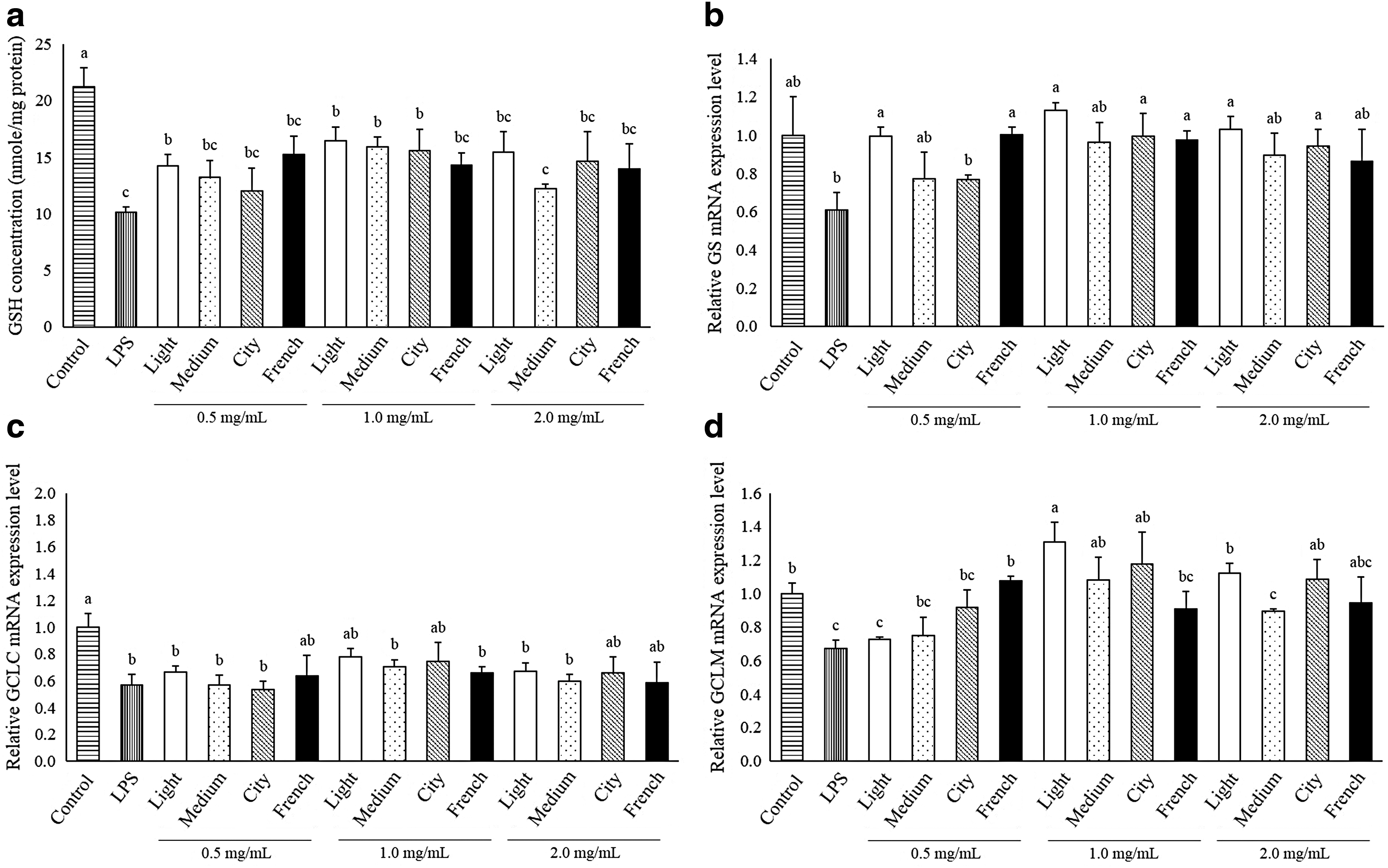
Effects of coffee extracts with different roasting levels on mRNA expression levels of GSH synthesizing enzymes in LPS-treated RAW 264.7 cells
To evaluate the mechanisms of increases in intracellular GSH concentration, qPCR was performed. The mRNA expression levels of the genes involved in GSH synthesis were significantly increased in some coffee extract-treated RAW 264.7 cells. Expression of GS mRNA, the key enzyme of GSH synthesis, in the Light roasted coffee extract-treated group was significantly increased at 0.5, 1, and 2 mg/mL compared with the LPS-treated group (Fig. 4b); however, it was decreased when the extracts of coffee with higher roasting levels were used. The mRNA expression levels of GCLC, which is rate-limiting enzymes of GSH synthesis and GCLM were also highest in cells treated with all three concentrations of Light roast coffee extract except for GCML mRNA expression treated with 0.5 mg/mL of Light roasted coffee extract (Fig. 4c, d).
Effects of coffee extracts with different roasting levels on mRNA expression levels of superoxide dismutase, catalase, and GSH peroxidase in LPS-treated RAW 264.7 cells
The mRNA expression levels of enzymes related to antioxidative defense systems were investigated to evaluate the effects of coffee extracts with different roasting levels. When the cells were treated with LPS, the mRNA expression levels of SOD1, SOD3, catalase, and GSH peroxidase decreased (Fig. 5). However, addition of coffee extracts increased the mRNA expression of those genes in the cells with LPS challenge. Especially, SOD3 and GSH peroxidase were significantly increased when Light roast coffee extract was used. However, the increased expression of both genes was negatively correlated with the roasting level.

Effect of coffee extracts obtained from various roasting levels on mRNA expression levels of antioxidant genes in LPS-treated RAW 264.7 cells.
Effects of coffee extracts with different roasting levels on mRNA expression levels of inflammatory factors in LPS-treated RAW 264.7 cells
To evaluate the anti-inflammatory effect of coffee extracts with different roasting levels, the mRNA expression levels of inflammatory factors were measured by qPCR. The mRNA expression level of TNF-α and interleukin 6 (IL-6) was increased in LPS-treated cells and decreased in the cells treated with all concentrations of Light roasted coffee extracts and some other different roasting levels of coffee extract (Fig. 6a, b). Light roasted coffee extract resulted in the lowest mRNA expression level of TNF-α and IL-6, and they increased as roasting levels were intensified. When RAW 264.7 cells were exposed to LPS, iNOS expression was increased compared with the control (Fig. 6c). When cells were treated with the coffee extracts, iNOS mRNA expression decreased usually in City and French degree of roasting. However, there was no difference among the coffee extracts, except for the treatments with 2.0 mg/mL Medium, City, and French roasting.
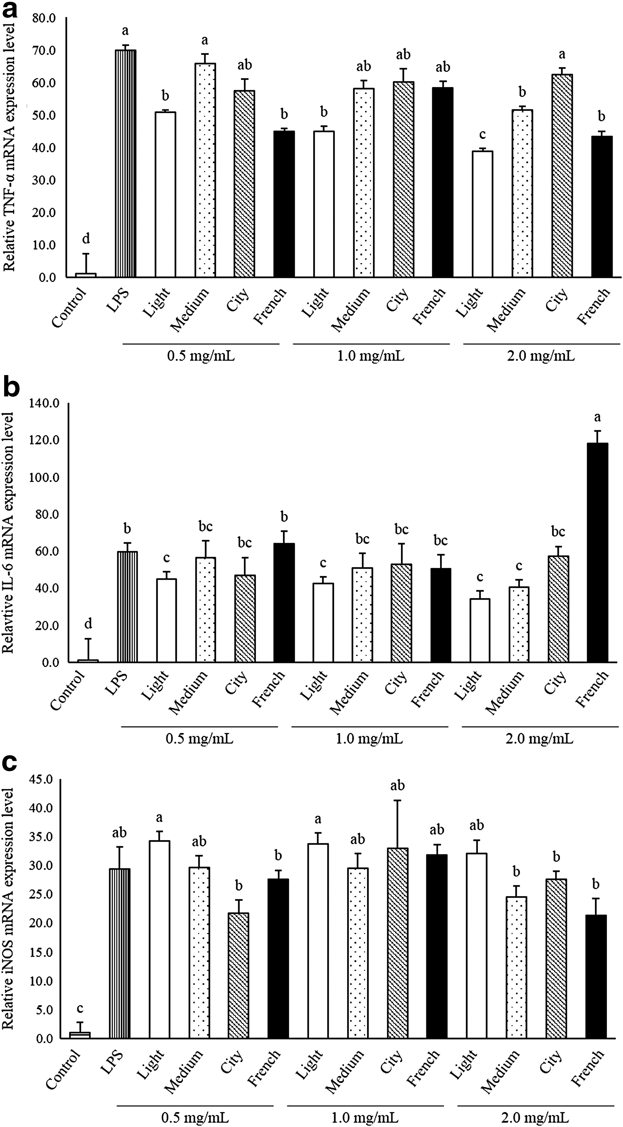
Effect of coffee extracts obtained from various roasting levels on mRNA expression levels of genes related to inflammatory system in RAW 264.7 cells.
Discussion
The antioxidant property of coffee extract prepared at different roasting levels has been studied. 26 –28 Relationship between levels of roasting and antioxidant property varies depending on the roasting conditions, extraction procedure, and antioxidant assay. The antioxidant property does not change linearly with the degree of roasting. 27 –29 Nonetheless, as the degree of roasting increases, the antioxidant property of coffee extract decreases. This study also investigated the antioxidant property of coffee extract with different roasting levels in vitro using a macrophage cell model. To the best of our knowledge, this is the first study to evaluate the antioxidant and anti-inflammatory properties of coffee extracts with different roasting degrees using actual cell models and will add valuable new knowledge about the balance between the biological benefits from coffee and the optimal taste from the proper level of roasting coffee beans.
Caffeine is the major component in coffee extract and has antioxidant property. Chlorogenic acid is another well-known efficient antioxidant in coffee extract; it was highest in Light roast coffee extract and highest with low roasting temperature and lowest in Dark roasted extract. Carbohydrates, protein, and chlorogenic acid are all decreased in coffee during the roasting process. 30 Currently, caffeine and chlorogenic acid content in coffee extracts with different roasting degrees were explored. Caffeine contents showed no differences among roasting levels, but chlorogenic acid content decreased as roasting degree increased (Table 2). As caffeine is thermostable, it is understandable that roasting degree was not influential, as has also been described before. 31,32 The decrease in chlorogenic acid during roasting could be explained by previous observations that polyphenolic compounds can be incorporated into melanoidins during roasting. 33 –35 The negative correlation between roasting level and chlorogenic acid concentration could be the result of the transformation of chlorogenic acid during roasting by the Maillard reaction.
Following the determination of caffeine and chlorogenic acid content, the antioxidant activity of each sample was evaluated. The antioxidant capacity of coffee is attributed to the presence of polyphenolic compounds, and it is well understood that roasting affects the antioxidant properties of coffee. It is well known that roasting greatly affects the chemical composition of coffee extract due to the high temperature during roasting process, 36,37 which can degrade compounds such as chlorogenic acid. 38,39 The effect of coffee roasting on the antioxidant properties of coffee extracts was investigated in several earlier studies; antioxidant capacity decreased in Dark roast coffee. 38,40,41 The antioxidant property of coffee extracts prepared with different roasting levels was also determined in this study. The best antioxidant activity was evident in Light roast coffee extract and the lowest in French roast coffee (Fig. 1a). These results indicate that the antioxidant property of coffee extracts is related to the amount of roasting, with a negative effect noted as the level of roasting increases. In the ABTS assay, no significant difference was observed between antioxidant properties of coffees affected by different roasting levels (Fig. 1b). The fluctuations in the antioxidant property imply that prolonged roasting at high temperature would cause a decrease in its antioxidant capacity. The differences between the DPPH and ABTS assay may be ascribed to the varying reactivity of radical to the components of coffee extracts. Our results are in agreement with previous studies, which observed that the antioxidant capacity of roasted coffee decreases compared to that of green coffee beans. 42,43
Due to the changes in antioxidant property of coffee extract by different processing, the biological antioxidant property of differently roasted coffee extracts in actual live system is important. In this study, we used different coffee extracts to treat AML-12 mouse hepatocyte cell line. There was no effect of coffee extracts on cell viability in AML-12 cells. Increase of intracellular GSH concentration was highest in Light roast coffee extract and decreased with more intensive roasting procedures (Fig. 3). To confirm the molecular pathway of intracellular GSH reduction, the genes related to GSH synthesis were analyzed. GS is the key enzyme of GSH synthesis, and GCLC and GCLM are rate-limiting enzymes of GSH synthesis. Although the antioxidant properties of coffee extracts decreased, roasting levels increased cellular GSH concentration, and gene expression levels involved in GSH synthesis were not significantly affected by the roasting level (Fig. 4). HPLC analysis for this study revealed that changes in chlorogenic acid concentration did not affect the biological antioxidant property (Table 2). In previous studies, caffeine, one of the most dominant compound in coffee extracts, was shown to contribute to protein relocalization of nuclear factor-like 2 (Nrf2) protein, a key transcription factor associated with antioxidant systems in cell models. 44,45 One of the enzymes whose expression is regulated by Nrf2 is GCLC, the first enzyme in the biosynthetic pathway of GSH. 46 Thus, our results suggest that the tested coffee extracts improved the cellular redox status by increasing the levels of GSH by caffeine. These results confirmed that intensified roasting negatively affects the antioxidant properties of coffee extracts and biological antioxidant properties in cells, not by decreasing cellular GSH-activating systems, but by decreasing the availability of other molecules with antioxidant properties, such as chlorogenic acid.
An epidemiological study 47 and this study indicate that coffee is associated with decreased oxidative stress. Inflammation is closely related to oxidative stress. Reactive oxygen and nitrogen species are involved in the redox regulation of cell functions. Oxidative stress is increasingly recognized as a major upstream component in the signaling cascade involved in inflammatory responses and stimulation of adhesion molecules and chemoattractant production. 48 Thus, antioxidant compounds have been accepted as anti-inflammatory agents. After the investigation of the biological antioxidant property of coffee extract with various roasting levels in AML-12 cells, we used LPS-treated RAW 264.7 cells to evaluate the effect of coffee extracts on an inflammatory model. A wide range of concentrations of LPS induced oxidative stress in macrophages of mice. There was no effect of coffee extracts on cell viability in LPS-treated RAW 264.7 cells. As GSH is a critical indicator of oxidative stress, GSH concentrations should be measured in LPS-challenged macrophage cells to determine oxidative stress and assess the anti-oxidative activity of coffee extracts. In this study, decreases in GSH concentration and mRNA expression of GSH-synthesizing genes were observed in LPS-treated cells (Fig. 4). Furthermore, coffee extracts attenuated the reduced intracellular GSH concentration and mRNA expressions of GSH-synthesizing enzymes. The increase in intracellular GSH concentration and mRNA expressions negatively correlated with the level of roasting, similar to a previous study using AML-12 cells. The antioxidant enzymes superoxide dismutase (SOD), catalse (CAT) and glutathione peroxidase (GPx) play important roles in maintaining the redox homeostasis in mammalian cells. These genes constitute a family of intracellular antioxidant enzymes that lower oxidative stress and play a critical protective role in detoxification of ROS produced during inflammation. 49 –51 Oxidative stress induced in RAW 264.7 cells by LPS depleted the mRNA expression levels of those genes; however, the effects were not dramatic compared with GSH concentration and mRNA expression levels of GSH-synthesizing genes (Fig. 5). This may reflect that compounds with antioxidant properties generally increase the mRNA expression of antioxidant-related enzymes, but these expressions can be downregulated in some antioxidant-treated cells because the compounds may have directly ameliorated the prevailing oxidative stress. 52,53 These results demonstrate that the antioxidant property of coffee extract is effective both in normal hepatocytes and LPS-challenged macrophages.
Previous studies have demonstrated anti-inflammatory effects of coffee in vitro and in vivo. However, little has been known of the relationships of roasting levels of coffee and its anti-inflammatory functions. Immunocytes stimulated with LPS are known to release cytokines as part of the immune response. Cytokines like TNF-α and IL-6 are increased not only in immunostimulatory responses but also when oxidative stress occurs. 54 –56 iNOS produces proinflammatory mediators, such as NO, at inflammatory sites. The most prominent phenomenon in the process of inflammation is the increase of NO production and proinflammatory cytokines. 57,58 In this study, we showed that coffee extracts ameliorated the inflammatory markers in LPS-treated cells (Fig. 6). This anti-inflammatory effect displayed a similar pattern as the antioxidant property, which was negatively correlated with the level of roasting. This means coffee extracts have anti-inflammatory effects in biological systems and this effect is decreased when coffee is highly roasted. Caffeine can decrease LPS-induced TNF-α and IL-6 release in a dose-dependent manner. 59 In addition, caffeine has an anti-inflammatory effect by the regulation of NFκB activation, which promotes the expression of inflammatory genes, including iNOS, COX2, and cytokines. 60 Chlorogenic acid, the major component in coffee extract, has a strong anti-inflammatory effect by decreasing the secretion of proinflammatory cytokine IL-6. 61 It also attenuates TNF-α production in LPS-stimulated RAW 264.7 macrophages by suppressing COX2 expression due to the attenuated activation of NFκB signaling pathways. 62 Moreover, not only its anti-inflammatory effect but also its antioxidant properties suppress the production of several cytokines and chemokines by suppressing COX2 expression by attenuating the activation of NFκB signaling pathways. 63 It seems that the differences in the anti-inflammatory effect of coffee extracts with different roasting levels are likely due to the differences of caffeine and chlorogenic acid contents in coffee extracts.
In conclusion, coffee extracts have antioxidant properties in AML-12 cells. This involves the activation of GSH synthesis and the antioxidative enzyme system. Further experiments using LPS-induced murine macrophages revealed that coffee extracts protect cells against oxidative stress by enhancing the content of the antioxidant GSH and stimulating expressions of the genes related with the cellular antioxidation system. Also, coffee extracts decreased the expression of proinflammatory cytokines and inflammatory mediators in LPS-stimulated RAW 264.7 cells. However, different roasting levels may dilute those effects by decreasing the concentrations of key compounds during the roasting procedures. Therefore, an appropriate method of roasting should be used to maintain the best coffee flavor with optimal beneficial functions of coffee.
Footnotes
Acknowledgments
This research was supported by a grant (714001-07) from the Center for Industrialization of Natural Neutraceuticals through the Agriculture, Food and Rural Affairs Research Center Support Program, Ministry of Agriculture, Food and Rural Affairs, Republic of Korea.
Author Disclosure Statement
No competing financial interests exist.