
Abstract
The number of diabetic patients worldwide is increasing, and complications such as stroke and cardiovascular disease are becoming a serious cause of death. Diabetes mellitus is classified into two types according to the etiopathogenic mechanism and insulin dependence. Type 1 diabetes (T1D), an insulin-dependent diabetes mellitus, is caused by damage and destruction of pancreatic β cells that produce insulin. It is a disease that is characterized by hyperglycemia and hypoinsulinemia. Aronia berry has been used as a medicinal food in Europe. Aronia contains a variety of ingredients such as polyphenols, anthocyanins, flavonoids, and tannins. Especially, anthocyanin content in aronia berry is known to be much higher than in other plants and berries. It is known for exerting antioxidant, anti-inflammation, and anti-aging effects. Therefore, this study was conducted to investigate the effects of aronia berry extract intake in multiple low-dose streptozotocin (STZ)-induced T1D and to confirm the functional properties of aronia berry. ICR mice (6-week male) were divided into four groups: control (normal control group), STZ (100 mg/kg of STZ-induced T1D group), AR 10 (STZ with oral administration of aronia 10 mg/kg), and AR 100 (STZ with oral administration of aronia 100 mg/kg). Afterward, STZ was injected in a single dose to induce T1D, and the extract was orally administered daily. Dietary intake and body weight were measured twice a week. We confirmed that aronia berry has the effect of decreasing the increase of blood glucose level and also has the protection effect of pancreas β cell (RINm5F cell). This study confirms the anti-diabetic activity of aronia berry, and it can be expected to increase the utilization according to the results.
Introduction
T
Diabetes is a metabolic disorder that is associated with impaired insulin secretion by pancreatic β cells. Also, hyperglycemia is caused by insulin resistance to peripheral tissues, as well as protein, lipid, and electrolyte metabolism. 3 Diabetes is caused by a combination of genetic factors and environmental factors, such as eating habits, lack of exercise, and stress. If the diabetes is long lasting or the treatment is not done properly, blood glucose cannot be taken up by body tissues, the glycogen in the organ is decomposed, and the energy metabolism of the carbohydrate, protein, and fat becomes abnormal. If symptoms of diabetes continue, numerous complications such as vascular disease and pulmonary tuberculosis will develop. 4 Further, it is also known that protein deficiency and decreasing lower hemoglobin, hematocrit, iron, and zinc statuses occur. 5 Currently, diabetes is the sixth leading cause of the death of Korean people. 6 Dietary therapy, exercise, and drug therapy can regulate blood glucose levels to prevent complications. However, there is no way to fundamentally cure diabetes, so interest in natural plant materials that can mitigate pathologies with fewer side effects is increasing. 7 Various drugs such as sulfonylureas, biguanides, α-glucosidase inhibitors, etc. are sold to treat diabetes. However, depending on the medicine, there are side effects such as hypoglycemia, hepatotoxicity, weight gain, abdominal bloating feeling, and diarrhea; thus, the use of these medicines can be limited. For this reason, research into natural vegetable materials with little side effects is actively under way.
Recently, there are various reports about natural plant material such as raw soy flour 8,9 and its proteins 10,11 and bioactive components, 12,13 bean sprouts extract, 14 ginseng and red ginseng extracts, 15 –17 and Allium hookeri root extract 18 to lower blood glucose levels and improve lipids. However, anti-diabetic effects of aronia berry have not been elucidated and tried.
Aronia berry (Aronia melanocarpa), also known as black choke berry, is a deciduous shrub of the Rosaceae family and grows naturally in North America. Recently, it is cultivated in Korea. Aronia berry mainly contains polyphenols such as anthocyanins, flavonoids, and tannins; it also contains a large amount of natural antioxidant and is mentioned as a functional food. In addition to antioxidative properties, aronia berry is known to have efficacy such as protection of the stomach, anti-inflammation, immunomodulation, etc. 19 –24 Also, the fruit juice of aronia berry has hypoglycemic effects in streptozotocin (STZ)-induced diabetic rats, such as regulating plasma glucose and plasma triglyceride. 25 However, aronia berry extract has not been studied in the STZ-induced type1 diabetes (T1D) in vivo model.
Therefore, in this study, aronia berry extract is used, which is mainly used in the food field. We evaluated whether aronia berry extract can protect pancreatic β cells and lower blood glucose level in vivo.
Materials and Methods
Reagents
STZ, (3-(4,5-Dimethylthiazol-2-yl)-2,5-diphenyltetrazolium bromide (MTT), sulfanilamide, sodium nitrate, eosin, and N-(1-naphthyl) ethylenediamine dihydrochloride (NEDHC) were purchased from Sigma-Aldrich (St. Louis, MO, USA). Phosphoric acid and xylene were purchased from JUNSEI Chemical Co. (Tokyo, Japan). Hematoxylin was purchased from MUTO PURE CHEMICALS (Tokyo, Japan). Easy-Blue was purchased from iNtRON Biotechnology (Seongnam, South Korea). Complementary DNA (cDNA) synthesis kit was purchased from BioFACT (Daejeon, South Korea). Inducible nitric oxide synthase (iNOS) antibody was purchased from Cell Signaling Technology (Danvers, MA, USA). β-actin antibody was purchased from Santa Cruz Biotech (Santa Cruz, CA, USA).
Preparation of aronia berry extract
Aronia berries were purchased from a joint market in Sunchang Province (Jeonbuk, South Korea). They were washed twice with distilled water and dried. Aronia berry extract was prepared by decocting with 70% ethanol for 1 h 30 min at 80°C. The solution in ethanol was then filtered and allowed to evaporate by using a rotary evaporator at a temperature of 40–45°C. The extract was diluted in 0.9% saline and filtered through a 0.22 μm syringe filter (HYUNDAI Micro, Seoul, South Korea).
Type 1 diabetes in vivo model
All experimental protocols (CBNU2016-0016) were approved by the Committee on the Care of Laboratory Animal Resources, Chonbuk National University. All experiments were conducted in accordance with the Guide for the Care and Use of Laboratory Animals. Five-week-old healthy male ICR mice were obtained from Samtako Experiment Animal (Samtako Bio, Gyeonggi, South Korea). Mice were acclimated for 1 week before the experiments and had free access to standard pellets (Samtako Bio) and water. They were housed under a 12 h light/dark cycle at a constant temperature of 23 ± 2°C and a relative humidity of 60 ± 5%. The mice were assigned to one of four groups (n = 8) and housed separately per group. Overnight-fasted mice (weighting 25–30 g) were injected with a single intraperitoneal dose of STZ (80 mg/kg), which was freshly dissolved in sodium citrate buffer (pH 4.5). Their fasting blood glucose levels were measured 7 days after the injection, by using an Accu-Chek glucometer (Roche Diagnostics GmbH, Germany). Mice with fasting blood glucose concentrations of 250 mg/dL and higher were considered diabetic and selected for the study.
Measurement of insulin in the serum
The serum insulin level was measured by Insulin ELISA Kit (EMD Millipore, Billerica, MA, USA). Briefly, mice blood serums were prepared at the end of the diabetes experiment. To prepare serum, whole blood was directly drawn into a 1.5 mL tube, allowed to blood clot at room temperature for 30 min., and promptly centrifuged at 3000 g for 15 min at 4°C. Pre-coated plate wells were washed three times with 300 μL of wash buffer. Next, 10 μL of assay buffer was added to each of the wells. Then, 10 μL of samples in duplicates was added to the wells. Next, 80 μL of detection antibody was added to all wells and incubated for 2 h at room temperature on an orbital microtiter plate shaker set to rotate at moderate speed. After the reaction, each well was washed three times, and 100 μL enzyme solution was added and held for 30 min at room temperature. Wells were washed six times, 100 μL of substrate solution was added, and absorbance was read at 450 nm in a plate reader.
Serum analysis
Mice blood serum was prepared at the end of the diabetes experiment. To prepare serum, whole blood was directly drawn into a 1.5 mL tube and blood was allowed to clot at room temperature for 30 min. Then, it was promptly centrifuged at 3000 g for 15 min at 4°C. Aspartate aminotransferase (AST), alanine aminotransferase (ALT), triglyceride (TG), high-density lipoprotein (HDL)-cholesterol, and low-density lipoprotein (LDL)-cholesterol were analyzed by Seoul Clinical Laboratories (Yongin, South Korea).
Cell culture
The RIN-m5F rat insulinoma pancreatic β-cells were purchased from ATCC Global Bioresource Center. RIN-m5F cells were grown in RPMI-1640 medium supplemented with 10% fetal bovine serum and penicillin/streptomycin (100 U/mL and 100 μg/mL, respectively). All cultures were maintained at 37°C in a 5% CO2 humidified cell culture incubator.
Measurement of nitric oxide level
5 × 10⁵ RIN-m5F cells were pre-treated with aronia berry extracts (0.01, 0.1, or 1 mg/mL) for 3 h. After that, they were treated with interleukine-1β (IL-1β) (1 U/mL) and interferon-γ (IFN-γ) (100 U/mL). After 24 h, the supernatants were collected for nitric oxide (NO) determination. To measure nitrite, an equal volume of Griess reagent (1% sulfanilamide/0.1% naphtylethyenediamine dihydrochloride in 2.5% H3PO4) was mixed with cell culture supernatant at room temperature for 10 min. Nitrite concentration was determined by measuring the absorbance at wavelength of 540 nm by using the VersaMax Microplate Reader. NaNO2 was used to obtain the standard curve.
RNA isolation and real-time RT-PCR
Total cellular RNA was isolated from RINm5F cells by using easy-BLUE reagent Kit (iNtRON Biotechnology). Total RNA was used as a template for first-strand cDNA synthesis by using a Power cDNA Synthesis Kit (Bio FACT, Daejeon, South Korea) according to the manufacturer's instructions. The transcription levels of genes were determined with a StepOnePlus Real-Time PCR System (Applied Biosystems, Foster City, CA, USA). The relative gene expression was calculated by using the comparative CT method with StepOne Software v2.1 (Applied Biosystems). The expression of glyceraldehyde 3-phosphate dehydrogenase (GAPDH) messenger RNA (mRNA) was used as an endogenous control. We used COX-2 forward primer 5′-TGCTCACTTTGTTGAGTCATTCAC-3′ and COX-2 reverse primer 5′-CATTCCTTCCCCCAGCAA-3′ for COX-2, iNOS forward primer 5′-TGTGCTAATGCGGAAGGTCAT-3′ and iNOS reverse primer 5′-CGACTTTCCTGTCTCAGTAGCAAA-3′ primer for iNOS, and 5′-TCACCACCATGGAGAAGGC-3′/5′-GCTAAGCAGTTGGTGGTGCA-3′ primers for GAPDH as an internal control.
Histological analysis
A cross-section of the liver (1 cm3) was fixed in 10% paraformaldehyde solution. It was then cut into small fragments, dehydrated in ethanol series (70–100%), cleared in xylene, and embedded in paraffin. The fragments were sliced into 4 μm-thick sections and stained with hematoxylin and eosin. The sections were analyzed by using an optical microscope coupled to a camera to capture images with a 40 × magnification. Images were analyzed with Image Pro Plus (Media Cybernetics, MA, USA).
Statistical analysis
Results are shown as a summary of data from at least three experiments. All results are presented as the mean ± standard error of the mean. Results were analyzed by using Graph Pad Prism version 5.0 program (Graph Pad Software, Inc, La Jolla, CA, USA). One-way analysis of variance with Tukey's post hoc test was used to determine statistically significant differences. P value <0.05 was considered significant.
Results
Effects of aronia berry extract on body weight and blood glucose in STZ-induced T1D mice
STZ-induced diabetic mice displayed a remarkable decrease in body weight compared with the control group on day 31 (32.71 ± 1.18 g in STZ group, 35.59 ± 1.88 g in control; P < .05). The decreased body weight was significantly increased in the group administered 100 mg/kg of aronia (34.34 ± 1.54 g in AR 100 mg/kg group, 32.71 ± 1.18 g in STZ; P < .05) (Fig. 1A). The serum glucose level in STZ-induced diabetic mice was markedly higher than the control group on the 31st day (486.60 ± 81.94 mg/dL in STZ group, 175.67 ± 10.60 in control; P < .05). But it is significantly decreased in the 100 mg/kg aronia berry administered group (208.60 ± 31.05 in AR 100 mg/kg group, 486.60 ± 81.94 mg/dL in STZ; P < .05) (Fig. 1B).
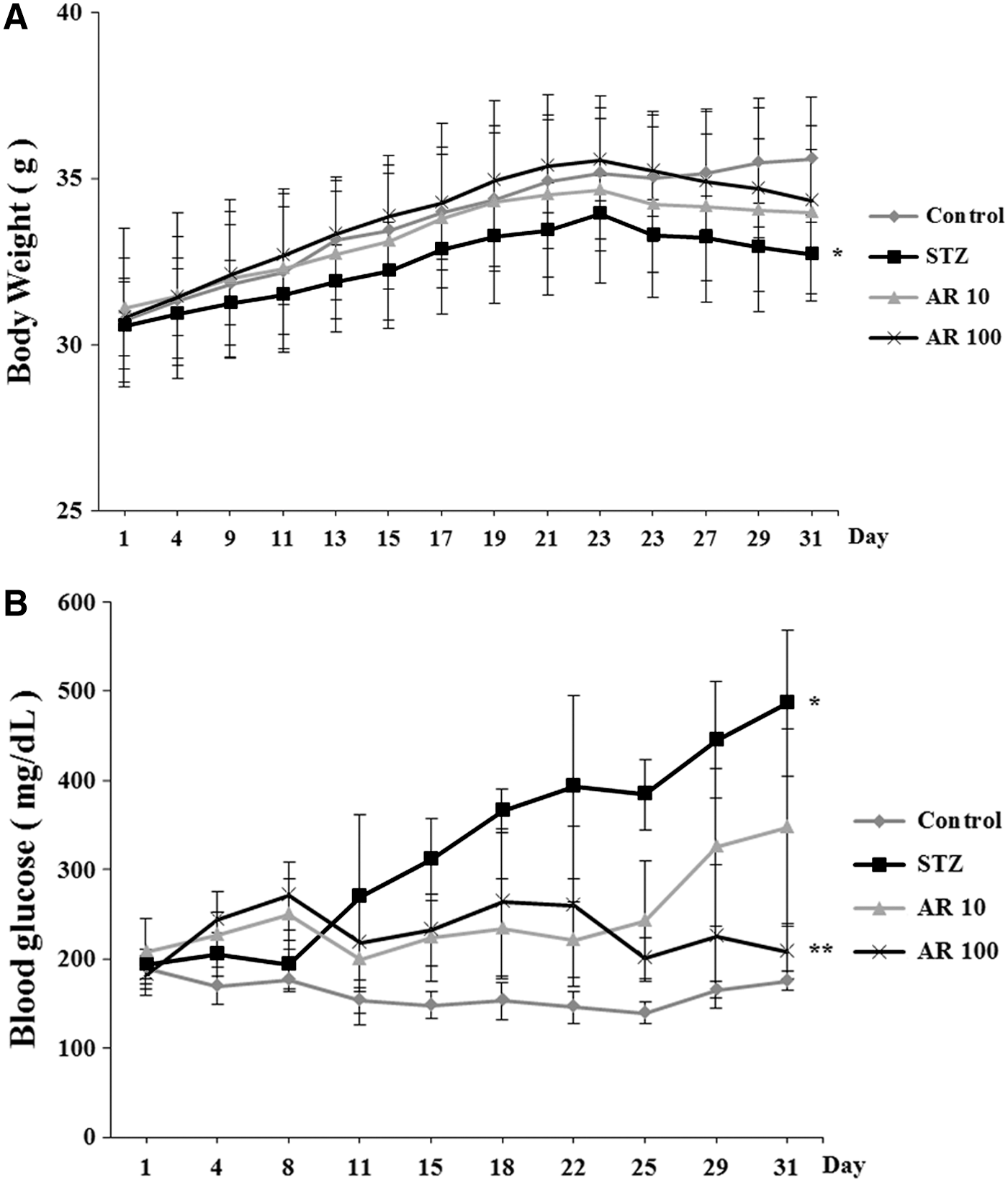
Effects of aronia extract on body weight and blood glucose in STZ-induced T1D mice. Diabetes was induced by an intraperitoneal injection of STZ (80 mg/kg) on the day 1. Control: untreated diabetic control, STZ: STZ (80 mg/kg)-induced diabetic group, AR 10: aronia extract (10 mg/kg)-treated diabetic group, and AR 100: aronia extract (100 mg/kg)-treated diabetic group.
Effect of aronia berry extract on insulin levels in STZ-induced T1D mice serum
The insulin level in mice serum was significantly lower in STZ-induced diabetic mice compared with non-treated mice (1.34 ± 0.54 ng/mL; STZ, 2.66 ± 0.36 ng/mL; control), but treatment with aronia berry extract significantly increased serum insulin levels, especially aronia berry 100 mg/kg dose (2.50 ± 0.39 ng/mL; AR 100 mg/kg, 1.34 ± 0.54 ng/mL; STZ) (Fig. 2).
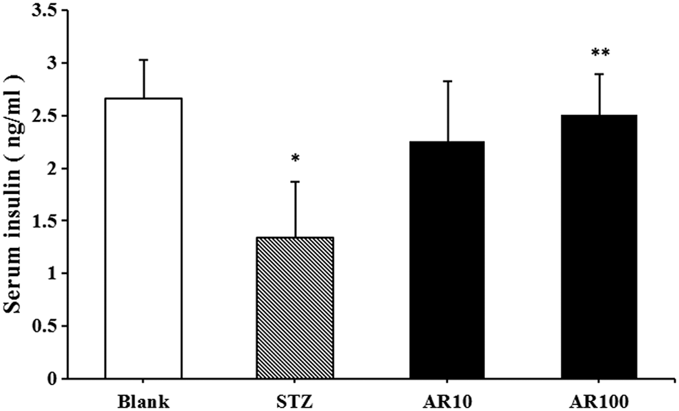
Effects of aronia extract on insulin level in STZ-induced T1D mice. Diabetes was induced by an intraperitoneal injection of STZ (80 mg/kg) on day 1. Control: untreated diabetic control, STZ: STZ (80 mg/kg)-induced diabetic group, AR 10: aronia extract (10 mg/kg)-treated diabetic group, and AR 100: aronia extract (100 mg/kg)-treated diabetic group. Serum insulin level was measured by Insulin ELISA Kit. Values represent mean ± SEM. n = 8. Data were analyzed by Tukey's post hoc test (*P < .05 vs. control and **P < .05 vs. STZ treatment).
Effects of aronia berry extract on lipid level in STZ-induced T1D mice
Diabetes leads to imbalances in lipid levels in serum. 26 Transaminases such as AST and ALT in mice serum were not affected by aronia berry extract administrations (Fig. 3A, B). HDL-cholesterol level in mice serum was decreased by STZ stimulation; however, it was increased to a normal level by aronia berry administration (Fig. 3C). The levels of LDL-cholesterol and TG increased in STZ-induced diabetes mice. But administration of aronia berry extract resulted in a decrease of LDL-cholesterol and TG normally (Fig. 3D, E).
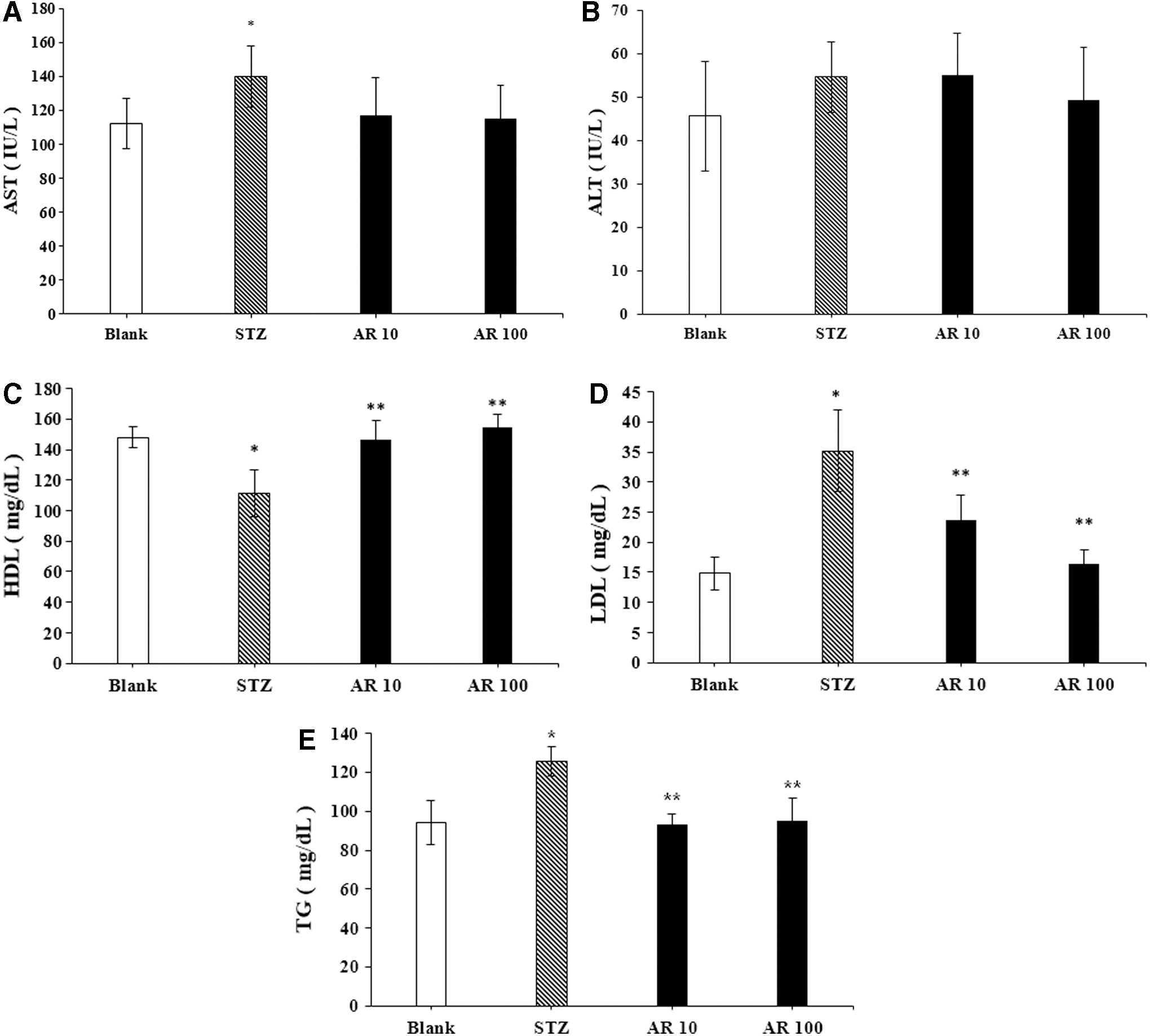
Effects of aronia extract on lipid level in STZ-induced T1D mice. Diabetes was induced by an intraperitoneal injection of STZ (80 mg/kg) on the day 1. Control: untreated diabetic control, STZ: STZ (80 mg/kg)-induced diabetic group, AR 10: aronia extract (10 mg/kg)-treated diabetic group, and AR 100: aronia extract (100 mg/kg)-treated diabetic group.
Effects of aronia berry extract on NO production in RINm5F cells
To determine the effects of aronia berry extract on pancreatic β cells, first we evaluated cell viability. MTT assay was performed to examine the effect of aronia berry extract in RINm5F cells. Cells were treated with aronia berry extract (0.001, 0.01, 0.1, or 1 mg/mL) for 24 h, and cell viability was measured. There was no difference in cell viability between non-treatment and aronia berry extract treatment groups (Fig. 4).
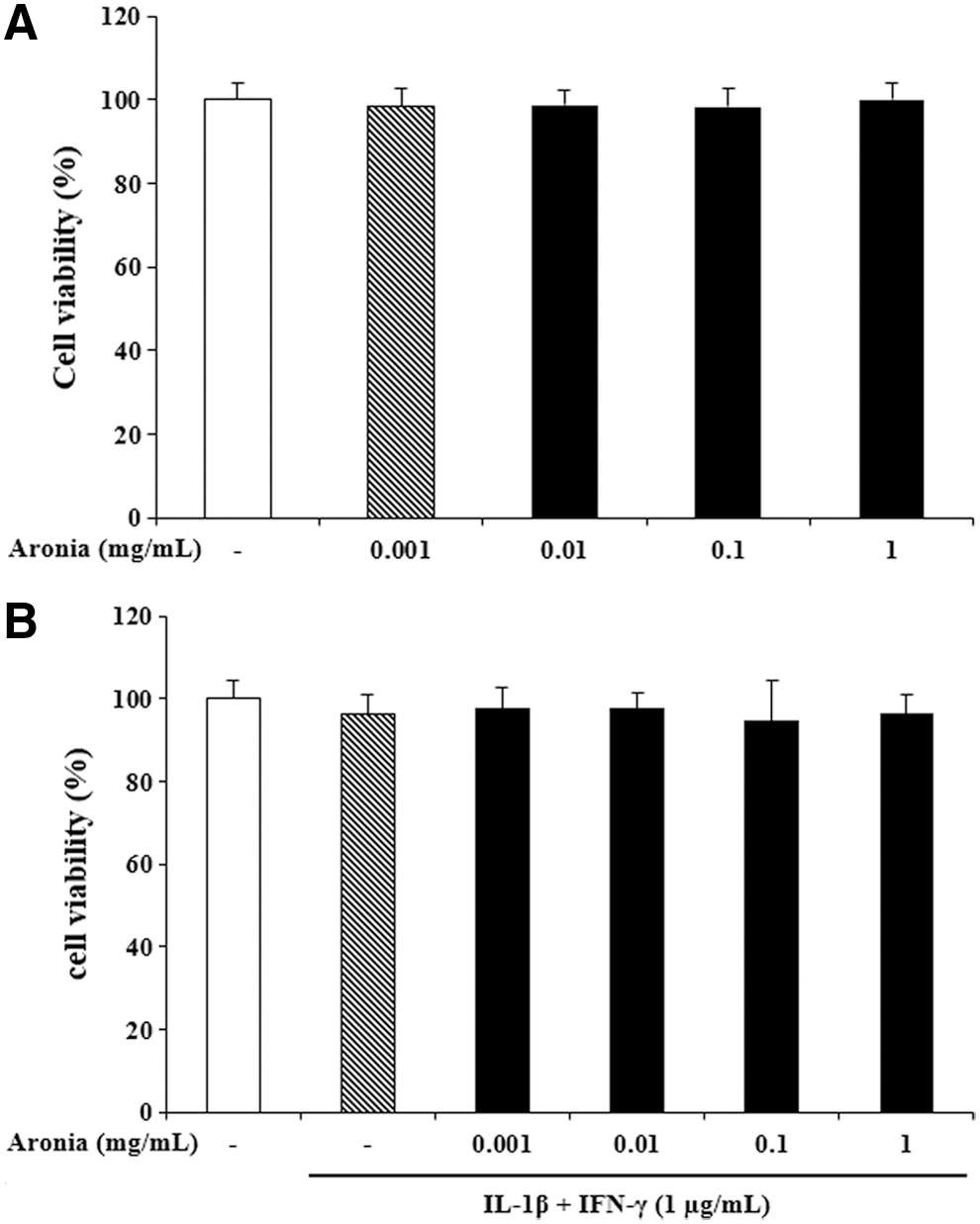
Effects of aronia extract on cell viability in RINm5F cells.
Excess formation of NO can lead to oxidative damage to cell function. A good balance of oxidative factors is required for maintaining a healthy pancreatic condition. 27 After RINm5F cells were pre-treated with aronia berry extract (0.001, 0.01, 0.1, or 1 mg/mL) for 4 h, the IL-1β and IFN-γ were costimulated for 48 h. NO production was significantly increased by cytokine stimulation compared with non-treatment (19.25 ± 0.39 μM; cytokine, 4.43 ± 0.60 μM; non-treatment). But increased NO production was decreased by aronia berry extract treatment, especially a 1 mg/mL dosage (19.25 ± 0.39 μM; cytokine, 17.15 ± 0.48 μM; aronia 1 μg/mL, 17.16 ± 0.36 μM; aronia 10 μg/mL, 14.91 ± 0.35 μM; and aronia 1 mg/mL) (Fig. 5).

Effects of aronia extract on NO production in RINm5F cells. NO production of aronia berry extract was determined by Griess assay in RINm5F. RINm5F cells were pre-treated with aronia berry extract for 4 h and then stimulated with cytokines for 24 h. Absorbance was measured at 540 nm. Values represent mean ± SEM. Data were analyzed by Tukey's post hoc test (*P < .05 vs. non-treatment and **P < .05 vs. cytokines treatment). IFN-γ, interferon-γ; IL-1β, interleukine-1 β; NO, nitric oxide.
Effect of aronia berry extract on tissue injury in STZ-induced T1D mice
Histological changes in liver tissue between STZ-injection and control group were observed. Then, we examined the effect of aronia berry treatment. Aronia berry extract treatment attenuated these effects induced by STZ-injection (Fig. 6A, B). In pancreatic tissue, it could also be observed that the STZ-induced group did not maintain pancreas morphology but the aronia berry-treated group maintained the round shape of the pancreas (Fig. 6C).
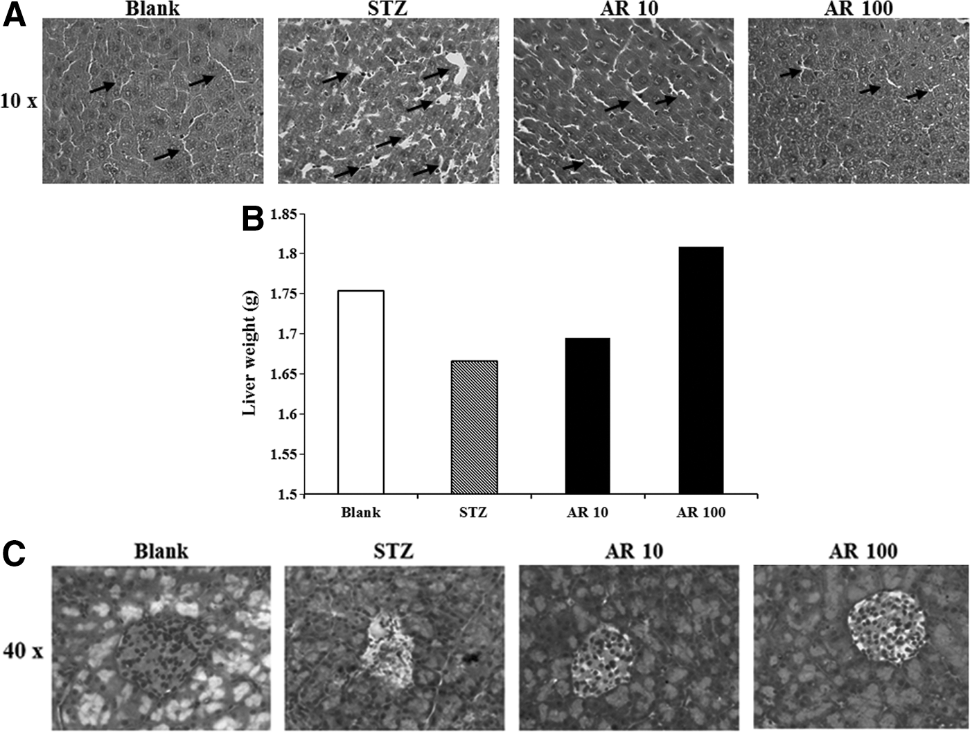
Effects of aronia extract on tissue injury in STZ-induced T1D mice. Diabetes was induced by an intraperitoneal injection of STZ (80 mg/kg) on day 1. Blank: untreated diabetic control, STZ: STZ (80 mg/kg)-induced diabetic group, AR 10: aronia extract (10 mg/kg)-treated diabetic group, and AR 100: aronia extract (100 mg/kg)-treated diabetic group.
Effects of aronia berry extract on inflammatory protein levels in RINm5F cells.
We investigated the inhibitory effects of aronia berry on cytokine-induced mitogen-activated protein kinases (MAPKs) and nuclear factor-κB (NF-κB) expression (Fig. 7). Cells pretreated with aronia berry for 1 h were stimulated with cytokines, and protein expression levels were measured by Western blot analyses. As shown in Figure 7, aronia berry significantly inhibited MAPKs and NF-κB protein expression in a dose-dependent manner in cytokine-stimulated RINm5F cells. These results support that aronia berry is correlated with inhibited MAPKs and NF-κB expression in cytokine-stimulated RINm5F cells. These results support the protective effect of aronia berry against cytokine-induced cell death.
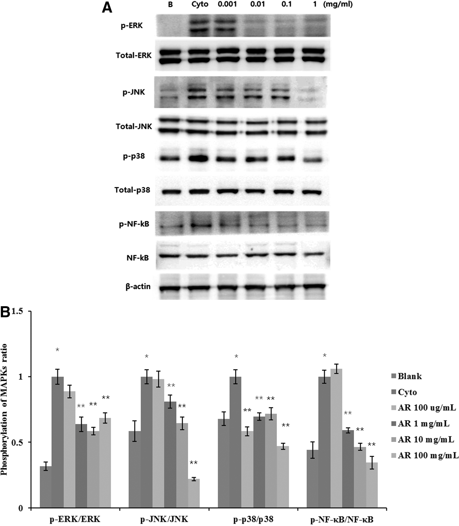
Effects of aronia extract on inflammatory protein levels in RINm5F cells.
Effects of aronia berry extract on inflammatory mRNA expression in RINm5F cell line
We determined the mRNA expression level of COX-2 and iNOS after aronia berry extract treatment by real-time RT-PCR. The results showed that aronia berry extract significantly suppressed the mRNA expression levels of COX-2 and iNOS in cytokine-induced RINm5F cells (Fig. 8).

Effects of aronia extract on inflammatory mRNA expression in RINm5F cells. mRNA expression was measured by real-time RT-PCR. RINm5F cells were pre-treated with aronia berry extract for 4 h and then stimulated with cytokines for 24 h.
HPLC analysis of aronia
To investigate the effective constituents of aronia berry, the HPLC fingerprint of aronia was analyzed. Gallic acid, chlorogenic acid, quercetin, and kaempferol were detected in aronia berry as constituents of aronia berry (Fig. 9).
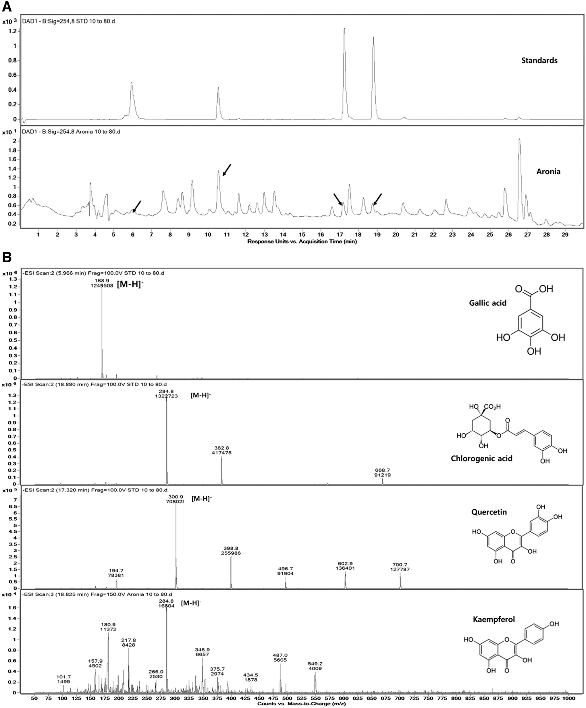
HPLC chromatogram of standards and aronia
Discussion
Type 1 diabetes (T1D), a chronic autoimmune disorder, is characterized by high blood glucose levels that cause serious complications. 28 The chronic high blood glucose levels are associated with serious damage, dysfunction, and failure of various organs. 29 The severity of T1D leads to microvascular disorders such as coronary artery and cerebrovascular diseases. 30 Thus, new types of remedies are needed to control blood glucose in T1D patients. This study was performed to elucidate the potential effect of aronia berry on the development of T1D. We used T1D models in vivo and in vitro.
STZ is a valuable agent for experimental induction of diabetes. Insulin, which selectively destroys pancreatic β cells, gradually decreases in STZ-administered mice and glucose in blood does not enter tissue. The glucose excreted into blood is increased by the action of glucose in the liver. 31 Insulin deficiency will lead to decreased activity of lipoprotein lipase and increased mobilization of free fatty acids from peripheral fat depots. The STZ-induced diabetic animal is, thus, considered an animal model of type I diabetes and hyperlipidemia. 32 Hyperglycemia is known to occur due to saccharides and other metabolic abnormalities. 33 The blood glucose levels increase up to 486.60 ± 81.94 mg/dL in STZ condition. At day 11th, the blood glucose level started becoming higher than the previous day; these results mean that it is about 8 or 10 days to induce T1D by STZ injection. Aronia berry extract treatment prevented rising of blood glucose level in the experimental period (Fig. 1). This result indicated that the administration of the aronia extract has an effect of reducing hyperglycemia in the STZ-induced diabetic animals.
Free oxygen radicals are the products of normal cell metabolism, but unbalanced free oxygen radical production causes the disruption of normal cellular function. 34 The overproduction of free oxygen radicals is related to protein, RNA, and DNA damage, furthermore, with aging and oxidative-related disease such as cancer, inflammation, and cardiovascular disease. 35 In addition, overproduction of free oxygen radicals could lead to apoptosis of pancreatic islet β cells and reduction of insulin secretion. For this reason, the reducible method of oxidant is important to treat diseases linked to free radicals. 36 In this study, we evaluated whether aronia berry extract had regulatory effects on insulin secretion. Injection of STZ suppressed insulin secretion significantly, but insulin secretion was restored by aronia berry extract administration during experimental days (Fig. 2).
Higher mortality is related to complications such as cardiovascular diseases and diabetes nephropathy in diabetes patients. Adverse effects of dyslipidemia such as lipoprotein oxidation, TG-rich very LDL particles, and changes in lipoprotein metabolism are the other reasons for diabetes complications. 37 Aronia berry extract normalized imbalanced blood lipid in STZ-induced diabetes condition (Fig. 3). These results indicated that aronia berry has the potential to prevent diabetes-related complications.
Overexpression of iNOS and the overproduction of NO in β-cells are observed at an early stage in STZ-treated mice. In this action, NO might react with superoxide anion to generate a strong oxidizer, peroxynitrite, and lead to aggressive oxidative stress. 38 In the early stage of diabetes, inflammatory cells secrete cytokines (IL-1β, tumor necrosis factor-α, and IFN-γ). The combination of IL-1β and others activates iNOS expression, and it causes the production of NO in pancreatic islets. 39 Aronia berry extract reduced NO production increased by cytokine (IFN-γ and IL-1β) cotreatment. Also, cytokine cotreatment increased mRNA expression of COX-2 and iNOS. This overexpression was decreased by aronia extract treatment at mRNA level.
A previous study reported that aronia extract can inhibit the inflammatory response in macrophages by directly blocking the expression of iNOS and COX-2, thereby suppressing uveitis induced in RAW264.7 macrophage cell line. 24 Aronia extract can also ameliorate inflammation caused by histamine and serotonin treatment. 40 It has also been shown that orally administered aronia extract has a protective effect on the liver after acute liver damage caused by carbon tetrachloride in mice. 41
In summary, we found that aronia berry extract suppressed the symptoms of STZ-induced T1D in a mice model. Aronia berry extract regulated blood glucose and serum insulin secretion. We also examined activities of aronia berry extract by using pancreatic β cells (RINm5F cell). Aronia berry extract was found to have beneficial effects for treating T1D. Given these results, aronia berry extract might be used as an effective factor to protect against apoptosis of pancreatic β cells related to diabetes. Therefore, aronia has the ability to remedy diabetes that can replace chemical drug therapies.
Footnotes
Acknowledgments
This research was supported by the Basic Science Research Program through the National Research Foundation of Korea (NRF) funded by the Ministry of Science, ICT and Future Planning (Grant No. 2015R1C1A1A01054675) and the Industrial Technology Research Infrastructure Program (Grant No. N0000004) funded by the Ministry of Trade, Industry and Energy (Sejong, Korea) and Sunchang Research Institute of Health and Longevity.
Author Disclosure Statement
No competing financial interests exist.