
Abstract
Atherosclerosis is a progressive disease that is characterized by accumulation of lipids and fibrous elements in large arteries. Its etiology is involved with pathophysiological factors such as lipoprotein oxidation, inflammation, and dyslipidemia. Kimchi is a Korean fermented vegetable side dish made with vegetables and kimchi condiments. To date, numerous in vitro, in vivo, and human studies have cited the health benefits of kimchi. 3-(4′-Hydroxyl-3′,5′-dimethoxyphenyl)propionic acid is one of the active compounds of kimchi, and its antioxidant and anti-atherosclerosclerotic effects have been reported. This review presents the laboratory and clinical evidence of the anti-atherosclerotic effects of kimchi based on its lipid-lowering, antioxidant, and anti-inflammatory activities.
Introduction
A
Kimchi, a type of Korean fermented vegetable, has abundant amounts of dietary fiber, vitamin C, lactic acid bacteria (LAB), minerals, and other compounds such as capsaicin and quercetin with assorted health benefits. 21,22 Accumulating evidence suggests that kimchi has plasma cholesterol-lowering effects in humans and animals. 23 –28 Moreover, kimchi was shown to exhibit anti-atherogenic effects in rabbits and apolipoprotein E-deficient (apoE KO) mice. 29 –31 3-(4′-Hydroxyl-3′,5′-dimethoxyphenyl)propionic acid (HDMPPA) has been isolated from dichloromethane fractions of fermented Korean cabbage kimchi (pH 4.4 ± 0.1) and was identified as an active compound responsible for inhibition of LDL oxidation and promotion of free radical scavenging activity (Fig. 1). 32 In addition, hypolipidemic, antioxidant, and anti-inflammatory activities of kimchi or kimchi ingredients such as garlic, ginger, onion, and red pepper are responsible for inhibition of atherosclerosis. 33 Kimchi was found to be effective in delaying atherogenesis in various animal models and correlated with various protective biological functions in human epidemiological studies (Tables 1 and 2). The objectives of this review are to summarize findings on the biological functions of kimchi for the prevention of atherosclerosis.
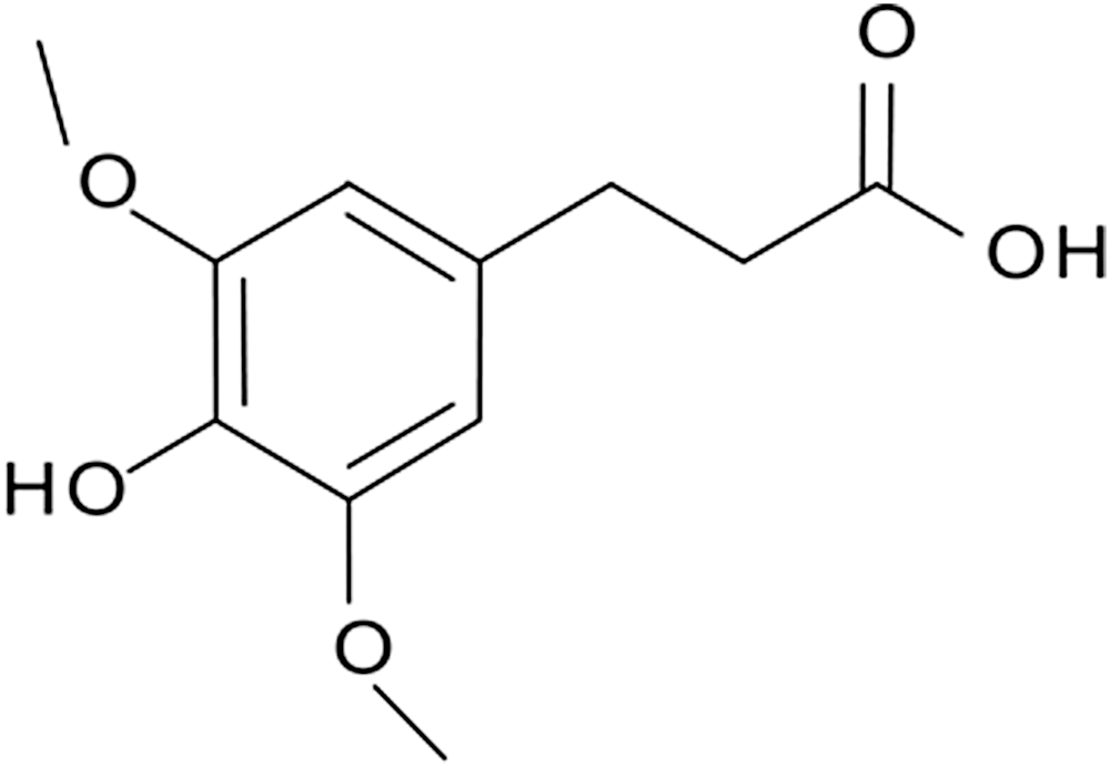
3-(4′-hydroxyl-3′,5′-dimethoxyphenyl)propionic acid.
ApoE, apolipoprotein E; CETP, cholesterol ester transfer protein; eNOS, endothelial nitric oxide synthase; GSH, glutathione; GSSG, glutathione disulfide; HDL-C, high-density lipoprotein cholesterol; HDMPPA, 3-(4′-Hydroxyl-3′,5′-dimethoxyphenyl)propionic acid; LDL-C, low-density lipoprotein cholesterol; NO, nitric oxide; POV, peroxide value; ROS, reactive oxygen species; TBARS, thiobarbituric acid reactive substances; TC, total cholesterol; TG, triglyceride; VLDL-C, very low-density lipoprotein cholesterol.
MCP-1, monocyte chemoattractant protein 1.
Development of Atherosclerosis and Its Related Pathophysiology
Atherosclerosis occurs in the arterial tree at sites of disrupted laminar flow. Atherogenic lipoproteins such as LDLs enter the endothelial intima and are then modified by oxidation or enzymes. As modified LDLs (mLDLs) aggregate into the extracellular intimal space, susceptibility to phagocytosis increases. Unregulated uptake of mLDLs by macrophages subsequently turns macrophages into foam cells, laden with lipids. Lipid accumulation in foam cells can lead to fatty streak formation in coronary arteries or peripheral vessels, which is considered the initial lesion of atherosclerosis leading to complex atherosclerotic lesions. 34 Vascular smooth muscle cells recruited from media into the intima or proliferated within the intima contribute to the atherosclerotic process via secretion of a large quantity of extracellular matrix components such as collagen. In addition to monocytes, other types of leukocytes, particularly T cells, are recruited to atherosclerotic lesions and perpetuate a state of chronic inflammation. As an atherosclerotic plaque grows, compensatory remodeling takes place in the aorta including an increase in overall diameter while the lumen size is maintained. After foam cells die, cellular debris and crystalline cholesterol are released. Moreover, smooth muscle cells form a fibrous cap beneath the endothelium, which walls off the plaque from the bloodstream. This process contributes to the formation of a necrotic core within the plaque and further promotes the recruitment of inflammatory cells, resulting in rupture of the nonobstructive plaque or erosion of the endothelium. These events allow the entrance of thrombogenic materials such as tissue factors into the lumen to promote thrombus formation. If the thrombus is sufficiently large to block the artery, acute coronary syndrome or myocardial infarction (heart attack) will occur. Otherwise, the plaque continues to grow without rupture and the lesion encroaches on the lumen; then, clinically relevant obstructive diseases will arise. 20,35 –38
Lipid-Lowering Effects of Kimchi
Hyperlipidemia is an important risk factor for the development of atherosclerosis and is characterized by increased plasma cholesterol and triglyceride (TG) levels. These lipids carried by “atherogenic” lipoproteins (chylomicrons, very LDL, their remnant lipoproteins, and LDL) can enter into arterial walls and accumulate inside to initiate early atherosclerosis. 39
Kimchi and its ingredients have been cited as being able to lower hyperlipidemia through regulation of lipid metabolism. In high-cholesterol diet-fed rats, dietary kimchi decreased the levels of TG, total cholesterol (TC), and LDL cholesterol (LDL-C) in the plasma and increased HDL cholesterol (HDL-C). 40 –42 In addition, lipid accumulation in aortic tissue of high-cholesterol diet-induced hypercholesterolemic rabbits was reduced by kimchi consumption via suppression of plasma TC and LDL-C levels. 43 –46 These hypolipidemic effects of kimchi were attributed to regulation of 3-hydroxy-3-methyl-glutaryl-CoA reductase (HMGCR), cholesterol ester transfer protein, and cholesterol acyltransferase activities in the liver of rats and rabbits. 45,46 A number of studies have reported that the cholesterol-lowering effects of kimchi and expression of cholesterol-related genes are comparable with statins, particularly simvastatin. 29,46 Kimchi has been demonstrated to lower hepatic cholesterol concentrations via inhibition of cholesterol biosynthesis mediated through regulation of HMGCR. 47 Moreover, kimchi was found to increase fecal excretion of lipids by preventing absorption of dietary lipids in the intestine due to its higher fiber content (24 g/100 g freeze-dried kimchi). 48 These results may contribute to the cholesterol-lowering and anti-atherosclerotic effects of kimchi.
The biologically active compounds in kimchi ingredients known to have lipid-lowering effects include β-sitosterol in cabbage, allicin in garlic, alliin in onion, and capsaicin in red pepper. 21 β-Sitosterol is a phyto-cholesterol that competes with cholesterol for absorption by the intestine. 49 Alliin stimulates plasma lecithin-cholesterol acyl transferase lipolytic enzymes and fecal excretion of sterols and bile acids. 50 Allicin inhibits cholesterol synthesis by inhibiting acetyl-CoA synthetase or HMGCR activity. 51,52 Capsaicin stimulates α-hydroxylase activity by converting cholesterol into bile acids, and it increases energy expenditure via thyroid hormone regulation. 53,54 LAB in kimchi is also known to have a lipid-lowering effect, especially cholesterol-lowering activity. Certain LAB strains such as Lactobacillus acidophilus and Lactobacillus plantarum can bind to cholesterol in cell walls, decompose cholesterol for assimilation, deconjugate bile acids, and facilitate the binding of bile by fiber, which is beneficial for increasing cholesterol excretion. 55 –57 Leuconostoc kimchii GJ2 producing bacteriocin has demonstrated ability to lower serum TC, TG, and LDL-C levels in rats fed a high-fat and high-cholesterol diet. 58 The direct feeding of L. acidophilus has shown the preventing effect of atherosclerosis via inhibition of intestinal cholesterol absorption and cholesterol transport to macrophages. 59
The lipid-lowering effect of kimchi has been studied in humans. Especially, TC and HDL-C levels in the blood are greatly influenced by kimchi consumption. A nutritional survey on kimchi intakes in 102 healthy Korean men between 40 and 64 years old combined with data from plasma lipid analysis demonstrated a positive correlation between the amount of daily kimchi consumption and HDL-C levels but a negative correlation between kimchi intake and LDL-C levels. 23 Moreover, a study on metabolic syndrome and food intake observed that greater consumption of kimchi with cooked rice was correlated with a lower risk of metabolic syndrome due to higher HDL-C and lower adiponectin levels. 27 In a study on the effects of kimchi pill supplementation on plasma lipid concentrations of middle school girls whose BMI was greater than 25 kg/m2 and healthy subjects in their mid-40s, daily intake of 3 g of freeze-dried kimchi as a supplement for 6 weeks, which is equivalent to 30 g of fresh kimchi, lowered serum TC and LDL-C levels and increased HDL-C levels compared with those of the placebo. 24,25 To evaluate the effect of kimchi amount on lipid profiles and other parameters in the blood, a randomized clinical trial in 100 healthy young adults in their mid-20s was conducted. A 7-day diet controlled intervention except that kimchi amount lowered fasting blood glucose (FBG) levels and improved serum lipid profiles and the total antioxidant status of subjects, regardless of the amount of kimchi intake. However, health benefits of kimchi on FBG and total serum cholesterol were found to be greater with the high kimchi intake (210 g/day) group than with the low kimchi intake (15 g/day) group. 28 In particular, high kimchi consumption by hypercholesterolemic young adults with serum TC concentration over 190 mg/dL exerted positive benefits on FBG and serum TC concentration. The lipid-lowering effects of kimchi were more profound in subjects with TC and LDL-C levels over 190 and 130 mg/dL, respectively, in both groups. 28
Antioxidant and Anti-Inflammatory Effects of Kimchi
The initiating events of atherosclerosis are considered a response to injury/inflammation, LDL retention, or oxidative modification of LDL. Especially, oxidation of LDL initiates recruitment of immune cells with significant secretion of inflammatory cytokines, chemokines, and adhesion molecules. In addition, lipid peroxidation products generated during LDL oxidation are chemotactic for both monocytes and T cells and inhibit motility of macrophages, thus trapping them in the intima. Consequently, in the initial stage of atherosclerosis, prevention of hypercholesterolemia and oxidative stress and reduction of LDL-C concentration can suppress the LDL oxidation and formation of foam cells.
The antioxidant properties of kimchi are derived the ingredients used to make kimchi and other biological compounds produced during fermentation. Carotenoids, flavonoids, polyphenols, vitamin C, vitamin E, and chlorophyll present in kimchi are known to be primary antioxidants. 21 Particularly, kimchi itself was shown to inhibit linoleic auto-oxidation 43 and LDL oxidation, 32 and scavenge free radicals. 32,43,60 –62 Antioxidant activities of fermented kimchi were found to be higher than those of fresh kimchi. 63 Moreover, optimally ripened kimchi (pH 4.4 ± 0.1) inhibited the expression of cyclooxygenase 2 (COX-2) and inducible nitric oxide (NO) synthase via the nuclear factor κB (NF-κB) pathway. 64 NF-κB, an oxidative stress-responsive transcription factor, plays a critical role in cellular signaling under oxidative stress conditions. 65 HDMPPA, an active compound of kimchi, also demonstrated anti-inflammatory activity in microglia by suppressing lipopolysaccharide-induced expression of pro-inflammatory mediators and cytokines through blockage of NF-κB, mitogen-activated protein kinases (MAPKs), and PI3K (phosphatidylinositol-3-kinase)-Akt signaling pathways. 66 Many researchers have suggested that the antioxidant properties of kimchi augment anti-mutagenecity/anti-cancer, anti-atherogenic, 30 and anti-aging activities. 30,60,61 These results demonstrate that kimchi intake may prevent atherosclerotic development by preventing auto-oxidation of unsaturated fatty acids, LDL oxidation, and inflammation responses.
The antioxidant effects of kimchi have been confirmed in cellular systems, in in vivo animal studies, and in humans. 64,66 –71 Kimchi and its ingredients are known to enhance activities of antioxidant enzymes such as superoxide dismutase, catalase, glutathione (GSH), and reductase/peroxidase. Plasma and hepatic vitamin E and carotene levels in high-cholesterol diet-fed rabbits were elevated by kimchi consumption. 68,69 These antioxidant effects were also observed in senescence accelerating mice (SAM). SAM that consumed kimchi for 1 year showed a decrease in free radical production in the brain, whereas antioxidant enzyme activities increased. 70
In a human study, the level of total free radicals and hydroxyl radicals in plasma of the elderly (143 subjects, mean age of 70) consuming more than 112 g of kimchi per day was lower than those who consumed less, whereas GSH concentration and GSH/glutathione disulfide (GSSG) were higher. Based on these evidences, kimchi might either inhibit production of free radicals or discard free radicals efficiently. 71
Anti-Atherogenic Effects of Kimchi and Active Principle Compound Hdmppa
Oxidized LDL (oxLDL) and hypercholesterolemia are major symptoms in the development of atherosclerosis. Rabbits fed a high-cholesterol diet for 12 weeks developed atherosclerosis; however, administration of kimchi ingredients retarded the progression of atherosclerosis. Lipid deposition in the aortic arch of rabbits fed kimchi, pepper powder, and garlic decreased. 45,68,72 The active compound HDMPPA isolated and identified from the dichloromethane fraction of Korean cabbage kimchi possesses antioxidant activity against LDL oxidation and free radical scavenging activities. 32 HDMPPA suppressed plasma TC and LDL-C elevation in rabbits and reduced the thickness of the aortic arch in rabbits fed a high-cholesterol diet, the effects of which were comparable to those of simvastatin, a prescribed drug for hypercholesterolemia. 29,46 The mechanisms of action of HDMPPA in the prevention of atherosclerosis are not fully understood. However, it is evident that HDMPPA inhibited fatty streak formation in the aortic sinus of apoE KO mice fed an atherogenic diet. 30 HDMPPA inhibited smooth muscle cell proliferation in the aorta by increasing NO synthesis and inhibition of COX-2 expression. 31 HDMPPA could maintain NO bioavailability via upregulation of endothelial nitric oxide synthase (eNOS) and prevention of NO degradation by reactive oxygen species. HDMPPA treatment in apoE KO mice inhibited eNOS uncoupling by increasing vascular tetrahydrobiopterin content and reducing serum asymmetric dimethylarginine levels. Moreover, HDMPPA diminished inflammation-related protein expression. 31 Therefore, HDMPPA, the active compound of kimchi, might exert its vascular protective effects through preservation of NO bioavailability and suppression of inflammatory responses. 32
Kimchi may regulate lipid metabolism and control immune homeostasis. In THP-1 macrophages, upregulation of LDL receptor, cluster of differentiation 36 (CD36) and aP2, induced by oxLDL facilitates formation of foam cells via acceleration of cholesterol and TG accumulation. 73 –76 HDMPPA inhibited lipid peroxidation induced by oxLDL in THP-1-derived macrophages. In addition, methanol extracts of kimchi (KME) and HDMPPA regulated cholesterol influx and efflux through suppressing CD36 and peroxisome proliferator activated receptor (PPAR)γ expression, and by augmenting liver X receptor (LXR)α, PPARα, and ATP-binding cassette transporter A1 (ABCA1) expression, which subsequently resulted in reduced lipid accumulation in cells. 21,77 These results suggest that kimchi could prevent conversion of monocytes/macrophages to foam cells by inhibiting cholesterol accumulation and lipid peroxidation.
Anti-Endoplasmic Reticulum Stress Effects of Kimchi
The endoplasmic reticulum (ER) is responsible for protein synthesis, folding, and transportation. The ER is known to integrate cellular responses to stress when excessive amounts of improperly folded proteins accumulate in the ER lumen. Under the condition of ER stress, the unfolded protein response (UPR) is activated to diminish ER stress through reduction of protein synthesis, increase in folding capacity, or terminal degradation of improperly folded proteins. 78 Three ER trans-membrane sensors, such as protein kinase-like ER kinase (PERK), inositol-requiring kinase/endonuclease 1 (IRE-1), and activating transcription factor (ATF)-6, are activated to initiate adaptive responses against ER stress. 79 PERK is a serine kinase that phosphorylates eukaryotic translation initiation factor 2α (eIF2α) to attenuate protein synthesis. IRE-1 activates the MAPK apoptosis signal-regulating kinase 1 through interaction with the adaptor protein tumor necrosis factor receptor-associated factor 2, which phosphorylates c-JUN N-terminal kinase. CCAAT/enhancer-binding protein homologous protein (CHOP) is a transcriptional factor for apoptosis regulated by the ATF-4- and ATF-6-dependent pathways in response to ER stress. Therefore, prolonged ER stress might enhance apoptosis. 80 ER stress is activated in macrophages of atherosclerotic lesions in mice and humans, and reduction of ER stress in macrophages alleviates atherosclerosis development in mice. 81 –83 These results suggest that the level of ER stress in macrophages might be a critical contributor to the pathogenesis of atherosclerosis, implying a link between macrophage apoptosis and plaque vulnerability. Association of elevated ER stress with several human diseases has been addressed. 84,85 In a mouse model of diet-induced insulin resistance and atherosclerosis, upregulated UPR markers were detected from macrophages in the early stages of vascular inflammation, even before formation of fatty streaks or atherosclerotic plaques. 86 In addition, persistent ER stress in macrophages accelerates uptake of modified cholesterol through activation of PPARγ signaling. 87
Oxysterols such as 7-ketocholesterol (7-KC) increase cytotoxicity of oxLDL and induce ER stress. 81 KME demonstrated inhibitory effects on aberrant ER stress and apoptosis in macrophages caused by 7-KC. ER stress markers such as X-box binding protein 1 (XBP-1), CHOP, and p-eIF2α were suppressed in a mouse model of diet-induced hypercholesterolemia by KME and indole-3-carbinol, capsaicin, and diallyl disulfide, which are present in sub-ingredients of kimchi. These results suggest that kimchi might have protective roles against oxLDL-induced ER stress and apoptosis. 67 Capsaicin in red pepper, a major ingredient for kimchi making, induced autophagy in MCF-7 and MDA-MB-231 cells where ER stress was inhibited via regulation of MAPK-p38 and extracellular signal-regulated kinases. 88
Conclusions
Kimchi, a traditional Korean food made by fermenting vegetables, has gained global popularity due to its beneficial and nutritional properties. Experimental and clinical studies on the effects of kimchi on atherosclerosis have produced surprising results (Fig. 2). Now genetic studies on atherosclerosis are increasingly performed. There is still no clear evidence that exposed individuals develop epigenetic alterations, which, in turn, increase the risk of atherosclerotic disease, and/or whether epigenetic changes in DNA repair are associated with changes in vascular cell functions. Basic research on kimchi has provided numerous data on the potential mechanisms of the protective effects, but, in fact, the identities of the protective agents and their mechanisms of action remain largely unknown and require further research.
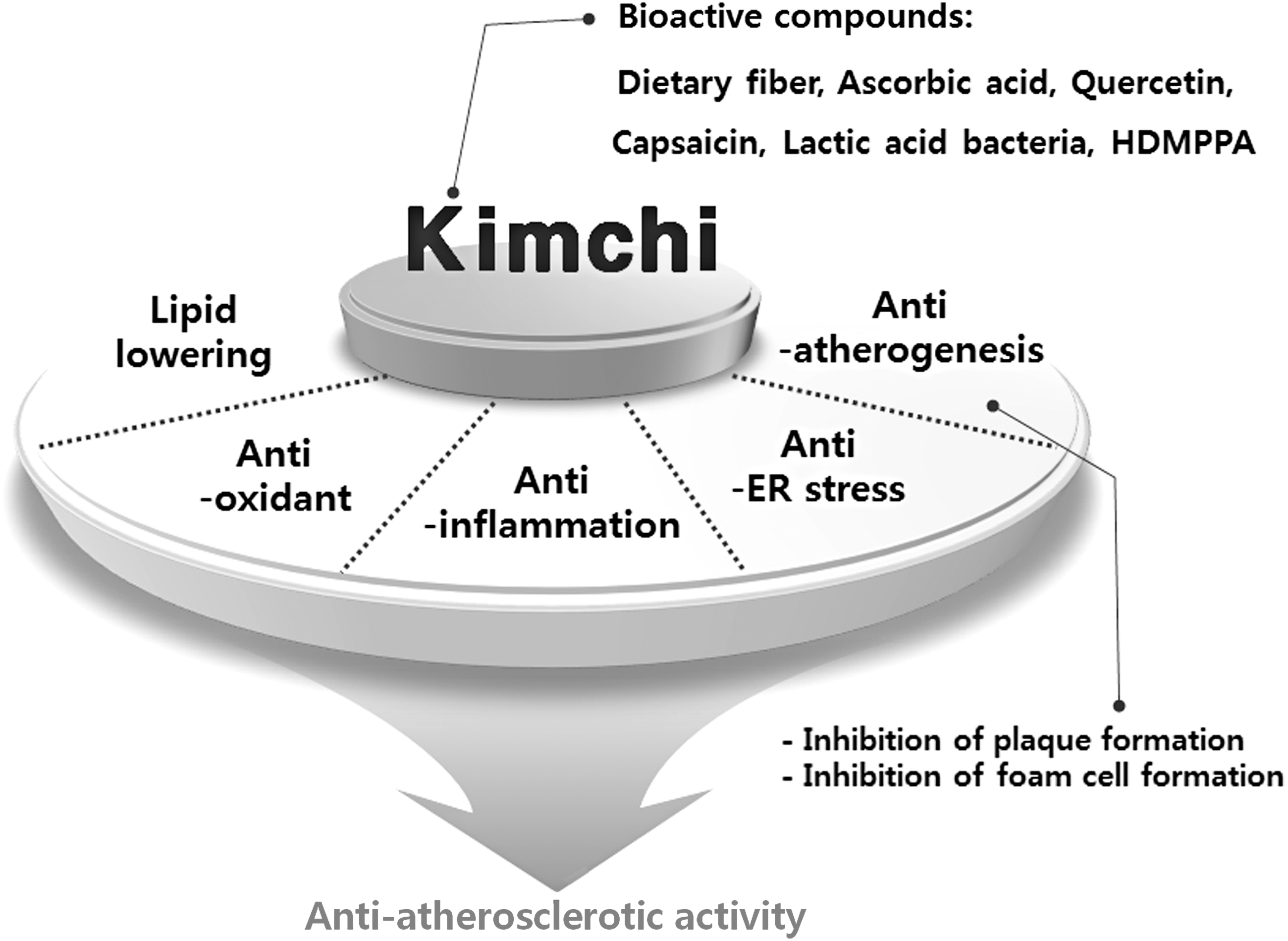
The beneficial effect of kimchi on preventing atherosclerosis.
Footnotes
Acknowledgment
This work was supported by a 2-year Research Grant of Pusan National University.
Author Disclosure Statement
No competing financial interests exist.