
Abstract
Probiotic complex, zinc, and coenzyme Q10 (CoQ10) are recognized dietary supplements with an anti-inflammatory role. Although these supplementations are individually known to benefit rheumatoid arthritis (RA), there is no evidence suggesting any synergic effect. The primary goal of this study is to determine whether probiotic complex, zinc, and CoQ10 attenuate the development of collagen-induced arthritis (CIA). The combination of probiotic complex, zinc, and CoQ10 reduced CIA severity by downregulating the levels of IgG, IgG1, and IgG2a in serum. Joint inflammation, bone destruction, and cartilage damage were also improved by the complex. There was a decrease in the expression of tumor necrosis factor (TNF)-α, interleukin (IL)-1β, IL-6, IL-17, and vascular endothelial growth factor (VEGF) in the joint synovium. The balance between helper T 17 (Th17) cells and regulatory T (Treg) cells was shown to be controlled reciprocally by the complex. These findings suggest that the combination of probiotic complex, zinc, and CoQ10 can ameliorate the development of CIA by inhibiting the expression of proinflammatory cytokines, and is thus an important therapeutic candidate for RA treatment.
Introduction
R
The progression of autoimmune diseases is related to T cells, 4,5 which is manifested in the pathogenesis of RA by the infiltration of CD4+ T cells into the inflamed synovium. 6 In RA, CD4+ T cells release IL-17 (helper T 17 [Th17]), which induces and maintains an exaggerated inflammatory response, 6,7 but regulatory T (Treg) cells secrete immunosuppressive cytokines such as IL-10 and inhibit the immune response in RA progression. 8 Recently, it has been suggested that a reciprocal balance between Th17 and Treg cells can be an important therapeutic strategy for RA. 9,10
Although there are several anti-inflammatory agents for RA, these drugs have many adverse effects in RA patients. It has been demonstrated that nonsteroidal anti-inflammatory drugs (NSAIDs) induce hepatic and renal toxicity. 11 It is also reported that NSAIDs can increase the relative risk of myocardial infarction. 12 Moreover, delayed bone healing, colitis, and ulcer complication are caused by NSAIDs. 11
Currently, the Ministry of Food and Drug Safety has approved 19 probiotics and 12 probiotics used as commercial products in Korea. Probiotics have been used as dietary supplements and exert a potent anti-inflammatory activity. For instance, lactobacillus reduced inflammatory cytokine levels and generated antimicrobial and anti-inflammatory factors. 13,14 Zinc and coenzyme Q10 (CoQ10) are also dietary supplements that possess anti-inflammatory properties. Several studies demonstrated the ability of CoQ10 and zinc to attenuate an inflammatory response by reducing proinflammatory cytokine expression. 15,16 These dietary supplements are also related to RA treatment and has been documented that lactobacillus supplementation alleviates joint swelling and disease activity. 17 Research has shown that CoQ10 can improve experimental arthritis, decreasing joint inflammation and Th17 cell differentiation. 18 The properties of zinc have been studied in clinical trials where there was a decrease of zinc among patients with RA compared to healthy subjects. 19 Although many reports suggest the effect of each individual supplement for use in treating RA, the combination of probiotic complex, zinc, and CoQ10 has not been applied in RA therapy.
We hypothesized that probiotic complex, zinc, and CoQ10 can exert antiarthritic and anti-inflammatory activity to improve RA. The purpose of this study was to identify whether the combination of probiotic complex, zinc, and CoQ10 decreases Th17 cell differentiation and an inflammatory response in an experimental arthritis. Thus, in vitro and in vivo tests using this complex were performed to determine whether the therapeutic function is capable of downregulating proinflammatory cytokines, including IL-6 and IL-17.
Materials and Methods
Combination of probiotic complex, zinc, and CoQ10
The probiotic complex was provided by CNS Pharm Korea Co., Ltd. The probiotic complex consists of Lactobacillus acidophilus (5%), Lactobacillus casei (9%), Lactobacillus fermentum (12%), Lactobacillus paracasei (5%), Streptococcus thermophilus (4%), Bifidobacterium longum (5%), Bifidobacterium bifidum (6%), Bifidobacterium breve (8%), Lactobacillus rhamnosus (17%), Lactobacillus plantarum (20%), Lactobacillus helveticus (3%), and Lactobacillus salivarius (6%). The probiotic complex was suspended in phosphate-buffered saline (PBS) at 5 mg/mL and killed by heating at 80°C for 30 min. Zinc and CoQ10 were purchased from Sigma Aldrich (St. Louis, MO, USA). The combination of probiotic complex (50 mg/kg), zinc (1 mg/kg), and CoQ10 (5 mg/kg) was administered orally to collagen-induced arthritis (CIA) mice.
Arthritis induction and probiotic complex treatment
To examine the effect of the probiotic complex on CIA, the probiotic complex was administered orally once on day 7 after CIA induction. To induce CIA, 100 μg of bovine type II collagen (CII) and complete Freund's adjuvant (Chondrex, Inc., Redmond, WA, USA) were injected intradermally into the base of the tail. Starting on the following day, three independent observers examined the severity of arthritis thrice a week.
CD4 T cell isolation
Splenocytes were prepared from normal DBA1/J mice. Anti-mouse microbeads were used, as recommended by the manufacturer (Miltenyi Biotec, Bergisch Gladbach, Germany). Briefly, cells were resuspended in 100 μL of MACS buffer (1% bovine serum albumin [BSA], 5 mM EDTA, and 0.01% sodium azide). CD4 microbeads (10 μL/1 × 107 cells) were added and incubated for 10 min at 4°C. Cells were diluted in 10 μL of MACS buffer, pelleted, resuspended in 500 μL, and separated magnetically in an AutoMACS magnet fitted with a MACS MS column (Miltenyi Biotec).
Clinical scoring for CIA
Mice were examined visually twice per week for the appearance of arthritis in the peripheral joints, which was graded using a previously reported index. 20 The final value represents the average index from all four legs, as recorded by two independent observers.
Histological assessment of arthritis
For histological assessments, the joints of each mouse were fixed in 10% formalin, decalcified in 10% EDTA, and embedded in paraffin wax. Hematoxylin and eosin (H&E)-stained sections were scored for inflammation, pannus invasion, and bone and cartilage damage. Scores were measured using published criteria. 21
Cell culture
Isolated CD4+ T cells (5 × 10⁵) were incubated with anti-mouse CD3 0.5 μg/mL (BD Biosciences, San Diego, CA), probiotics 100 μg/mL, zinc 50 μM, and CoQ10 μM for 3 days.
Enzyme-linked immunosorbent assay
The concentration of IL-17, IL-6, IFN-γ, and IL-10 in culture supernatant was measured by sandwich enzyme-linked immunosorbent assay (ELISA). All antibodies were purchased from R&D Systems (Minneapolis, MN, USA). Anti-mouse IL-17, IL-6, IFN-γ, and IL-10 were added to a 96-well plate (Nunc, Roskilde, Denmark) and incubated overnight at 4°C, respectively. After blocking (blocking solution; PBS containing 1% BSA and 0.05% Tween 20), the culture supernatant samples and the standard recombinant IL-17, IL-6, IFN-γ, and IL-10 (R&D Systems) were added to the 96-well plate, and the plate was incubated. Biotinylated IL-17, IL-6, IFN-γ, and IL-10 polyclonal antibody (Ab; R&D Systems) was added, and the reaction was allowed to proceed. The plate was washed, 1:2000 diluted ExtrAvidin–alkaline phosphatase (Sigma-Aldrich) was added, and the reaction was allowed to proceed. The plate was washed and 50 μL of p-nitrophenyl phosphate disodium salt (Pierce Chemical Company, Rockford, IL, USA) diluted in diethanolamine buffer was applied.
Measurement of Ig concentrations
The serum concentrations of IgG, IgG1, and IgG2a were measured using mouse IgG, IgG1, and IgG2a ELISA quantitation kits (Bethyl Laboratories, Montgomery, TX).
Immunohistochemistry
Mouse joint tissues were obtained 8 weeks after immunization. Joint tissues were sectioned at 7-μm thickness, dewaxed using xylene, dehydrated through a gradient of alcohol, and then stained with H&E, toluidine blue, Safranin O, and tartrate-resistant acid phosphatase to detect proteoglycans. Immunohistochemistry was performed using the Vectastain ABC kit (Vector Laboratories, Burlingame, CA). Joint tissues were incubated with the first primary monoclonal Abs (mAbs) at 4°C; the antibodies were goat anti-mouse TNF-α mAb, rabbit anti-mouse IL-17, rabbit anti-mouse IL-1β mAb, rabbit anti-mouse IL-6 mAb, and rabbit anti-mouse vascular endothelial growth factor (VEGF). The primary antibodies were detected with a biotinylated secondary linking Ab, followed by incubation with streptavidin–peroxidase complex for 1 h. The final color product was developed using 3,3′-diaminobenzidine chromogen (Dako, Carpinteria, CA). Positive cells were counted and results are expressed as mean ± standard deviation (SD).
Staining for confocal microscopy
Spleen tissues were obtained 8 weeks after primary immunization. The various cell populations were identified with specific antibodies (all from eBioscience). To examine the populations of T cells, the tissues were stained with anti-CD4–fluorescein isothiocyanate, anti-CD4–phycoerythrin (PE), anti-IL-17–PE, anti-CD25–allophycocyanin (APC), and anti-phospho STAT3 705–PE. The stained sections were analyzed using a confocal microscopy system (LSM 510 Meta; Carl Zeiss). All images were analyzed with the MetaMorph software (Molecular Devices, PA, USA).
Statistical analysis
All data were expressed as the mean ± SD. Statistical analysis was performed using SPSS 10.0 for Windows (IBM Corp., Armonk, NY, USA). Comparing numerical data between groups was performed with nonparametric Mann-Whitney tests. Statistical analysis was performed using SPSS 10.0 for Windows (SPSS, Chicago, IL, USA). P values <.05 were considered significant.
Results
The complex reduced the levels of IL-6, IL-17, and IFN-γ cytokines, but increased IL-10 expression
Proinflammatory cytokines such as IL-6, IL-17, and IFN-γ cause systemic inflammatory response. We treated mice splenocytes stimulated by anti-CD3 with the complex and found that the probiotic complex downregulated the expression of IL-6, IL-17, and IFN-γ significantly in culture media. In contrast, the expression of IL-10, which is representative of anti-inflammatory cytokine in media cultured with mice splenocytes stimulated by anti-CD3, was enhanced significantly by the probiotic complex (Fig. 1). These data demonstrate the anti-inflammatory efficacy of the combination of probiotic complex, zinc, and CoQ10.

The complex reduced the levels of IL-6, IL-17, and IFN-γ cytokines, whereas it increased IL-10 expression. Isolated CD4+ T cells were cultured with anti-mouse CD3 0.5 μg/mL, probiotic complex 100 μg/mL, zinc 50 μM, and CoQ10 μM for 3 days. IL-17, IL-6, IFN-γ, and IL-10 levels were determined by ELISA. Data represent mean ± SD of three independent experiments (*P < .05, ***P < .001). CoQ10, coenzyme Q10; ELISA, enzyme-linked immunosorbent assay; IL, interleukin; n.s., not significant; SD, standard deviation.
The complex improved the progression of CIA
We administered the complex once a day starting from day 7 after the first immunization to examine the effect of probiotic complex, zinc, and CoQ10 on the progression of arthritis. The complex significantly decreased the arthritis index compared to the vehicle-treated CIA during the entire observation period (Fig. 2A). Eight weeks after immunization with CII, the complex treatment significantly decreased the concentrations of IgG, IgG1, and IgG2a (Fig. 2B). These results demonstrate that the complex improved CIA development through inhibition of immune response.
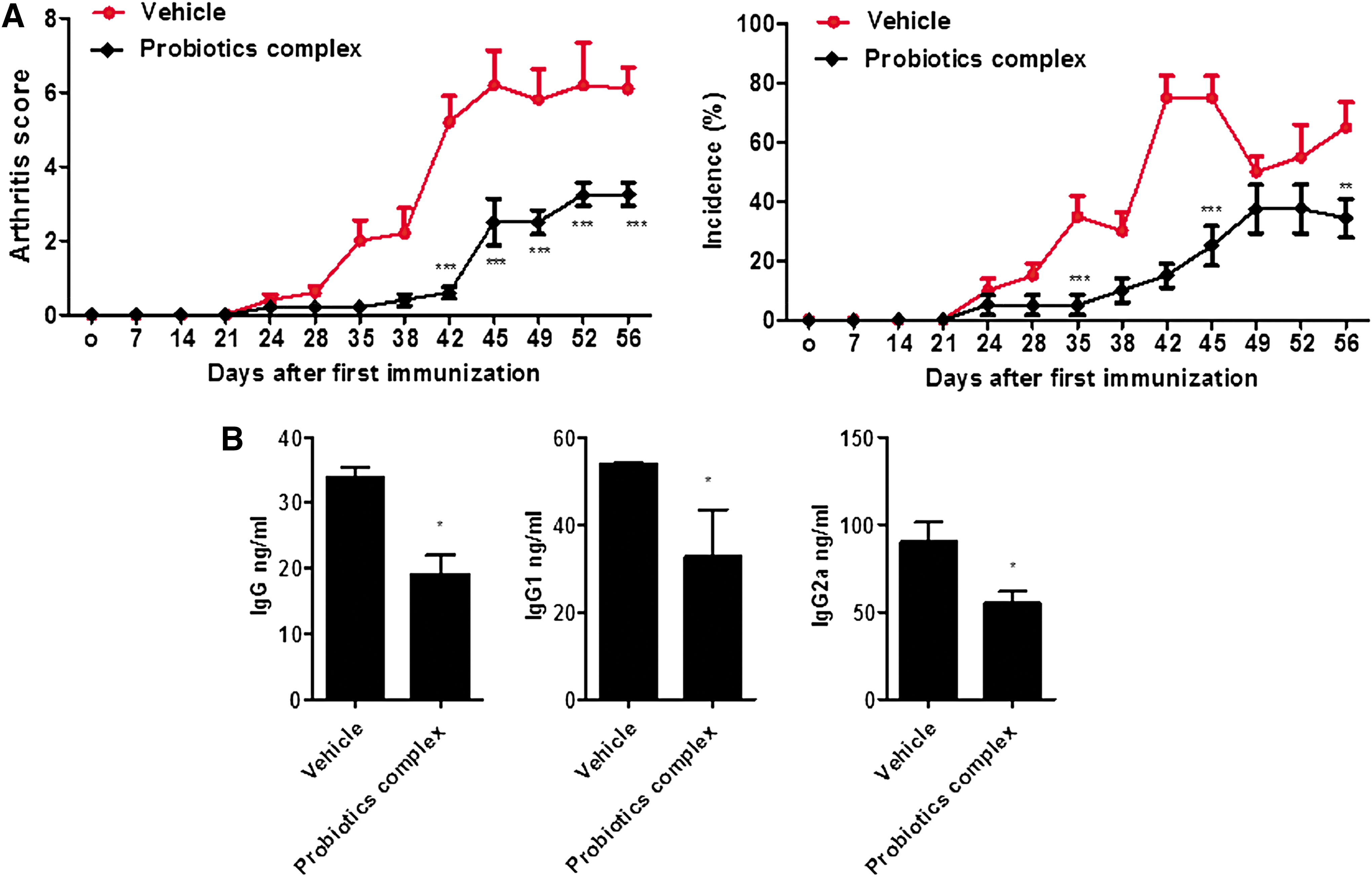
The complex suppressed the progression of CIA.
The complex protected joint devastation in CIA
Compatible with the arthritis score, histological analysis showed protective properties of the complex. Severe immune cell infiltration and cartilage destruction were observed in vehicle-treated CIA mice, whereas the complex treatment decreased signs of inflammation and bone and cartilage damage among the complex-treated CIA mice (Fig. 3). These results suggest that the combination of probiotic complex, zinc, and CoQ10 possesses therapeutic efficacy in joint inflammation.

The complex protected joint devastation in CIA. Ankle joint tissues were obtained from each group. The joint tissues were stained with H&E and Safranin O. Images were obtained from each mouse (n = 5), and here we show the representative images and bar graphs. Data represent mean ± SD of experiments (**P < .005). H&E, hematoxylin and eosin.
The complex downregulated proinflammatory cytokine production in an arthritic joint
Because we detected that the combination of probiotic complex, zinc, and CoQ10 decreased joint inflammation and destruction, we next studied whether the complex suppresses the expression of proinflammatory cytokines in the joint. The complex treatment significantly reduced the expression of IL-1β, IL-6, IL-17, TNF-α, and VEGF in the arthritic joint synovium (Fig. 4). The data suggest that the complex decreased inflammatory cytokine production in the arthritic joint.
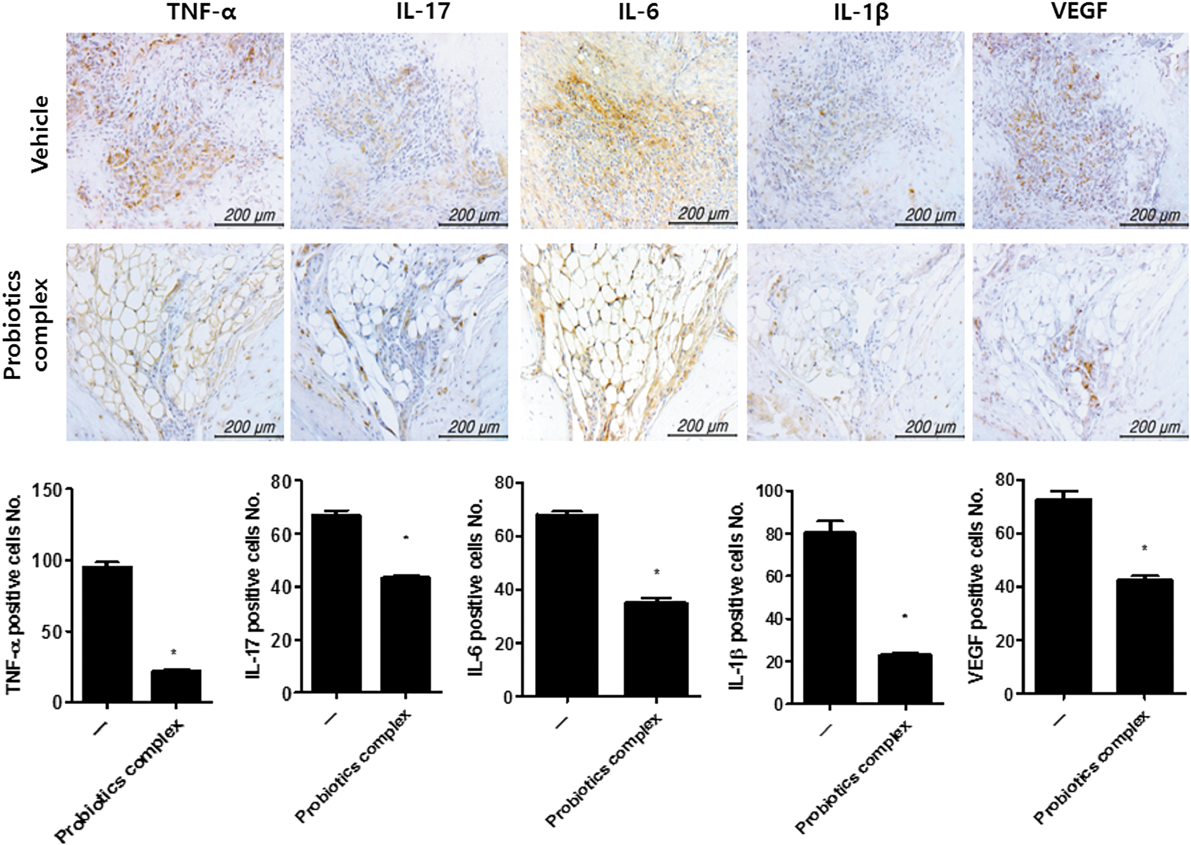
The complex downregulated proinflammatory cytokine production in arthritic joints. Ankle joint tissues were stained to show TNF-α, IL-17, IL-6, IL-1β, and VEGF antibodies. Images were obtained and analyzed one slide per mouse (n = 5), and the representative images are shown (scale bar = 200 μm). Positive cells were indicated in bar graphs. Data represent mean ± SD of experiments (*P < .05). TNF-α, tumor necrosis factor-α; VEGF, vascular endothelial growth factor.
The complex controlled reciprocal balance between Th17 and Treg in CIA
Th17 cell differentiation and CD4+pSTAT3 tyr705+ cell differentiation decreased significantly in splenic tissue from the complex-treated CIA mice. However, Treg cell differentiation markedly increased with the complex treatment (Fig. 5).
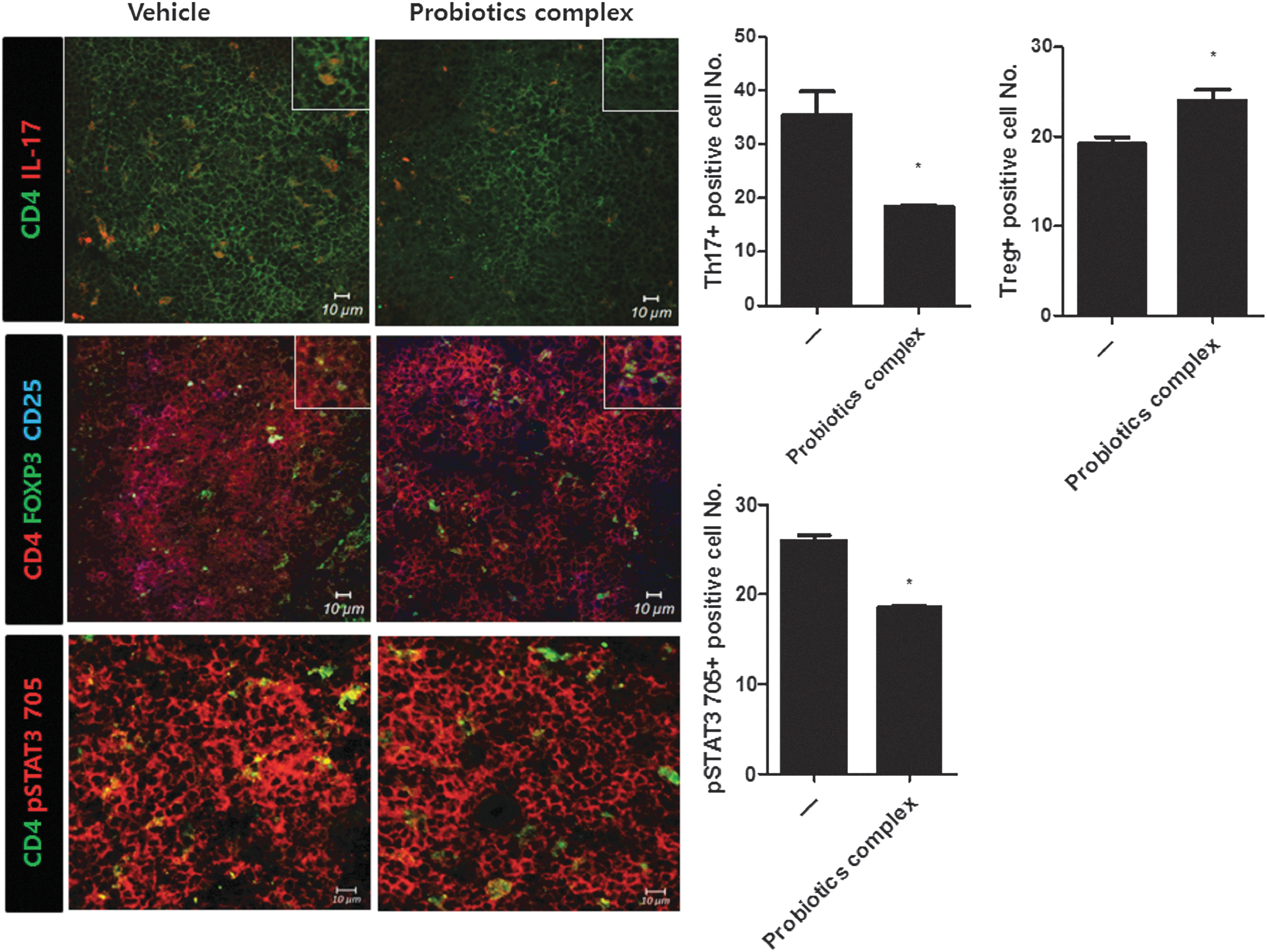
The complex controlled reciprocal balance between Th17 and Treg in CIA. Splenic tissues were immunostained for Th17 and Treg cell analysis. Confocal microscopy images were obtained for each mouse (n = 5), and here we show the representative images and bar graphs. Mean ± SD of experiments (*P < .05). The white boxes are large images of positive cells. Treg, regulatory T.
Discussion
Although probiotics, zinc, and CoQ10 have been used as anti-inflammatory dietary supplements, little is known regarding the effect of the complex on autoimmune arthritis. To our knowledge, there is no previous evidence that the complex shows antiarthritic effects by suppression of the inflammatory response. This study showed that the anti-inflammatory activity of the probiotic complex, zinc, and CoQ10 improved experimental autoimmune arthritis.
In this study, the complex decreased the development of CIA through the downregulation of proinflammatory cytokine expression and Th17 cell differentiation. In the pathogenesis of RA, many proinflammatory cytokines cause an excessive immune inflammatory response in the synovium. There are several reports that proinflammatory cytokines such as IL-6, IL-17, and TNF-α levels increase within the synovium and synovial fluid among RA patients. 22 –24 Th17 cells also act as pathogenic factors for RA patients. It has been suggested that Th17 cells lead to chronic inflammation in RA patients. 25 Thus, blocking proinflammatory cytokines in the synovium and Th17 cell differentiation reveals a therapeutic effect in experimental autoimmune arthritis. 9,10,26 In this study, the complex decreased the expression of proinflammatory cytokines such as IL-6, IL-17, and TNF-α in the inflamed synovium. Moreover, the combination of probiotic complex, zinc, and CoQ10 reduced Th17 cell differentiation. Therefore, the complex can conduct a significant function in autoimmune arthritis by inhibiting inflammation.
Since RA is a chronic autoimmune disease, there is an increase in the immune response, bone erosion, and cartilage damage. It has been demonstrated that an upregulation of serum IgG is a direct risk factor of RA and is related with RA progression. 27 In addition, bone and cartilage destruction is an important characteristic of RA and related to the development of the disease. 1 There are several proofs that the inhibition of serum IgG and devastation of bone and cartilage are mediated by a therapeutic molecule activity in experimental autoimmune arthritis. 18,28,29 The combination of probiotic complex, zinc, and CoQ10 reduced the concentration of IgG, IgG1, and IgG2a in serum as well as the destruction of bone and cartilage in inflamed joint, improving CIA severity. These results suggest that the complex can be used to treat RA.
Since Treg cells that release IL-10 are immunosuppressive factors, enhancement of Treg cell differentiation has been shown to induce a therapeutic effect in an inflammatory disease model. 30 –32 On the contrary, STAT3 activation is a target for autoimmune arthritis because STAT3 is an important transcription factor for Th17 cells. 9,10,26 Reciprocal balance between Th17 and Treg is a key factor to improve CIA progression. The upregulation of Treg cell differentiation shows an anti-inflammatory function in experimental arthritis. 9,26 In this study, the complex increased IL-10 levels in vitro and Treg cell differentiation in vivo. These results demonstrate that the complex can attenuate CIA by promoting IL-10 expression and Treg cell differentiation.
Our observations of the therapeutic activity of probiotic complex, zinc, and CoQ10 suggest the possibility of therapeutic efficacy against RA. In a previous study, CoQ10 attenuated experimental arthritis severity. 18 Probiotic complex, zinc, and CoQ10 are used as supplementation and can also be used clinically to improve autoimmune arthritis without any harmful side effects. Therefore, this investigation suggests that the complex is a potential candidate for treating RA by downregulating an inflammatory response and Th17 cell differentiation.
Footnotes
Acknowledgments
This study was supported by a grant of the Korean Health Technology R&D Project, Ministry for Health & Welfare, Republic of Korea (HI14C3417). This research was supported by a grant of the Korea Health Technology R&D Project through the Korea Health Industry Development Institute (KHIDI), funded by the Ministry of Health & Welfare, Republic of Korea (HI15C1062).
Author Disclosure Statement
No competing financial interests exist.