
Abstract
Notoginseng Radix and Rehmanniae Radix Preparata have been widely used traditionally for treating inflammatory diseases. This research studies the therapeutic effects of YH23537, the extracts of Notoginseng Radix and Rehmanniae Radix Preparata, on pain and cartilage degeneration in an experimental osteoarthritis (OA) model. Male Wistar rats were inoculated intra-articularly with 3 mg of monosodium iodoacetate (MIA) in the right intra-articular. Four days later, the animals were administrated orally with YH23537 daily for 24 days. Tactile allodynia and weight bearing were measured. Macroscopic and microscopic observations for articular cartilage were performed at the end of the experiment. Protein expression in the joint was determined by immunohistochemistry. The effects of YH23537 on mRNA levels in chondrocytes stimulated with interleukin (IL)-1β were analyzed using random polymerase chain reaction. OA induction was confirmed by significant decrease of paw withdrawal latency, paw withdrawal threshold, and weight bearing compared with the normal group at 3 days after MIA injection. The YH23537-treated groups displayed significant increases in pain thresholds and weight bearing throughout the observation period. The damage to articular cartilage was significantly lessened visually and histopathologically by YH23537 treatment. YH23537 suppressed the expression of metalloproteinase-3, nitrotyrosine, IL-1β and IL-6 increased in OA joints. YH23537 upregulated tissue inhibitor of metalloproteinase (TIMP)-1 and TIMP-3 in IL-1β-stimulated human OA chondrocytes. The protein levels of the NF-κBp65 and HIF-2α in the joint tissues were reduced by YH23537. YH23537 exerted antinociceptive effects and cartilage protective effects in experimental OA rats by suppressing oxidative injury, inflammatory mediators, and inducing anabolic factors. We suggest that YH23537 may have efficacy for treating OA in humans.
Introduction
O
The current conception of OA has altered to an inflammatory joint disease from cartilage “wear and tear.” 3 Cartilage degradation in OA joints is induced by the expression of inflammatory mediators, including chemokines, cytokines, reactive oxygen species (ROS), and matrix metalloproteinases (MMPs). 4,5 Moreover, interleukin (IL)-1β has been revealed to generate chondrocytes to generate inflammatory mediators such as nitrotyroine and IL-6, which further amplifies harmful cellular reactions. 6 In addition, elevated IL-1β results in a downregulation of cartilage extracellular matrix (ECM) components by suppressing anabolic factors and increasing catabolic factors in chondrocytes, resulting in a degradation of cartilage in ECM, the hallmark of OA. Disproportion of tissue inhibitor of metalloproteinases (TIMPs) and MMPs is involved in a decisive role in cartilage degradation. 7 Furthermore, the combination of elevated inflammatory mediators with raised production of ROS has been observed in the joints of patients with OA such as the subchondral bone, cartilage, and synovium. Oxidative stress can cause mitochondrial damage and lipid peroxidation. 8,9
Recently, research has assessed herbal agents and their anti-inflammatory effects at the cellular level as an alternative way of suppressing the induction of proinflammatory cytokines. 10,11 Herbal medicines, GCSB-5 (Shinbaro™) and PG201 (Layla™), were recently approved as anti-OA medicines by Korean Ministry of Food and Drug Safety. Their anti-inflammatory effects in animal experiments and beneficial effects in patients with OA in randomized clinical study have been well demonstrated. 12 –15 Furthermore, these medicines showed not only similar efficacy but also fewer gastrointestinal adverse effects than celecoxib. 13,15 Gastrointestinal adverse effects are associated with poor adherence to nonsteroidal anti-inflammatory agents and additional prescription of gastroprotective drugs. 16
Panax notoginseng is one of the 13 species of ginseng identified. 17 The biologically active compounds of ginseng are considered to be the saponins of which the ginsenosides Rg1, Rb1, and the notoginsenoside R1 are the major compounds of Notoginseng Radix. 17 The steamed root of Rehmannia glutinosa Libosch (Rehmanniae Radix Preparata), a traditional Chinese agent, has been accustomed to alleviate inflammatory circumstances in Asian clinics. This herb is known to have anti-inflammatory properties 18 and wound healing effects. 19 As OA pathogenesis involves inflammation, we hypothesize that Notoginseng Radix and Rehmanniae Radix will have a therapeutic effect. In this article, we evaluate the effects of the extracts of Notoginseng Radix and Rehmanniae Radix Preparata on pain and cartilage degradation in an OA rat experimental model and its mechanisms.
Materials and Methods
Chemicals and materials
HPLC-grade acetonitrile and methanol were acquired from Burdick & Jackson, USA. Analytical grade ethanol was from Duksan (Gyeonggi, Korea). All aqueous solutions were prepared with ultrapure water made by Milli-Q system (18.2 MΩ; Millipore Bedford, MA, USA). Notoginsenoside R1 and ginsenosides Rg1, Rb1, Rd, 20(S)-Rh1, Rh4, 20(R)-Rg3, 20(S)-Rg3, Rg5, Rk1, and Rk3 standards were purchased from Chromadex (CA, USA). Notoginseng Radix was purchased from MyohyangSanqi (Wenshan, Yunnan Province, China). Rehmanniae Radix Preparata was purchased from Bolak (Gyeonggi, Korea).
Preparation of YH23537
Notoginseng Radix and Rehmanniae Radix Preparata were identified using the Korean Herbal Pharmacopoeia and the Korean Pharmacopoeia. YH23537 was manufactured according to bulk good manufacturing practices by Bolak. In brief, a mixture of 320 kg of Notoginseng Radix powder and 40 kg of Rehmanniae Radix Preparata (8:1, w/w) was extracted twice in 2520 and 1800 L of 50% ethanol for 4 and 2 h, respectively. The extracted solution was filtered and concentrated to remove the ethanol. The resultant solution was boiled for 8 h, filtered, and mixed with dextrin at a 1:1 ratio of solid content before drying.
YH23537 was tested for the presence of heavy metals (total heavy metal ≤30 ppm, AS ≤3 ppm, and Pb ≤5 ppm), pesticide residues (BHC [sum of α, β, γ, and δ-BHC] ≤0.2 ppm, DDT [sum of o,p-DDE, p,p-DDE, o,p-DDT, p,p-DDT] ≤0.1 ppm, Aldrin+Dieldrin+Endrin ≤0.01 ppm), general bacteria (≤1 × 10⁵ CFU/g), fungi (≤100 CFU/g), and specific pathogens (negative for Escherichia coli, Pseudomonas aeruginosa, Staphylococcus aureus, and Salmonella spp.), and were also subjected to quantitation tests (G. Rb1 ≥ 9.0 mg/g in extract).
HPLC-based fingerprinting of YH23537
Five hundred milligrams of YH23537 was placed into a 50-mL volumetric flask and then 40 mL of 50% methanol was added into the flask. After sonication for 1 h, 50% methanol was added up to 50 mL and the prepared sample was filtered by way of a 0.45 μm of regenerated cellulose membrane filter. HPLC was used for determining the ginsenoside profile with a Waters Alliance LC (Waters Corporation, Milford, MA, USA) equipped with a quaternary gradient pump and a sample manager. An HPLC process was progressed using a reverse-phase column (Agilent Zorbax SB C18, 4.6 × 150 mm, 3.5 μm). The column was maintained at 30°C. A 20 μL aliquot was injected onto the column. The gradient elution system consisted of 5% of acetonitrile (A) and 95% acetonitrile (B) and separation was fulfilled using the following gradient: 0–3 min, 5% B; 3–18 min, 5–50% B; 18–28 min, 50–90% B; 28–40 min, 90% B; 40–41 min, 90–5% B; and 41–45 min, 5% B. The flow rate was 1.0 mL/min and the eluate was checked at 203 nm by an ultraviolet detector. The peaks were confirmed by comparing the retention times of the peaks with the reference compounds eluted underneath the equal states. The studied saponins were well separated (Fig. 1) and the extracts contained 0.68 mg Notoginsenoside R1, 4.70 mg Ginsenoside Rg1, 18.97 mg Ginsenoside Rb1, 0.81 mg 20(S)-Ginsenoside Rh1, 15.60 mg Ginsenoside Rd, 16.42 mg Ginsenoside Rh4, 28.41 mg Ginsenoside Rk3, 7.67 mg 20(S)-Ginsenoside Rg3, 7.75 mg 20(R)-Ginsenoside Rg3, 10.64 mg Ginsenoside Rg5, and 14.68 mg Ginsenoside Rk1 per gram.

Chemical fingerprint chromatograms of YH23537. For identifying profile of ginsenosides, YH23537 was dissolved in 50% methanol and filtered. And then analysis was performed with RP column under the gradient elution system consisted of 5% of acetonitrile and 95% acetonitrile. Notoginsenoside R1 (1), ginsenosides Rg1 (2), Rb1 (3), 20(S)-Rh1 (4), Rd (5), Rh4 (6), Rk3 (7), 20(S)-Rg3 (8), 20(R)-Rg3 (9), Rg5 (10), and Rk1 (11). Five hundred milligrams of YH23537 was placed in a 50-mL flask and then 40 mL of 50% methanol was added into the flask. After sonication for 1 h, 50% methanol was added up to 50 mL and the prepared sample was filtered by 0.45 μm of regenerated cellulose membrane filter. HPLC method was performed using a Waters Alliance LC (Waters Corporation) equipped with a quaternary gradient pump and a sample manager. An HPLC method was developed using RP column (Agilent Zorbax SB C18, 4.6 × 150 mm, 3.5 μm). The column was maintained at 30°C. A 20 μL aliquot was injected onto the column. The gradient elution system consisted of 5% of acetonitrile (A) and 95% acetonitrile (B) and separation was achieved using the following gradient: 0–3 min, 5% B; 3–18 min, 5–50% B; 18–28 min, 50–90% B; 28–40 min, 90% B; 40–41 min, 90–5% B; and 41–45 min, 5% B. The flow rate was 1.0 mL/min and the eluate was monitored at 203 nm by an ultraviolet detector. The peaks were identified by comparing the retention times of the peaks with those of the reference compounds eluted under the same conditions. The investigated saponins were well separated and comparison of chemical profile between YH23537 was performed (Fig. 1). Color images available online at
Animals
Male Wistar rats were purchased from Central Lab. Animal, Inc. (Seoul, South Korea). The rats (weight 140–230 g, age 8–12 weeks) were housed under standard laboratory states (in a temperature-controlled room [21°C −22°C], controlled lighting [12-h dark/12-h light cycle]), and had free access to sterile water and food. All animal processes were certified by the Animal Research Ethics Committee of the Catholic University of Korea (Approval Number: 2014-0139-02).
OA induction and YH23537 treatment
Rats were randomized and allocated to groups before the initiation of the research. Monosodium iodoacetate (MIA) (Sigma, Poole, UK) was weighed and dissolved in saline at 60 mg/mL on the day of injection. After 4 days, the rats received oral administration of YH23537, celecoxib (CIPLA, Mumbai, India), or carboxymethycellulose (CMC; Sigma) solution as control. YH23537 and celecoxib were kindly supplied by Yuhan Corporation (Seoul, Korea). YH23537 and celecoxib were dissolved in 0.5% CMC solution (vehicle) or with vehicle only by oral administration. YH23537 (100 and 300 mg/kg) or celecoxib (50 mg/kg) was administered once daily for 24 days.
Measurement of pain behavior
Nociceptive analysis was performed by operating a dynamic plantar esthesiometer (Ugo Basile, Comerio, Italy), an automated model of the von Frey hair analysis process, before OA induction (day 0). Pain behavior scores were measured using published criteria. 20
Weight bearing measurement
Weight bearing was evaluated using an incapacitance tester (Linton Instrumentation, Norfolk, UK) that included a dual-channel weight mean value. The rats were attentively positioned in a plastic chamber. The strength applied by an individual hind limb was averaged more than a 3-sec time. Individual data point was the average of three measurements. The percentage of weight divided onto the handled (ipsilateral) hind limb was calculated utilizing the following equation: (weight on right leg/weight on right leg and left leg) × 100.
Joint histology assessment
Histological differences were tested to define the impact of YH23537 on cartilage degeneration in the knee joints of OA rats. The knee joints of each rat were fixed in 10% formalin, decalcified in 5% formic acid, and embedded in paraffin. The serial sections (7μm) were acquired at the medial and lateral midcondylar level in the sagittal plane and were stained with hematoxylin and eosin (H&E), and Safranin O, enabling the evaluation of proteoglycan content. An adapted Mankin's histological score was used to score histological injuries of the articular cartilage as references. 21 Joint degeneration was assessed histologically with the Osteoarthritis Research Society International (OARSI) cartilage degeneration score. 22
Immunohistochemistry
Immunohistochemistry slides were deparaffinized and rehydrated using a graded ethanol series. The sections were incubated overnight at 4°C with antibodies to IL-1β (Santa Cruz Biotechnology, Santa Cruz, CA, USA), MMP-3 (Abcam, Cambridge, UK), IL-6 (Abcam), and nitrotyrosine (Santa Cruz). The slides then responded to each secondary antibodies, biotinylated antimouse IgG for 20min, conjugated to a streptavidine peroxidase complex (Vector Laboratories, Burlingame, CA, USA) for 1 h, and at last with 3,30-diaminobenzidine (Dako, Glostrup, Denmark). The slides were counterstained with Mayer's hematoxylin and photographed using Olympus photomicroscope (Olympus, Tokyo, Japan).
Microscopic measurement of OA joints
Tibial and femoral bones were isolated and the total surplus smooth tissue was cautiously dissected under an anatomical microscope. The articular face of respective sample was washed with phosphate-buffered saline. The cartilage face of the femoral condyle and the tibial plateau were investigated using a microscope.
Human OA chondrocyte separation and primary culture
The chondrocytes used in this study were accumulated with the approval of the Bucheon St. Mary's Hospital IRB (HC17TESI0106) that was fulfilled in accordance with the Declaration of Helsinki. All patients were informed about the purpose and procedures of the experiment and written informed agreement was acquired. All patients with OA fulfilled the American College of Rheumatology criteria for this disease. 20 The chondrocytes were isolated using the published method. 23 After 24 h, the cells were pretreated with YH23537 for 2 h and then stimulated with or without recombinant human IL-1β (20 ng/mL; R&D Systems) for 48 h.
Real-time polymerase chain reaction
Total RNA was extracted using TRI Reagent (Molecular Research Center) according to the manufacturer's instructions. Complementary DNA (cDNA) was prepared using reverse transcription of the single-stranded RNA according to the manufacturer's instructions included in the High Capacity cDNA Reverse Transcription Kit (Applied Biosystems). A LightCycler 2.0 instrument (software version 4.0; Roche Diagnostics) was utilized for polymerase chain reaction amplification. All responses were executed with LightCycler FastStart DNA Master SYBR Green I (Takara) according to the manufacturer's directions. The primer pairs utilized in these responses were as follows: for control human β-actin, forward: 5′-GGA-CTT-CGA-GCA-AGA-GAT-GG-3′, reverse: 5′-TGT-GTT-GGG-GTA-CAG-GTC-TTT-G-3′; for human TIMP-1, forward: 5′-AAT-TCC-GAC-CTC-GTC-ATC-AG-3′, reverse: 5′-TGC-AGT-TTT-CCA-GCA-ATG-AG-3′; for human TIMP-3, forward: 5′-CTG-ACA-GGT-CGC-GTC-TAT-GA-3′, reverse: 5′-GGC-GTA-GTG-TTT-CTG-GT-3′.
Statistical analysis
Data are shown as means ± standard error of the mean. Statistical analysis of the data was carried out using one-way ANOVA (analysis of variance) and Dunnett's multiple comparison test or for index data; if there was significance in the Kruskal–Wallis one-way analysis, significant difference among the groups was analyzed using Dunn's multiple comparison test (GraphPad Prism 5.0; GraphPad software, CA, USA). P < .05, P < .01, and P < .001 were considered statistically significant.
Results
Effects of YH23537 on pain in the MIA-induced OA model
To assess the capability of YH23537 to ameliorate the pain in OA, tactile allodynia was assessed in MIA-induced OA compared with celecoxib. The oral administration with YH23537 was started on day 4 after MIA injection and maintained for 24 days. In the von Frey hair assessment test, paw withdrawal latency and paw withdrawal threshold were extended significantly in the rats given oral YH23537 (100 and 300 mg/kg) compared with those in the vehicle group (Fig. 2A). In addition, YH23537 significantly improved weight bearing tolerance (Fig. 2B).
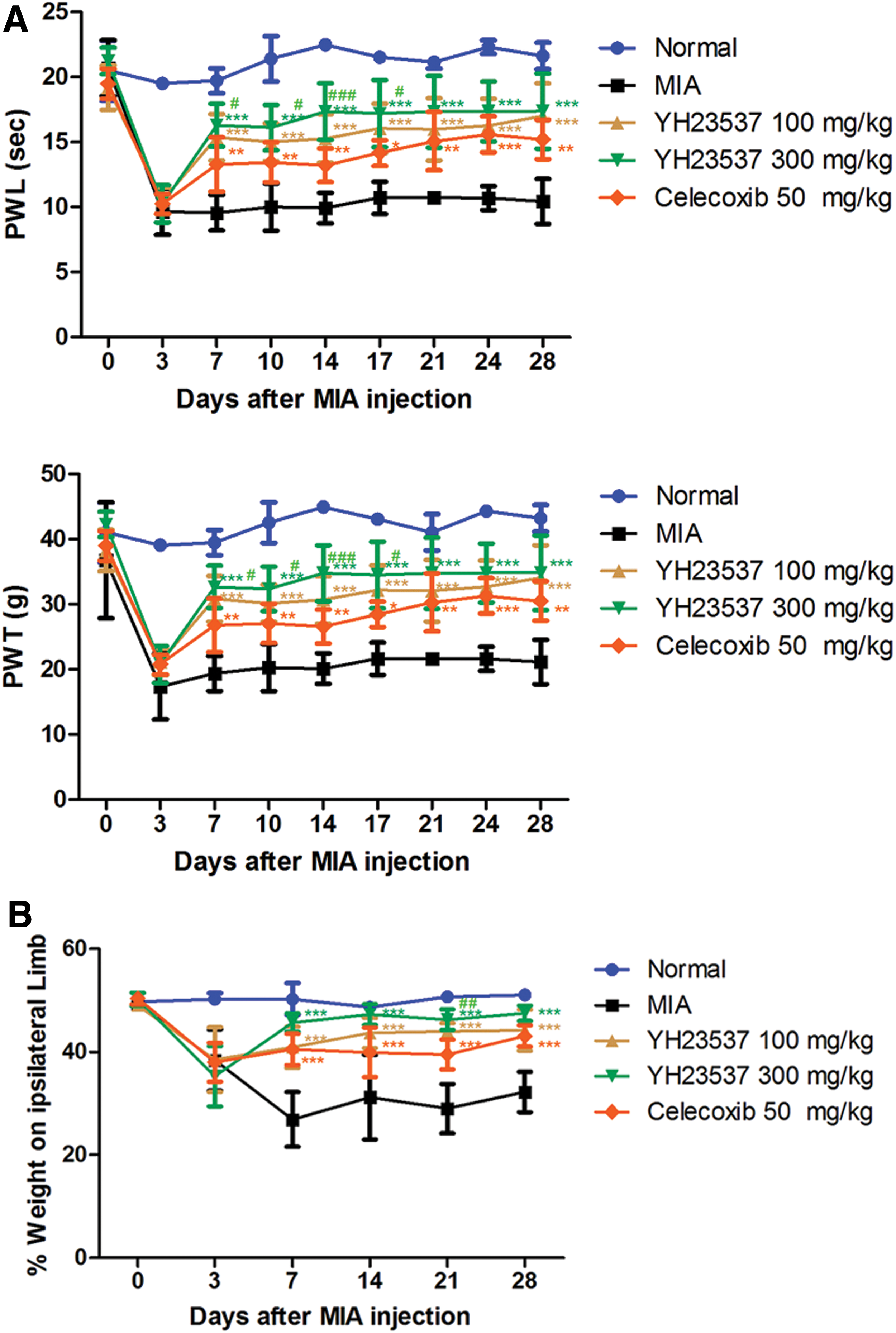
Effects of YH23537 on mechanical hyperalgesia in a model of MIA-induced OA rats. Rats were injected with 3 mg of MIA into the right knee. YH23537 and celecoxib were administered every day for 4–28 days after the MIA injection.
Attenuated joint damage by oral administration of YH23537 in MIA-induced OA rats
The damage to the articular cartilage exterior surface was assessed by using India Ink 28 days after MIA injection. Anatomically, the normal control joints showed gleamingly articular surfaces and round shape. The joints of the MIA-treated group revealed irregular abrasion on the articular cartilage faces of the femoral condyle and the tibia plateau, but the YH23537 100 and 300 mg/kg and celecoxib-treated rats had shinier articular surfaces than the vehicle administration group. The degree of articular cartilage in YH23537 300 mg/kg-treated OA rats was most comparable with those of normal group (Fig. 3).
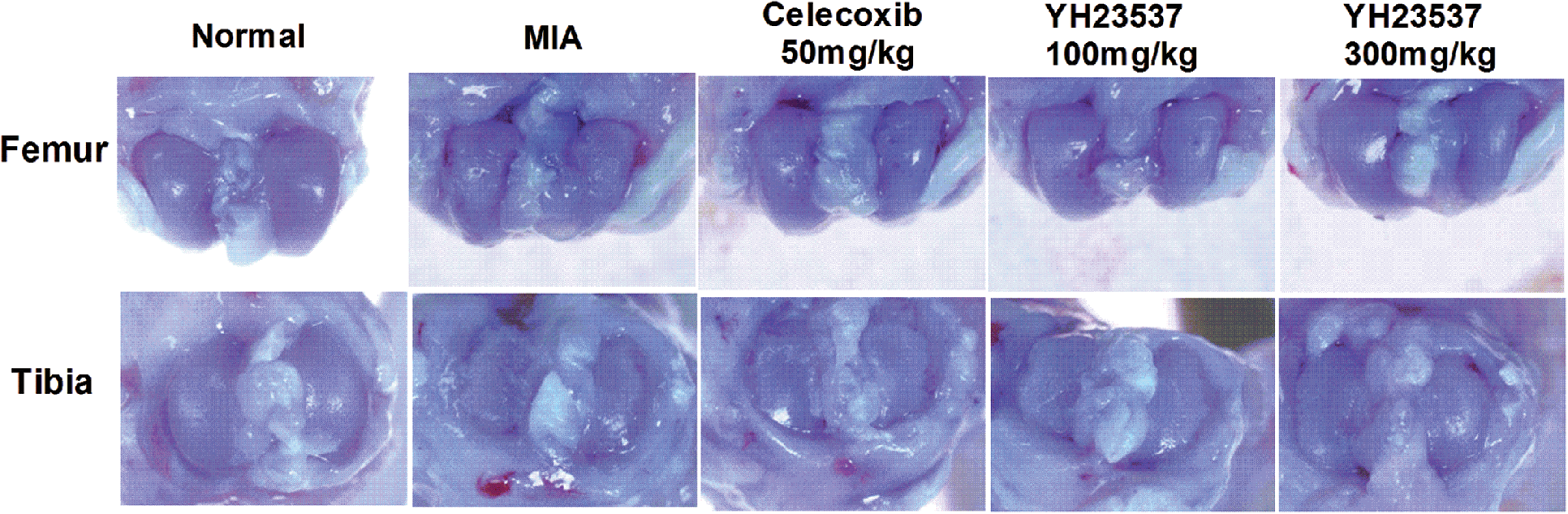
Macroscopic photographs of the damaged articular cartilage after treatment with YH23537 in MIA-induced OA rats. Rats were injected with 3 mg of MIA in the right knee. YH23537 was administrated orally for 28 days. The gross morphological changes of the femoral condyles and tibial plateau were photographed using a microscope. Color images available online at
Cartilage degradation reduced by treatment of YH23537 in OA rats
As the cartilage degradation is the central feature of OA joints, the enhancement of cartilage degradation of OA joints was studied. On day 28 after MIA injections, the knees were excised and the cartilages were stained with H&E and Safranin O after routine tissue processing and slide preparation. The cartilage thickness and the ingredient of proteoglycan in joints of OA rats were significantly increased than the vehicle group. In the YH23537 oral administration group, cartilage degradation was inhibited in a dose-dependent manner until 28 days after MIA injection. The stage of cartilage degradation was evaluated using Mankin's score system. This system scores structure damage, cellular irregularity, and matrix staining. High-dose group of YH23537 indicated significantly lower Mankin's scores than the vehicle-treated group (Fig. 4A, B). In addition, the high-dose group of YH23537-treated rats all revealed lower OARSI scores than the vehicle-treated and celecoxib-treated rats (Fig. 4C).
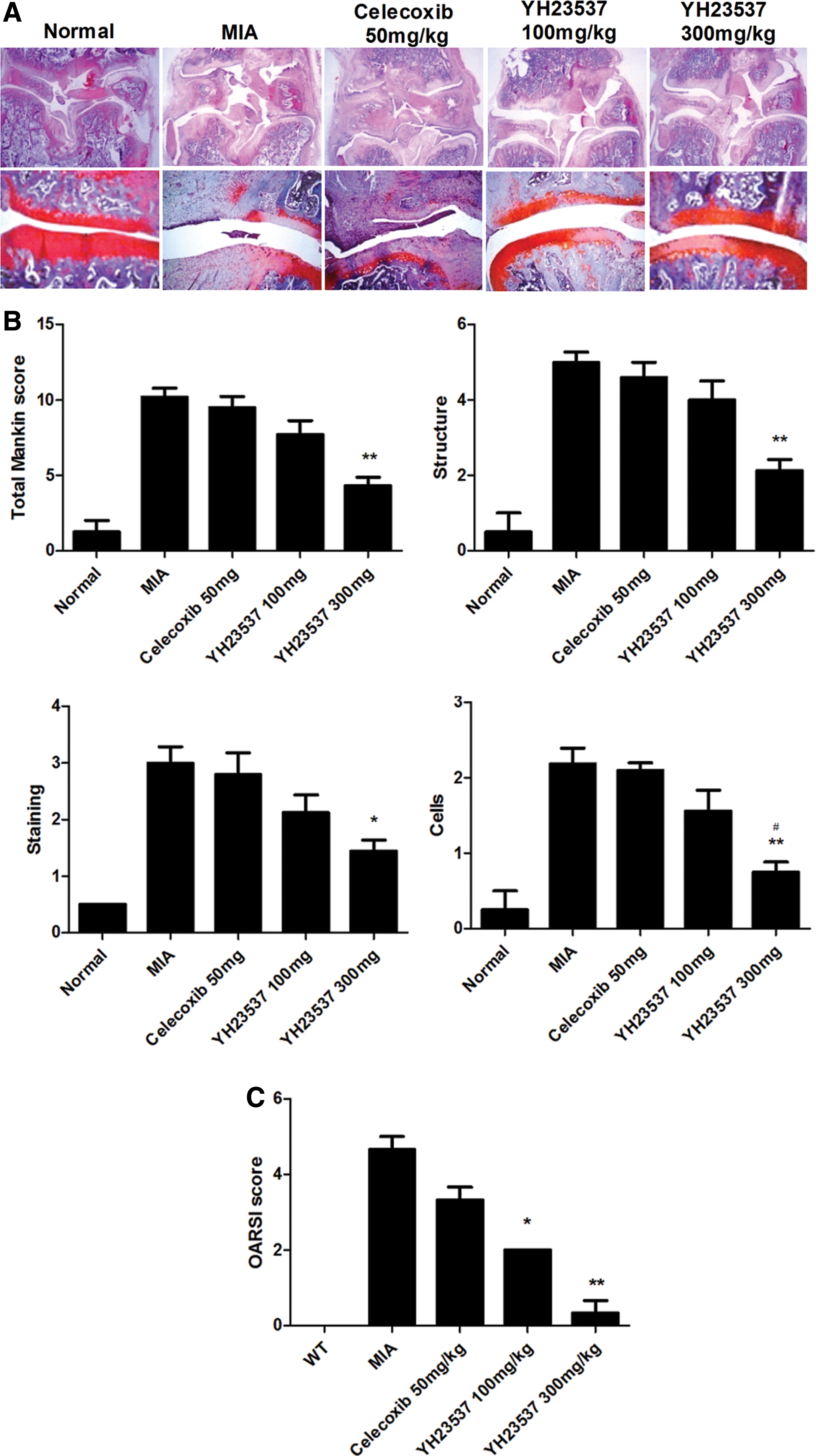
Histological evaluation of joints after treatment with YH23537 in MIA-induced OA. Rats were injected with 3 mg of MIA in the right knee. YH23537 and celecoxib were administered every day for 4 to 28 days after the MIA injection. The knee joints were resected on day 28 after MIA injection.
Ameliorated expression of MMP-3, nitrotyrosine, IL-1β, and IL-6 in the cartilage of YH23537 oral administration OA rats
Expression of the matrix degrading enzymes, MMP-3, was measured to analyze the chondroprotective effect of YH23537. Upregulated MMP-3 expression by MIA injection was downregulated by YH23537 treatment in a dose-dependent manner.
Furthermore, to assess the ability of YH23537 to inhibit oxidative stress related to the pathogenesis of OA, the expression of nitrotyrosine in the joints of OA rats was examined. The nitrotyrosine production was increased by MIA injection; however, it was suppressed by YH23537 administration. Furthermore, proinflammatory cytokines IL-1β and IL-6 expression levels were also determined. Results also showed that cytokines IL-1β and IL-6 were upregulated in the OA joints and expression levels were reduced by YH23537 treatment (Fig. 5).
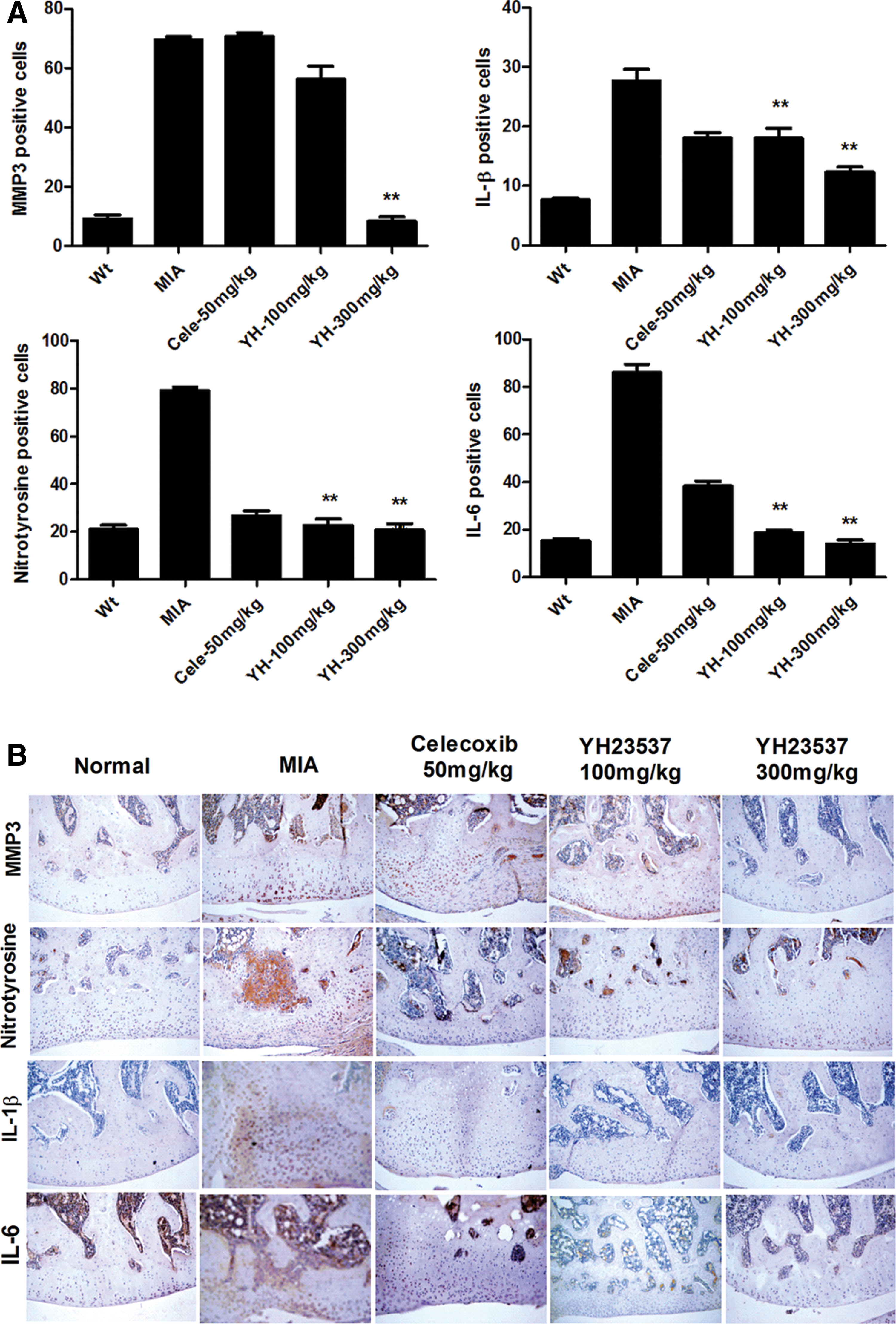
Effects of YH23537 on the expression of MMP3, IL-1β, iNOS, and nitrotyrosine in OA joints.
Increased anabolic activity in human OA chondrocytes by YH23537 treatment
To know the mechanism of YH23537 on chondroprotective properties demonstrated in animal study, we next studied the effects of YH23537 on the expression levels of anabolic factors in IL-1β-stimulated human OA chondrocytes. The induction levels of anabolic mediator TIMP1 and TIMP3 in IL-1β-induced OA chondrocytes were developed significantly after YH23537 oral administration in a dose-dependent response (Fig. 6).
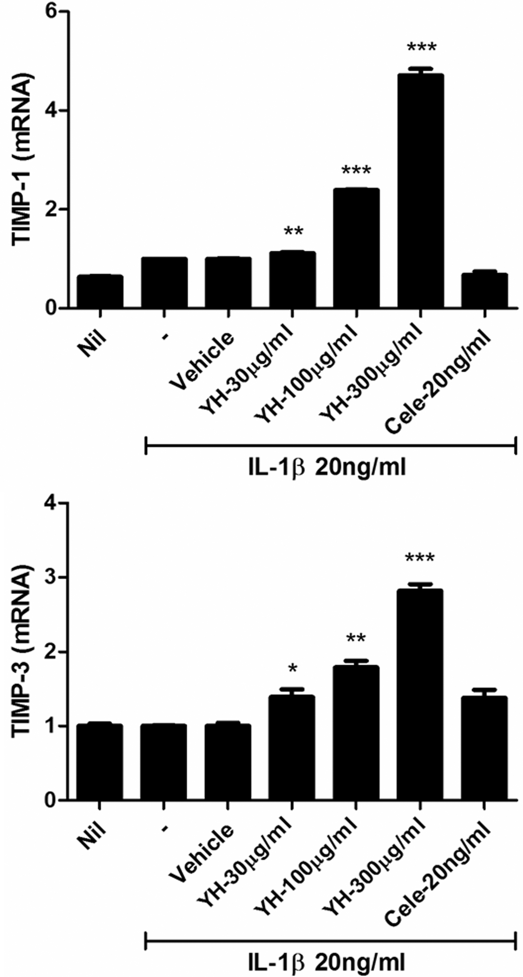
Recovery of anabolic activities in human OA chondrocytes after the treatment with YH23537. Human articular chondrocytes from patients with OA were cultured with IL-1β in the presence or absence of YH23537 for 48 h after 24 h in serum-free medium. The mRNA expression of anabolic factors (TIMP-1, 3) was measured by quantitative RT-PCR; β-actin was used as the internal control. The data are expressed as mean ± SEM from three independent experiments. *P < .05, **P < .01, and ***P < .001 compared with the IL-1β stimulated chondrocytes. RT-PCR, real-time polymerase chain reaction; TIMP, tissue inhibitor of metalloproteinase.
Immunohistochemical assessment of NF-κB and HIF-2α expressions after oral administration with YH23537 in the OA rat model
To understand the molecular signal mechanisms that were efficient to YH23537, we next studied whether YH23537 regulates NF-κB and HIF-2α signaling. NF-κB and HIF-2α are major mediators of mechanical stress to OA progression in joint cartilage. The expression of NF-κB and HIF-2α was increased in OA joint's chondrocyte. YH23537 treatment abrogated the elevated expression (Fig. 7).
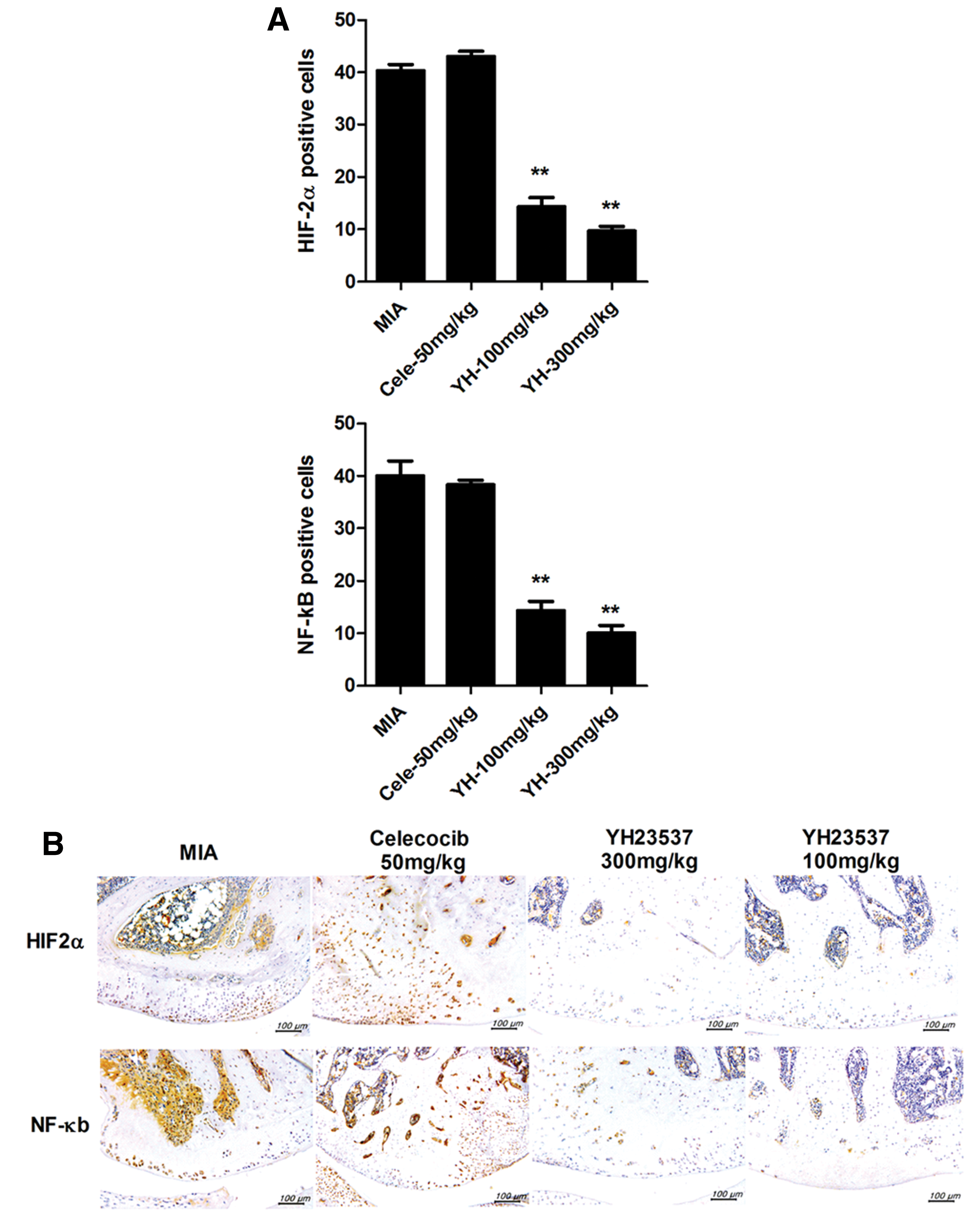
Attenuated induced NF-κB and HIF-2α in YH23537-treated OA rats.
Discussion
In this research, we evaluated the effects of a mixed extracts of Notoginseng Radix and Rehmanniae Radix Preparata (YH23537) in an OA model caused by intra-articular injection of MIA in rats. Data revealed that YH23537 significantly reduced the pain severity as well as the gross and histopathological changes caused by intra-articular injection of MIA in rats.
OA is not solely a degenerative disease, but a multiconstituent inflammatory disease concerning inflammatory mediators, including IL-6, IL-1β, and nitrotyrosine. 24 In this study, we showed that treatment of YH23537 reduced cartilage damage as well as pain severity and attenuated oxidative stress in the subchondral bone and the articular cartilage region in an experimental OA rat model.
It is now recognized that maintained low-grade inflammation plays a major role in OA pathogenesis. 25 We showed that the expression of IL-1β, IL-6, and MMP-3 develops in the MIA-induced OA cartilage. IL-1β is known to attenuate type II collagen expression in chondrocytes 26 and raise MMP. 27 IL-6 has been reported to be increased in knee OA joints. 28 In cooperation with IL-1β, IL-6 expressed MMP-1 and MMP-13 in human cartilage extract culture. 29 The decreased level of these cytokines demonstrates an anticartilage degenerative activity of YH23537. These chondroprotective effects were correlated with the induction of anabolism of the articular cartilage matrix. The degeneration of articular cartilage is the major clinical features of OA. 21 Cartilage degeneration is one of the most general components of joint dysfunction and pain. 30 In addition, a study has demonstrated that OA leads to the deformation of the femur and tibia. 31 The inducer of TIMPs reveals a protective role in the pathogenesis of OA and plays a key role in the development of OA. 32 In this study, we reveal that YH23537 induced the mRNA expression of TIMP-1 and TIMP-3, suggesting that YH23537 may inhibit cartilage degradation, thus alleviating the pain of OA.
Our study showed the effects of YH23537 on NF-κB and HIF-2α expression in the joints. They were decreased with YH23537 oral administration in a dose-dependent response. YH23537 oral administration modulates the protein induction levels of upstream mediators, leading to the production of inflammatory cytokines. These results proposed that the anti-inflammatory effects of YH23537 administration are associated with inhibition of NF-κB and HIF-2α activation. Further molecular studies are required to understand whether each interaction needs factors, are required to understand.
In conclusion, YH23537 oral administration reduced the seriousness of pain and articular cartilage degradation in an MIA-induced OA model. YH23537 diminished oxidative stress and inflammatory cytokine release in the OA joint tissues. We propose that YH23537 may be a viable candidate for the administration of OA.
Footnotes
Acknowledgments
This research was supported by a grant of the Korea Health Technology R&D Project through the Korea Health Industry Development Institute (KHIDI), funded by the Ministry of Health & Welfare, Republic of Korea (grant number: HI14C1002) and by Basic Science Research Program through the National Research Foundation of Korea (NRF) funded by the Ministry of Education (NRF-2015R1D1A1A01057072).
Author Disclosure Statement
Jang-Woo Shin, Hyun-Je Park, and Se-Woog Oh are employees and stock holders of Yuhan Corporation, and Jang-Woo Shin and Hyun-Je Park are included as inventors on a patent filing for YH23537.
The authors have no financial conflict of interest.