
Abstract
Agrimonia pilosa Ledeb has been previously reported to produce an anti-nociceptive effect in ICR mice in both tail-flick and hot-plate tests. Studies have shown that Salvia miltiorrhiza Bunge, also renowned in traditional Chinese medicine, is an effective anti-inflammatory treatment. Among the extraction solvents investigated, a 50% ethanol (EtOH) extract of A. pilosa produced the highest anti-nociceptive effect in monosodium uric acid-induced gout pain models and the greatest yield. The 80% EtOH extract of S. miltiorrhiza moderately inhibited lipopolysaccharide-induced nitric oxide release from RAW 264.7 murine macrophages and exhibited outstanding yield. The mixture of optimized A. pilosa and S. miltiorrhiza extracts were evaluated for enhanced anti-nociceptive effects in gout arthritis treatment. To control extract quality, four marker compounds were determined using an HPLC-DAD method. A 1:1 mixture of A. pilosa 50 and S. miltiorrhiza 80% EtOH extracts of produced better results than when the extracts were administered individually.
Introduction
P
Agrimonia pilosa Ledeb (AP) is a well-known traditional Chinese medicine and is listed in Pharmacopeia of the People's Republic of China. 4 AP has been used traditionally to treat abdominal pain, sore throat, headaches, bloody discharge, eczema, and parasitic infections. Modern pharmacological studies have shown that this medicinal plant has antihemorrhagic, antioxidant, hepatoprotective, and anticancer properties. 5,6 Also, AP was already well known for its anti-nociceptive properties 7 and polyphenols, flavonols and their derivatives have been identified. Moreover, AP's constituents were reported to contain active substances that contribute to its anti-inflammatory and anti-nociceptive properties. 8,9
Salvia miltiorrhiza bunge (SM) is a well-known traditional Chinese medicine used for the treatment of hemorrhages, menstrual disorders, miscarriage, swelling, inflammation, and pyretic arthralgia. 10,11 The major active constituents of SM are tanshinones (including tanshinone I, tanshinone IIA (TSA), and cryptotanshinone, which belong to a group of lipid-soluble diterpenoids), and water-soluble polyphenolic compounds (including caffeic acid, salvianolic acid A, salvianolic acid B (SAB), salvianolic acid D, etc.). 12 These constituents in SM have anti-inflammatory and antioxidative properties. 13,14
We hypothesized that the symptoms of gout arthritis could be treated using the anti-nociceptive properties of AP and anti-inflammatory properties of SM. The objective of this study was to optimize extraction conditions and mixed ratios of extracts of AP and SM for anti-nociceptive activity. Therefore, we first investigated the inhibitory effects of various ethanol (EtOH) ratios (%) on anti-nociceptive effects of AP and the anti-inflammatory activity of SM, respectively; to increase the anti-nociceptive effect, mixtures of AP and SM extracts were evaluated at different ratios for anti-nociceptive activity in gout arthritis treatment model. In addition, four marker compounds were simultaneously validated for quality control and development of functional food source from mixtures of the AP and SM extract using a simple and reliable HPLC-DAD method.
Materials and Methods
Plant materials and reagents
AP and SM specimens were collected in Yeongcheon-si and Yeongyang-gun in Gyeongsangbuk-do Province, South Korea and the voucher specimens (No. 2015-RIC-APYC-03 and 2015-RIC-DSYY-03) were deposited at the Regional Innovation Center, Hallym University, South Korea. The specimens were authenticated by Emeritus Prof. H. J. Chi, Seoul National University. Standard compounds, including rutin (RT) and apigenin-7-O-glucuronide (A-O-G), were isolated from AP in our laboratory. 15 Reagents and solvents were purchased from Sigma-Aldrich Co., unless stated otherwise.
Extract and standard solution preparation
Dry AP (10 kg) and SM (10 kg) were extracted for 24 h twice at room temperature from 0% to 100% EtOH, respectively. The solvent was evaporated under reduced pressure at 40°C, dissolved in methanol, and then filtered through a 0.45-μm syringe filter before analysis. All samples were analyzed in triplicate. Stock solutions of the RT, A-O-G, TSA, and SAB were prepared by dissolving in methanol. Then, the stock solutions were diluted to five concentrations for the construction of calibration curves in the ranges 9.8–490, 89–1780, 9.8–490, and 89–1780 μg/mL, respectively. Stock and working solutions were all stored at 4°C.
HPLC analysis
The HPLC equipment used was a Dionex series instrument system (Dionex, CA, USA), consisting of a vacuum degasser, quaternary pump, auto-sampler, thermostatted column compartment, and a variable wavelength detector system.
HPLC was conducted using an Agilent Eclipse XDB-Phenyl column (150 × 4.6 mm i.d., 3.5 μm particle size; Agilent). Elution solvents used were 0.1% aqueous trifluoroacetic acid (A), and acetonitrile (B), with a gradient elution as follows: 0–20% B (0–3 min), 20–30% B (3–12 min), 30–30% B (12–17 min), 30–87% B (17–23 min), 87–90% B (23–32 min), 90–100% B (32–35 min), 100–0% B (35–38 min), and 0–0% B (38–48 min). The flow rate was 0.7 mL/min and the injection volume was 10 μL at a sample concentration of 5 mg/mL. RT and A-O-G were detected at 334 nm, and TSA and SAB were detected at 254 nm.
Method validation
Linearity, limits of detection (LOD), limits of quantification (LOQ), intra- and inter-day precisions, and recovery were conducted to validate the HPLC method using the formula in ICH guideline Q2B. 16 LOD and LOQ were calculated at signal-to-noise ratios of 3 and 10, respectively, with the standard deviation (SD) of the response and slopes of the concentration obtained from the calibration curves. Intra-day precision was evaluated in three replications during the same day, whereas inter-day precision was assessed by comparing the assays in six replications over 3 different days. Three replicate extractives at each level were used to determine the extraction recovery rates as mean values ± SD, and then the relative standard deviation (RSD) was calculated.
Animal care
These experiments were approved by the University of Hallym Animal Care and Use Committee (Registration No.: Hallym 2015-06-08). All procedures were conducted in accordance with the “Guide for Care and Use of Laboratory Animals” published by the National Institutes of Health and the ethical guidelines of the International Association for the Study of Pain. 4
Production of monosodium uric acid-induced gout pain model
Monosodium uric acid (MSU) crystals were prepared as described previously. 17 One gram of uric acid in 180 mL of 0.01 M sodium hydroxide (NaOH) was heated to 70°C. NaOH was added as required to maintain the pH between 7.1 and 7.2, and the solution was filtered and incubated at room temperature, with slow and continuous stirring, for 24 h. MSU crystals were kept sterile, washed with EtOH, dried, autoclaved, and resuspended in phosphate-buffered saline (PBS) by sonication. MSU crystals contained <0.005 endotoxin U/mL of endotoxin (Limulus Amebocyte Lysate Endotoxin Assay; GenScript).
In most experiments (and unless stated otherwise), 0.5 mg of MSU crystals in 10 μL of PBS was injected intra-articularly (IA) in one ankle joint, and PBS alone was injected in the contralateral ankle joint. We used Microliter #705 syringes (Hamilton, Whittier, CA, USA) with 27-gauge needles for all IA injections. Injections were performed with the mice under isoflurane anesthesia, and the quality of IA injection was controlled by assessing the location of MSU crystal deposits histologically on ankle tissues collected 24 h after the injection.
Cell culture and treatment
RAW 264.7 murine macrophages were cultured and maintained at 37°C under a 5% CO2 atmosphere in DMEM containing 10% heat-inactivated FBS, 100 U/mL penicillin, and 100 μg/mL streptomycin, as previously described. 10 RAW macrophages 1 × 10⁶/mL were cultured in DMEM media supplemented with 5% FBS in a 24-well plate. Cells were first pretreated for 1 h using a variety of extract concentrations, followed by lipopolysaccharide (LPS) (5 μg/mL) treatment, and then the supernatant was collected after 24 h.
Measurement of nitric oxide
After treatment with extracts and LPS for 24 h, the RAW 264.7 murine macrophage culture media was collected and centrifuged at 12,000 × g RPM for 5 min, and the supernatant was saved for the measurement of nitrite, an indicator of nitric oxide (NO) production. The supernatant (100 μL) was incubated with 100 μL of Griess reagent (0.1% napthalethylenediamine dihydrochloride, 1% sulfanilamine, and 2.5% H2PO4) at room temperature for 20 min in the dark. The absorbance was read at 540 nm wavelength, using a Spectramax M2/e fluorescence microplate reader (Molecular Devices). Nitrite concentration was calculated using sodium nitrite as the standard, as previously described. 18
Drug administration
Oral administration was performed with gavage in a volume of 1 mL/kg body weight. Mice were administered orally with either vehicle or mixtures of the two extracts at ratios including 5:1, 2:1, 1:1, or 1:2, and each solvent extracts of AP.
Statistical analysis
Statistical analysis was carried out by student t-test GraphPad Prism Version 4.0 for Windows (GraphPad Software, San Diego, CA, USA). P values less than .05 were considered to indicate statistical significance. All values were expressed as the mean ± SEM.
Results
Optimization of HPLC-DAD condition
All four components in the mixture of AP and SM extract were completely separated by HPLC. To develop an accurate, valid, and optimal HPLC analysis method, various influencing factors, including analytical column, temperature, mobile phase, flow rate, and detection wavelength were investigated. Results indicated that good separations were carried out on Agilent Eclipse XDB-Phenyl column using the mobile phase composed of 0.1% aqueous trifluoroacetic acid and acetonitrile. Additionally, acetonitrile was found to have a better analytic resolution than methanol. 19 These results indicated that this method exhibits satisfactory selectivity and specificity (Fig. 1).
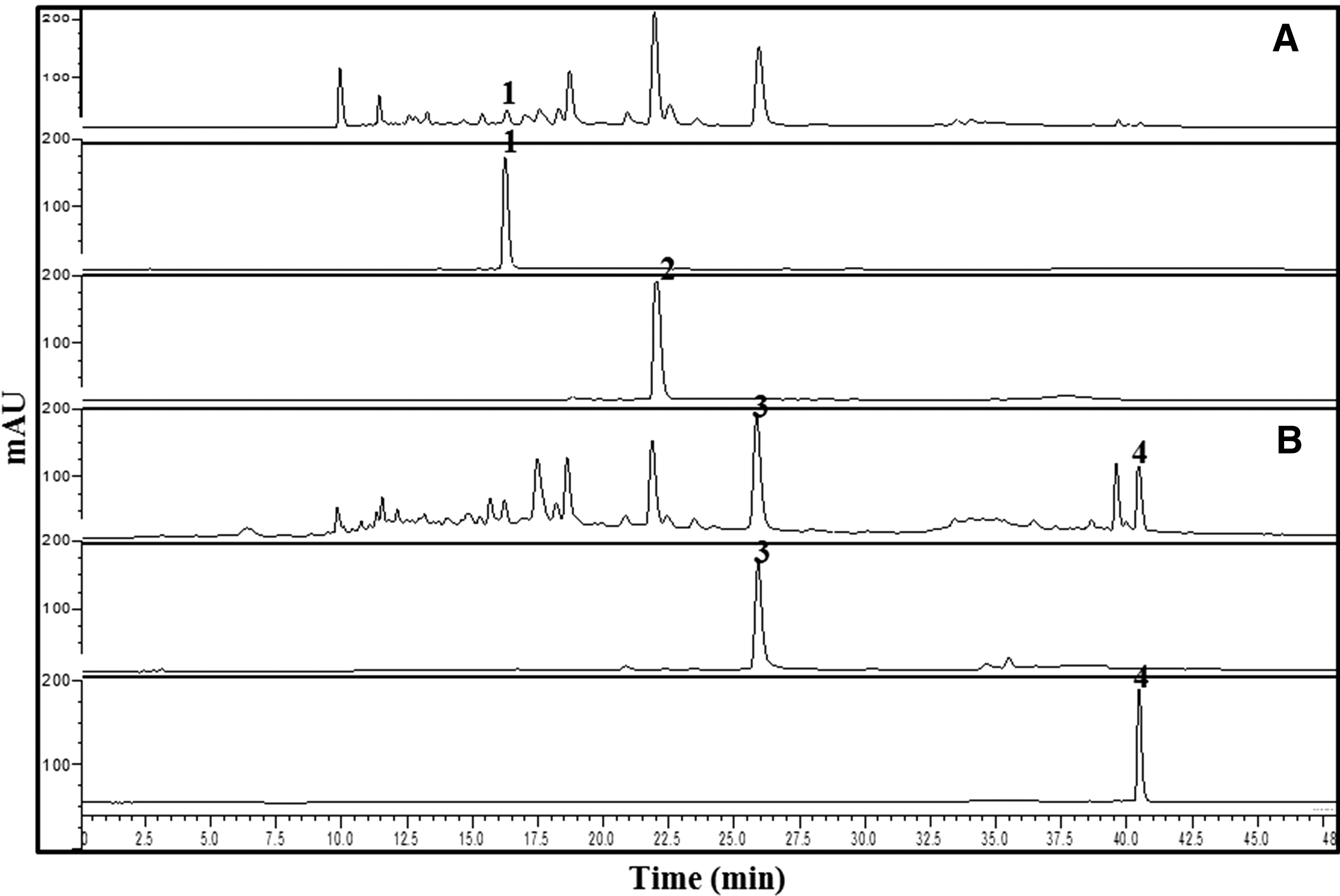
Representative HPLC chromatograms of mixture extract from 50% ethanol extract of Agrimonia pilosa Ledeb and 80% ethanol extract of Salvia miltiorrhiza Bunge at
Linearity, LOQ, and LOD
Method linearity for each of the four compounds was analyzed at five concentration levels in triplicate. The calibration curves were prepared from peak area versus concentration of the standards, and showed that the developed method was linear across ranges of 9.8–490, 89–1780, 9.8–490, and 89–1780 μg/mL, for compounds (RT, A-O-G, TSA and SAB, respectively, with a good correlation coefficient (r 2 ≥ 0.999) (Table 1). As shown in Table 1, LOQ and LOD under the present chromatographic conditions were expressed as signal-to-noise ratios of 3 and 10, respectively. LOQ and LOD for four compounds were found to be 0.01 and 0.02, 0.04 and 0.13, 0.01 and 0.02, and 0.02 and 0.06 μg/mL, respectively.
LOD: 3.3 × (standard deviation of the response/slope of calibration curve).
LOQ: 10 × (standard deviation of the response/slope of calibration curve).
LOD, limits of detection; LOQ, limits of quantification.
Precision, accuracy, and recovery
Intra- and inter-day variations were chosen to validate method precision. Intra-day precision was determined three times in 1 day using three concentrations of mixed standard solutions under optimized conditions. Inter-day precision was determined once a day for three consecutive days using the same mixed standard solutions as described above. RSD (%) values for intra- and inter-day tests were within the ranges of 0.18–1.62% and 0.35–2.34%, as shown in Table 2. These results indicated that the HPLC method enabled quantitative determinations of RT, A-O-G, TSA, and SAB in AP and SM extracts with a high degree of precision. The recovery test was conducted using three different concentrations of the four standards added to the known sample.
Intra-day precision tested three times on 1 day.
Inter-day precision tested on 3 different days.
RSD (%), relative standard deviation.
The average recovery (%) of each analysis showed the accuracy of this analytical method. 20 The average recovery of RT, A-O-G, TSA, and SAB ranged from 102.54% to 109.48%, 97.71% to 109.80%, 104.36% to 109.46%, and 107.22% to 109.82%, respectively, and all RSD values were <2.00%, as shown in Table 3.
Sample analysis and anti-nociceptive effect from A. pilosa extracts
In this study, we found that AP extract administered orally produces anti-nociception in mechanical pain models. Several previous studies have demonstrated that MSU can be used in gout pain models. 17,21 To determine which EtOH percentage extract produced the greatest anti-nociceptive response (Fig. 2), MSU was injected into the ankle joints of mice. Twenty-four hours after MSU treatment, mechanical pain thresholds were measured using the von Frey test. MSU treatment caused a significant reduction in mechanical pain thresholds as measured by von Frey testing. AP extracts (0%, 10%, 30%, 50%, 60%, 80%, or 100% EtOH) were administered orally and pain thresholds were measured 30, 60, 90, and 120 min after oral administration. As shown in Figure 3, a 50% EtOH extract from AP effectively reduced MSU-induced pain thresholds.
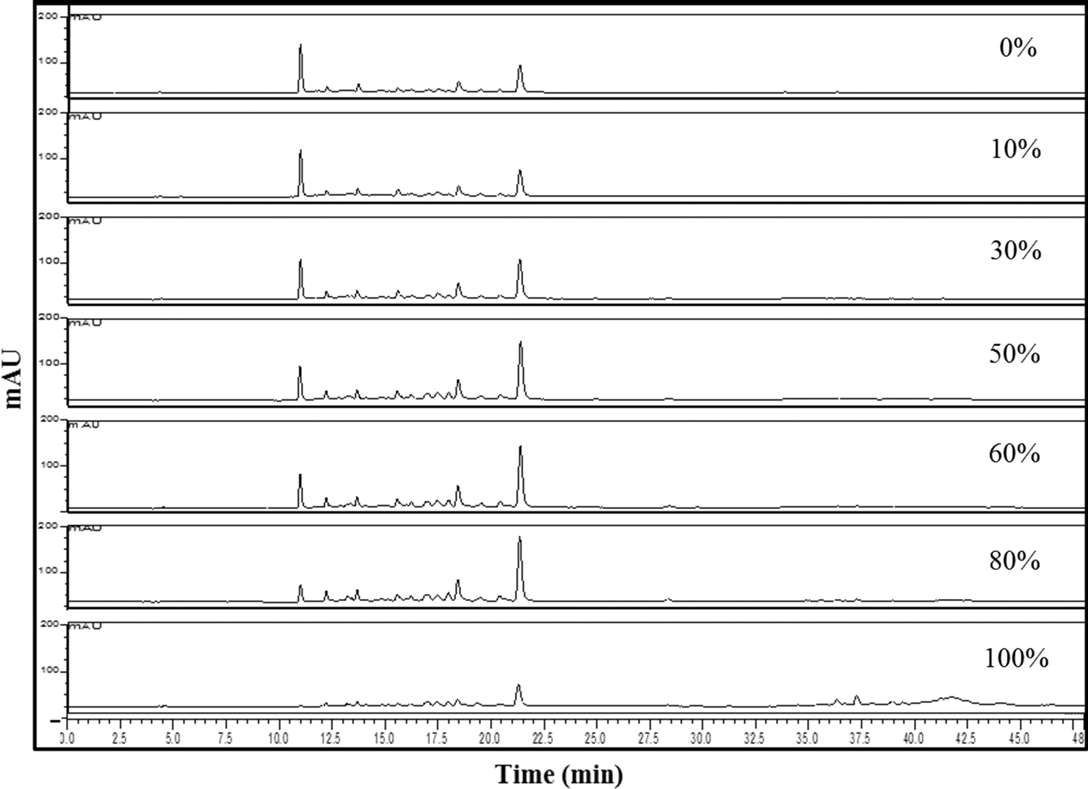
HPLC chromatograms of A. pilosa Ledeb extracts along with different ethanol percentage (0%, 10%, 30%, 50%, 60%, 80%, and 100% ethanol).
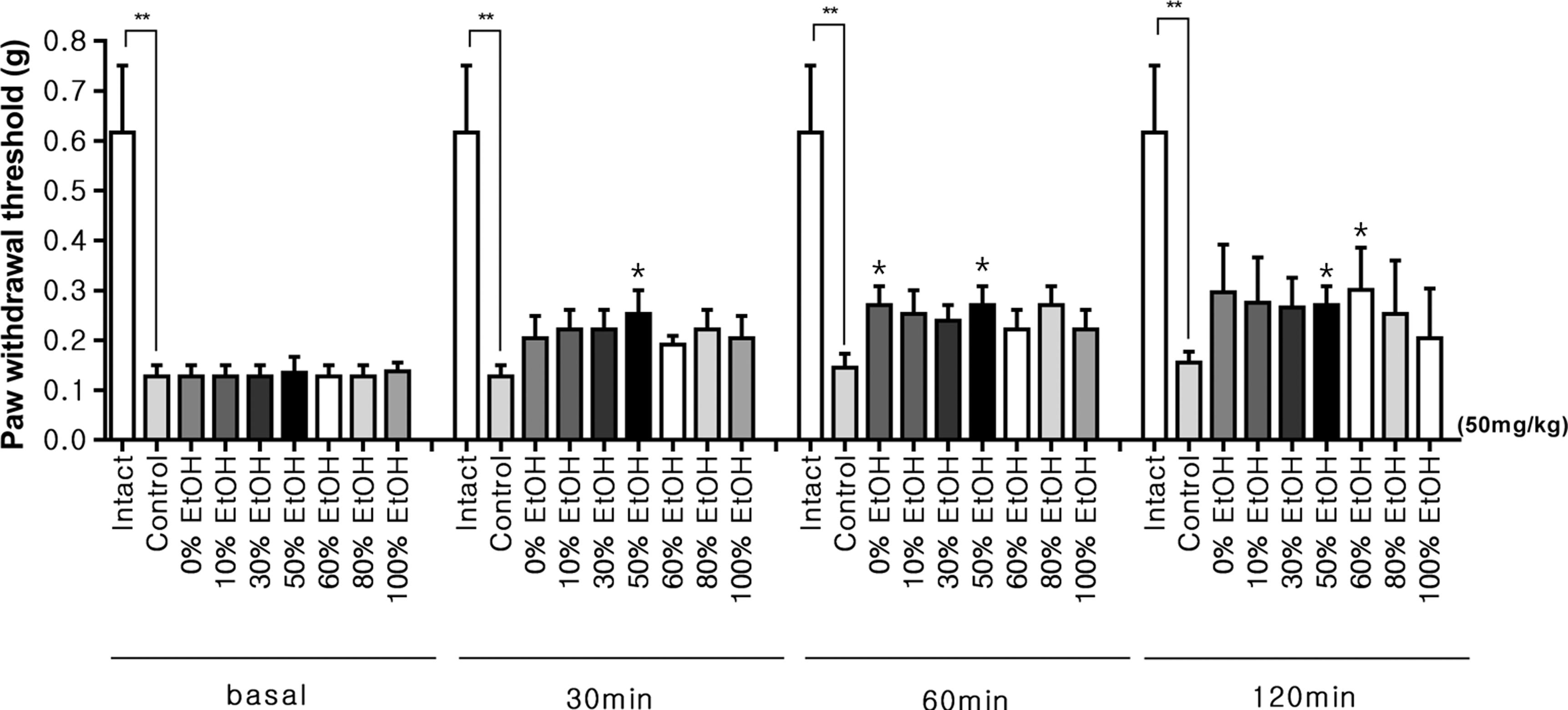
Antinociceptive effect of A. pilosa Ledeb extracts administered orally in MSU-induced pain model. Mice were administered orally with either vehicle or A. pilosa Ledeb extracts (0%, 10%, 30%, 50%, 60%, 80%, and 100% EtOH) and the pain threshold was measured at 30, 60, 90, and 120 min after treatment using the von Frey test. Vertical bars denote the standard error of the mean. The number of animals used for each group was 8–10 (*P < .05, **P < .01 compared to the vehicle-treated control group of mice). The glucosamine is the positive control for MSU-induced pain model. The control (negative) means in MSU-induced pain model without test samples. EtOH, ethanol; MSU, monosodium uric acid.
The proposed HPLC-DAD method was applied for quantitative analysis of the contents of RT and A-O-G from different extracts along with EtOH percentage. As indicated in Table 4, an 80% EtOH extract from AP produced significant amounts of RT and A-O-G, at 1.47 mg/g, and 37.12 mg/g, respectively. In contrast the amount of RT and A-O-G found in a 50% EtOH extract from AP was more moderate, at 1.85 mg/g and 27.85 mg/g, respectively. These findings led us to believe that RT and A-O-G, along with other related components, influence anti-nociception. A 60% EtOH extract from AP produced the highest overall yield (7.91%), and a 50% EtOH extract from AP had the second-highest yield (7.64%). Based on these results, we selected a 50% EtOH extract from AP in anticipation of producing an increased anti-nociceptive effect when blended with other extracts.
EtOH, ethanol.
Sample analysis and anti-inflammatory activity from S. miltiorrhiza extracts
In this study, we found that SM extract exerts an inhibitory effect against LPS-induced NO release in RAW cells. This effect suggests that SM possesses anti-inflammatory properties. The results of this study are partially consistent with those of previous studies in that several types of inflammatory responses are inhibited by SM. 22,23 We used LPS-induced RAW cells to select the most effective extract according to EtOH percentage. RAW cells treated with LPS (5 μg/mL) for 24 h caused a profound release of NO, as shown in Figure 3. Several concentrations (100–300 μg/mL) of SM extracts (0%, 20%, 50%, 80%, or 100% EtOH) were added to the media 1 h before LPS treatment.
As indicated in Figure 4, the 50%, 80%, and 100% EtOH extracts of SM attenuated an LPS-induced NO release from the RAW cells in a concentration-dependent manner. The treatment of RAW cells with 50%, 80%, and 100% EtOH extract of SM did not affect basal NO levels.
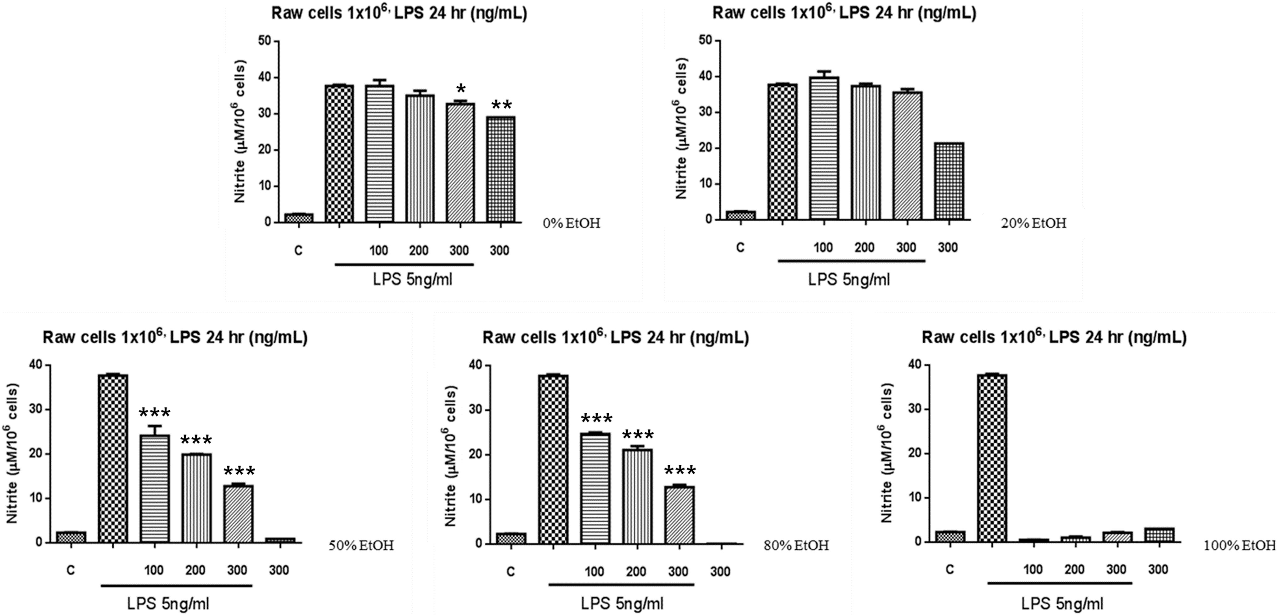
Effect of S. miltiorrhiza Bunge extract on LPS-induced NO release from cultured RAW cells. Several concentrations (from 100 to 300 μg/mL) of S. miltiorrhiza Bunge extract (0%, 20%, 50%, 80%, and 100% ethanol) were added to the media 1 h before LPS treatment. NO level in the medium was measured after RAW cells were incubated with LPS for 24 h. Vertical bars denote the standard error of the mean. (*P < .05, **P < .01, ***P < .001 compared to the vehicle-treated control group of mice). LPS, lipopolysaccharide; NO, nitric oxide.
The proposed HPLC-DAD method was applied for quantitative analysis of the contents of SAB and TSA from different extracts along with EtOH percentage. As shown in Table 5, a 50% EtOH extract from SM contained the greatest amounts of SAB at 69.12 mg/g, but also contained 1.89 mg/g of TSA (Fig. 5), an 80% EtOH extract had 109.08 of TSA and 53.03 mg/g of SAB, and an 100% EtOH extract had the highest 38.94 of SAB and 94.59 mg/g of TSA (Fig. 5). Therefore, although the 50%, 80%, and 80% EtOH extracts from SM produced similar anti-inflammatory responses, we selected the 80% EtOH extract because its yield (40.52%) was more than the 50% and 100% EtOH extracts (20.71% and 2.32%).

HPLC chromatograms of S. miltiorrhiza Bunge extracts along with ethanol percentage (0%, 20%, 50%, 80%, and 100% ethanol).
Anti-nociceptive effect of mixtures of A. pilosa and S. miltiorrhiza extracts on mechanical pain thresholds in MSU-induced pain model
A 50% EtOH extract of AP produced the greatest anti-nociceptive response in MSU-induced pain models, whereas an 80% EtOH extract of SM produced an inhibitory effect against LPS-induced NO release from RAW cells. To create a potentially increased anti-nociceptive effect, these two percentage extracts were mixed according to several blend ratios of AP:SM, (5:1, 2:1, 1:1, or 1:2), and administered against pain thresholds at 30, 60, and 120 min after treatment using the von Frey test. As shown in Figure 6, a 50% EtOH extract of AP (A) combined with an 80% EtOH extract of SM (D) was potent enough to be highly effective in MSU-induced pain models. Although no synergistic effect occurred, greater additive results were produced than when AP and SM were administered individually.
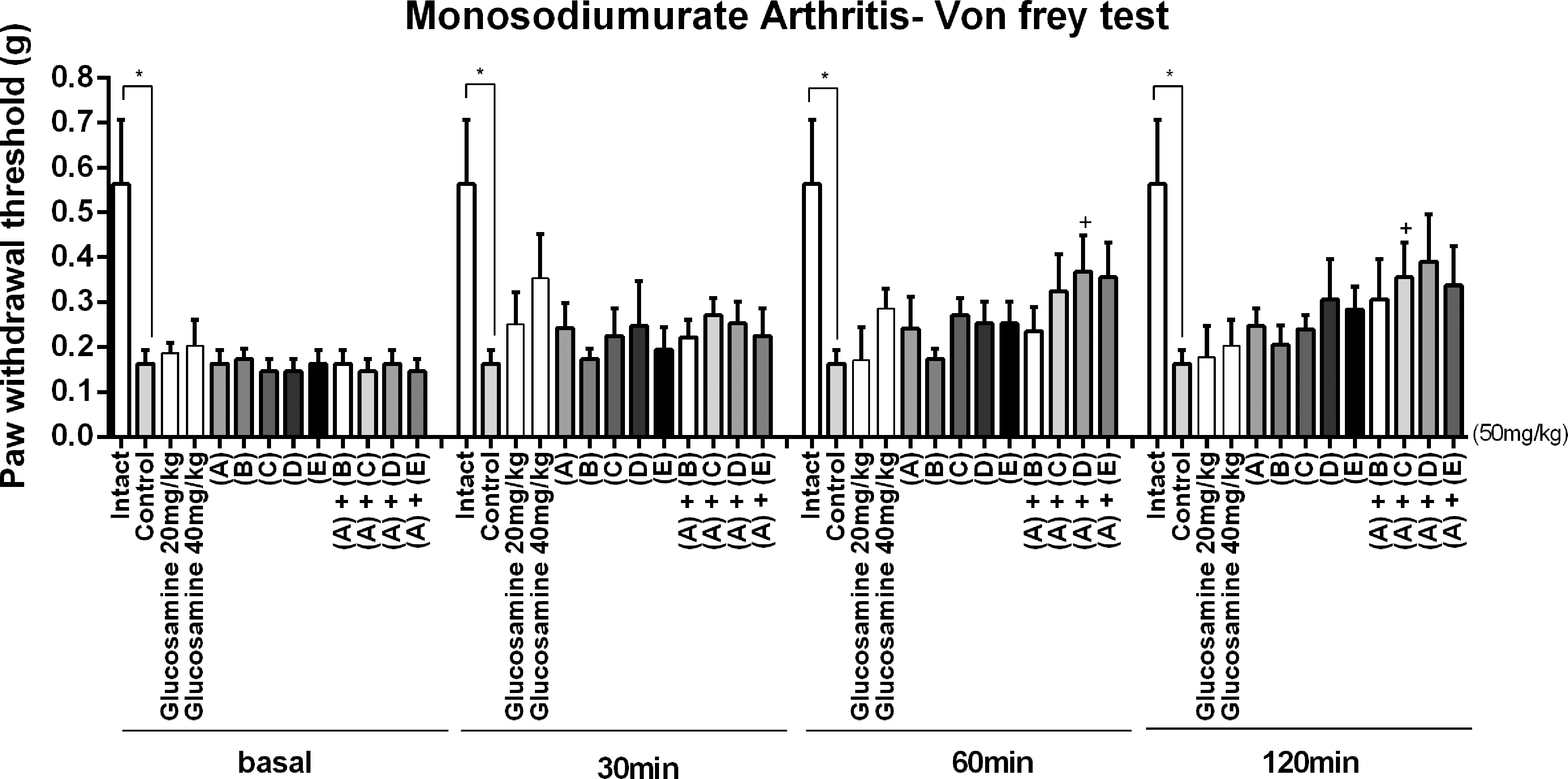
Anti-nociceptive effect of mixture extracts from A. pilosa Ledeb and S. miltiorrhiza Bunge administered orally in MSU-induced pain model. Mice were administered orally with either vehicle or complex extracts (5:1, 2:1, 1:1, or 1:2) and the pain threshold was measured at 30, 60, and 120 min after treatment using the von Frey test. Vertical bars denote the standard error of the mean. The number of animal used for each group was 8–10 (*P < .05 compared to the vehicle-treated control group of mice). Extracts are grouped as follows: (A) 50% EtOH extract of A. pilosa Ledeb (50 mg/mL), (B) 80% EtOH extract of S. miltiorrhiza Bunge (10 mg/mL), (C) 80% EtOH extract of S. miltiorrhiza Bunge (25 mg/mL), (D) 80% EtOH extract of S. miltiorrhiza Bunge (50 mg/mL), (E) 80% EtOH extract of S. miltiorrhiza Bunge (100 mg/mL). The glucosamine is the positive control for MSU-induced pain model. The control (negative) means in MSU-induced pain model without test samples.
Therefore, we were simultaneously determined for quantitative analysis of four compounds using HPLC-DAD from mixture of AP and SM extracts. As shown in Table 6, an extract ratio of 1:1 (AP: SM, v/v) contained 4.03 mg/g, 17.36 mg/g, 4.62 mg/g, and 33.70 mg/mg of RT, A-O-G, TSA, and SAB, respectively. Findings indicated that greater proportions of AP content resulted in higher amounts of RT and A-O-G being determined; similarly, greater proportions of SM content resulted in higher amounts of TSA and SAB being determined.
Discussion
Our research team reported and suggested that AP 80% EtOH extract administered orally produces anti-nociception in various pain models and administration of AP 80% EtOH extract in ICR mice may exert their anti-nociceptive effect via the central sites, possibly spinally mediated mechanisms. 2 In addition, AP 30% EtOH extract shown to significantly decrease LPS-induced NO production in RAW 264.7 cells by Hwang et al., 24 and was reported to inhibit mRNA expressions of iNOS, COX-2, TNF-α, IL-1β, and IL-6 in LPS-stimulated cells in a dose-dependent manner by Chen et al. 25 Furthermore, Kim et al. reported that AG extract inhibited the OVA-induced airway inflammation 26 and may provide a valuable therapeutic strategy in treating various inflammatory diseases. Salvia species have been used for the treatment of various inflammatory ailments in traditional medicine. 27
Preliminary results demonstrated that three Salvia species exert anti-inflammatory activities on carrageenan-induced inflammatory paw edema model. Also, SM and it constituents could inhibit RAW 264.7 cells, inducible NO synthase and cyclooxygenase-2-mediated PGE2 production. 28 TSA in SM reduced persistent spontaneous nociception in both phases 1 and 2 in the formalin test, the number of writhes in the acetic acid writhing test and mechanical hypersensitivity in bee venom model. 29 Based on the previous reports and our results, the evidence suggests that administration of a mixture of AP and SM extracts can inhibit gout pain and enhances anti-inflammatory activity in the body, and implies that administration of a mixture of AP and SM extracts in a certain ratio can more effective than single administration of either AP and SM alone.
In summary, a simple, accurate, and rapid HPLC has been developed to quantify these four polyphenols in mixed extract and was successfully validated. To the best of our knowledge, this is the first time that a HPLC gradient method has been applied to the simultaneous determination of four polyphenols in a mixed extract. Our results suggested that the HPLC separation combined with an adequate efficiency described in this work allows the application of the method for routine and QA/QC analysis from mixed and complex extracts. In addition, taking into consideration both yield and response, the 50% EtOH extract of AP was shown to produce both the highest anti-nociceptive effect in MSU-induced gout pain models and the greatest yield. The 80% EtOH extract of SM produced a moderate inhibitory response against LPS-induced NO release from RAW 264.7 murine macrophages, and exhibited superior yield that was as much as two times that of the 50% EtOH extract. Based on anti-nociceptive response, we selected the most effective ratio among mixture extracts. The results shown in this study suggest that a mixture extract consisting of a 1:1 ratio comprised of a 50% EtOH extract of AP and an 80% EtOH extract of SM has significant therapeutic potential for treating gout pain and the mixed extract may potentially be used as an herbal drug to treat pain.
Footnotes
Acknowledgments
This work was supported by Korea Institute of Planning and Evaluation for Technology in Food, Agriculture, Forestry and Fisheries (IPET) through High Value-Added Food Technology Development Program, funded by the Ministry of Agriculture, Food and Rural Affairs (MAFRA) (115001-3) and the Basic Science Research Program through the National Research Foundation of Korea (NRF) funded by the Ministry of Education (2015R1D1A1A01059199).
Author Disclosure Statement
No competing financial interests exist.