
Abstract
In this study, the effects of Humulus japonicus (HJ) aqueous extract on 3T3-L1 preadipocytes and HepG2 cells (in vitro model) as well as on C57BL/6 mice fed on high-fat diet (HFD) (in vivo model) were evaluated. Mice fed on HFD for 12-weeks were taken the HJ water extract (HJW) at various doses, 50, 150, and 250 mg/kg, orally for 8 weeks. We have noticed the accumulation of fat globules in preadipocytes and HepG2 cells using Oil Red O staining. In addition, supplementation with HJW considerably inhibited the weight gain, lipid accumulation, and adipogenesis and decreased the size of subcutaneous adipocytes in 3T3-L1 adipocytes. Furthermore, treatment with HJW improved hyperlipidemia via decreasing the levels of serum triglyceride (TG) and low-density lipoproteins as well as the atherogenic index. Supplementation with HJW could attenuate HFD-induced lipid accumulation, increase the mRNA expressions of fatty acid synthase (FAS) and stearoyl-CoA desaturase (SCD1), and would elevate the levels of serum aspartate aminotransferase and alanine aminotransferase in mice liver. The levels of TG and FAS mRNA in HepG2 cells treated with palmitate were reduced in a dose-dependent manner. In sum, HJW could alleviate the HFD-induced obesity and decrease the dyslipidemia profiles; the keys that could contribute to cardiovascular and nonalcoholic liver diseases.
Introduction
O
Hyperlipidemia is a disorder characterized by elevation of plasma cholesterol and triglyceride (TG) levels. 4 Hyperlipidemia is among the highest potential risk factors that could contribute to the development of atherosclerosis, which increases the incidence of cardiovascular disease. 3,5 Generally, the managemental panels of hyperlipidemia are associated with the upregulation of high-density lipoprotein (HDL) or the downregulation of low-density lipoprotein (LDL), total cholesterol (TC), or TG. 6 Excessive caloric intake with obesity is often associated with NAFLD. Furthermore, NAFLD is associated with overall higher liver-related mortality. 7,8 Increases of adipose-related hormone (adipokine) level is considered as a leading cause in the progression of diseases linked to NAFLD. 9 Notably, the protective effects of various compounds and/or bioactive compounds on obesity or hyperlipidemia in NAFLD have been previously investigated. 3,10,11
Humulus japonicus (HJ), ornamental plant native to eastern Asia, exerts antimutagenic, antimicrobial, antimycobacterial, antioxidant, anticancer, and anti-inflammatory effects. 12 –16 Alcoholic extract of HJ could suppress atherosclerosis through inhibiting the proatherogenic factors and lipid accumulation in the aortic endothelium in apolipoprotein E-deficient mice. 17 However, studies on the pharmacological activity of HJ as an antihyperlipidemic and antiobesity agent are very scarce. Therefore, we aimed to determine the effects of HJ water extract (HJW) on the differentiation of 3T3-L1 preadipocytes through analysis of lipid accumulation and gene expression to reveal the antiobesity activity. Furthermore, we target to evaluate various lipid parameters and histological changes using an animal model of obesity and HepG2 cells.
Materials and Methods
Chemicals
HJ extract was generously offered by I-woo Agricultural Company (Seoul, Republic of Korea) and chemical agents used throughout the experimental protocol were acquired from Sigma-Aldrich (St. Louis, MO, USA)
Sample preparation
The lyophilized water extract was prepared from dried HJ that was obtained from Woori Oriental Medicine Materials and authenticated by Dr. Yuan Lu Sun of Solvit P&F (Seoul, Republic of Korea). Dried HJ (650 g) was mixed with 15 L distilled water and boiled for 4 h at 100°C in duplicate. The extract was filtered and the filtrate was evaporated up to 15.6% and finally, lyophilized using freeze-drying lyophilizer (Labconco; Freezone 1 L) at 5 mmHg and −50°C. The lyophilized powder was stored at −30°C pending analysis.
Ultra performance liquid chromatography high-resolution mass spectrometer analysis
Ultra performance liquid chromatography and high-resolution mass spectrometer analysis was performed with a Dionex 3000 RSLC and Thermo Scientific Q-Exactive™ (Hybrid Quadrupole-Orbitrap Mass Spectrometer, Bremen, Germany). The chromatographic separation was carried out on Acquity BEH C18 column (2.1 × 100 mm, 1.7 μm). The mobile phase consisted of 0.1% formic acid in water, and acetonitrile was flowed at a rate of 0.35 mL/min; 10 μL of HJ (10 mg/mL) was injected. Compounds were identified using pure standards of vitexin, luteolin, luteolin 7-O-β-d-glucoside, apigenin, and chlorogenic acid. Negative ion mode electrospray ionization mass spectrometry was used for detection of all analytes. An ion spray voltage of 3.5 kV and S-lens level of 50 were used. Capillary and heater temperatures were set at 350°C and 100°C, respectively.
Animals
Four-week-old male C57BL/6 mice were used (Nara Biotechnology, Seoul, Republic of Korea) for in vivo model study. The animals have free access to food and ad libitum access to water. The animals were placed in cages at temperature of 20°C ± 2°C, relative humidity of 45% ± 10%, and a 12 h light/dark cycle.
High-fat diet and treatment
Ten C57BL/6 mice were randomly assigned to control diet (CD, D12450B; Research Diets, New Brunswick, NJ, USA) or high-fat diet (HFD) (D12492; Research Diets) group and fed for 12 weeks. Four weeks later, the HFD group was treated with saline or HJW (orally administered at 50, 150, and 250 mg/kg/day) for 8 weeks. Body weight gain and food intake were measured twice a week. Finally, all groups were anesthetized with urethane, and serum was harvested for biochemical analysis. Some organs and tissues, such as liver, epididymal fat, inguinal subcutaneous fat, and mesenteric fat were surgically isolated. Immediately, they were weighed and rapidly frozen in liquid nitrogen. This experimental design was approved by the Animal Care and Ethical Use Committee of Chung-Ang University, Republic of Korea (Approval date: 02/11/2016, clearance no. 2015-00074).
Adipocyte morphology
The subcutaneous adipose tissue of control, HFD, and HJW (250 mg/kg) groups was fixed in 4% paraformaldehyde solution. The tissues were sectioned at 4 μm-thicknesses, fixed on slide, stained with hematoxylin and eosin, and mounted on microscope at a final magnification of 200 × . 18
Biochemical analysis
Plasma TG, TC, HDL-cholesterol (HDL-C), LDL-cholesterol (LDL-C), aspartate aminotransferase (AST), and alanine aminotransferase (ALT) levels were run on AU5421 apparatus (Beckman Coulter, Palo Alto, CA,USA). Total lipids were extracted with chloroform/methanol (2:1, v/v) mixture and the extracted TG was measured colorimetrically using TG assay kit (Biovision, Milpitas, CA, USA).
Liver histopathology
The livers of control, HFD, and HJW (250 mg/kg) group were harvested in 10% formalin solution. They were sectioned at 10 μm-thicknesses and stained with Oil Red O. These sections were examined to detect lipid deposition as described elsewhere. 19
RNA extraction and quantitative real-time PCR
TRIzol reagent (Invitrogen, Carlsbad, CA, USA) was used for isolation of total RNAs from liver tissue. Gene expression analysis was performed using quantitative PCR (qPCR). The fluorescent TaqMan 5′ nuclease assay on an Applied Biosystems 7000 sequence detection system (Foster City, CA, USA) was used for qPCR. This process was carried out at 95°C for 15 sec and 60°C for 1 min 40 times and then for 10 min at 95°C. Mouse fatty acid synthase (FAS) (F-TTGCTGGCACTACAGAATGC, R-AACAGCCTCAGAGCGACAAT), stearoyl-CoA desaturase (SCD1) (F-CATCGCCTGCTCTACCCTTT, and R-GAACTGCGCTTGGAAACCTG) mRNA were expressed by normalization to that of mouse beta-actin (Mm00607939_sl; Applied Biosystems).
Cell culture
Dulbecco's modified Eagle's medium (DMEM) high glucose with 10% calf serum was used for culturing of mouse 3T3-L1 preadipocytes. Cell culture maintained at 37°C in a humidified atmosphere with 5% CO2. A mixture of 0.5 mM 3-isobutyl-1-methylxanthine, 100 μM indomethason, 0.25 μM dexamethasone, and 100 mM insulin (DMII) in DMEM containing 10% fetal bovine serum was used for initiation of cell differentiation. The medium was replaced every 48 h. After supplementation with 0, 1, 5, or 10 μg/mL of HJW for 4 days, the treated 3T3-L1 adipocytes were prepared for western blot analysis. The HepG2 cells (ATCC, Manassas, VA, USA) were cultured on DMEM (Invitrogen) supplemented with 10% fetal bovine serum (Invitrogen), 100 U/mL penicillin, and 100 μg/mL streptomycin (Invitrogen).
Oil Red O staining assay
Oil red stained cells were prepared with filtered Oil Red O solution (60% isopropanol and 40% water). The staining was carried for 30 min. After the lipid droplets stained red, the plates were washed and dried. The stained droplets were observed through an Olympus microscope (Tokyo, Japan).
Statistical analyses
Data are expressed as mean ± standard error of the mean. Student's t-test or Tukey's multiple comparison method was used for statistical differences following analysis of variance (ANOVA).
Results
Chemical profile of the HJW
Chemical profile of HJW was compared with ethanolic extract (Fig. 1). Unexpectedly, HJW contained some unknown compounds (Fig. 1A) and their MS/MS data are not shown. The chromatographic profile of ethanolic extract contains the chemical constituents we used as chemical markers, including main components, luteolin-7-D-glucose derivatives (Fig. 1B).

Total ion chromatograms of Humulus Japonicus extract
Effects of HJW on body weight gain and fat pad weight
Food intake amounts were not changed significantly in HFD groups during 12 weeks (Fig. 2A). After 12 weeks, the HFD group exhibited a significantly higher body weight gain than control group (Fig. 2B). All HJW regimens inhibit weight gain in HFD mice. Group treated with 250 mg/kg HJW prevented weight gain by 17.63% and held the highest significance.

Effects of HJW on food intake
Figure 3 shows the weight changes in epididymal as well as mesenteric fat pads of all groups. It has to be noted that the HFD group had a significantly higher fat pads weight than control group. Groups treated with various concentrations of HJW have shown significant decrease in epididymal (Fig. 3A) and mesenteric (Fig. 3B) fat pad weights in a dose-dependent way.
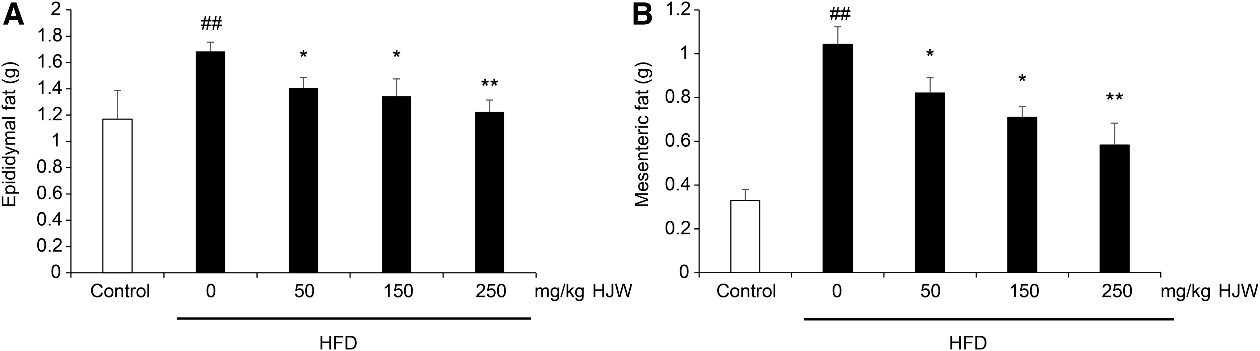
Effects of HJW on epididymal
Effects of HJW on lipid accumulation in subcutaneous adipose tissue
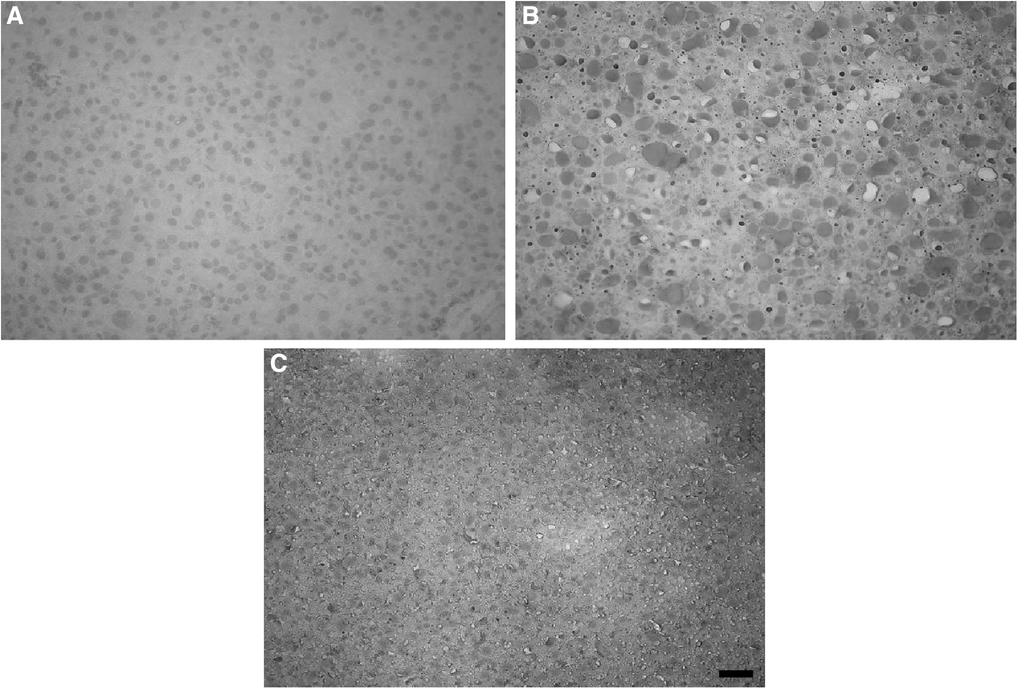
A prominent accumulation of fat globules was observed in adipocytes of HFD group when compared with control mice. This would result in substantial increments in the sizes of subcutaneous adipocytes (Fig. 4B). The HJW-treated group (250 mg/kg) significantly decreases the cell sizes compared with HFD group (Fig. 4C).

Effects of HJW on morphological changes of subcutaneous adipose tissue. Micrographs of adipose tissue sections from control
Effects of HJW on plasma lipid parameters
The serum levels of TC, TG, HDL-C, and LDL-C of the HFD group were significantly higher than control group (Table 1). The levels of TG, TC, and LDL-C were significantly decreased in HJW (250 mg/kg)-treated group. However, the serum HDL-C level of HJW-treated group was not significantly changed. The effect of HJW on the atherogenic index was remarkable (Table 2). The atherogenic index (TG to HDL-C rate) is a reliable marker to assess whether cholesterol is accumulated, metabolized, or excreted. The atherogenic index was significantly reduced in the HJW (250 mg/kg)-treated group. Furthermore, HDL-C/TC and LDL-C/TC were normalized in HJW groups, although the difference was not significant.
Mean ± SEM.
P < .05 or ## P < .01 versus control; * P < .05 or ** P < .01 versus HFD.
HDL-C, high-density lipoprotein cholesterol; HFD, high-fat diet; HJW, Humulus japonicus water extract; LDL-C, low-density lipoprotein cholesterol; SEM, standard error of the mean; TG, triglyceride.
Atherogenic index: TG/HDL-C. Mean ± SEM.
P < .01 versus Control; ** P < .01 versus HFD.
TC, total cholesterol.
Effects of HJW on morphological, biochemical, and functional changes in the liver
In this study, liver histology and function were analyzed in control, HFD, and HJW (250 mg/kg) groups. In HFD group, significant lipid droplets were accumulated in hepatic tissue (Fig. 5A, B), and treatment with HJW would decrease the HFD-induced lipid deposition (Fig. 5C). While TG was significantly increased and accumulated in HFD group, it is significantly decreased in the HJW-treated group (Fig. 6A). The results of quantitative real-time PCR have showed increased mRNA expressions of FAS and SCD1 in HFD group. These increments were significantly attenuated in HJW (250 mg/kg)-treated group (Fig. 6B, C). The AST and ALT levels in HFD group were much higher than control group. Notably, liver enzymes of HJW-treated group were lower than those of HFD group (Fig. 6D, E).
Effects of HJW on the liver histopathological changes in control

Effects of HJW on biochemical and functional changes in the liver.
Effects of HJW on lipid accumulation in 3T3-L1 cells
In preliminary experiments, HJW did not show any cytotoxic effects on 3T3-L1 adipocytes and HepG2 cells at a concentration rate of 20, 100, and 500 μg/mL (Fig. 7). Subsequently, different concentrations of HJW (1–100 μg/mL) were selected.

Cytotoxicity of HJW in 3T3-L1 adipocytes
To investigate the effects of HJW on the differentiation of adipocytes, we have noticed that 3T3-L1 preadipocytes were differentiated into adipocytes with DMII mixture and treated with HJW for 6 days. Oil Red O staining was used to assess the lipid accumulation (Fig. 8A). It has to be noted that treatment with HJW would decrease the degree of Oil Red O staining in a dose-dependent manner (Fig. 8B).
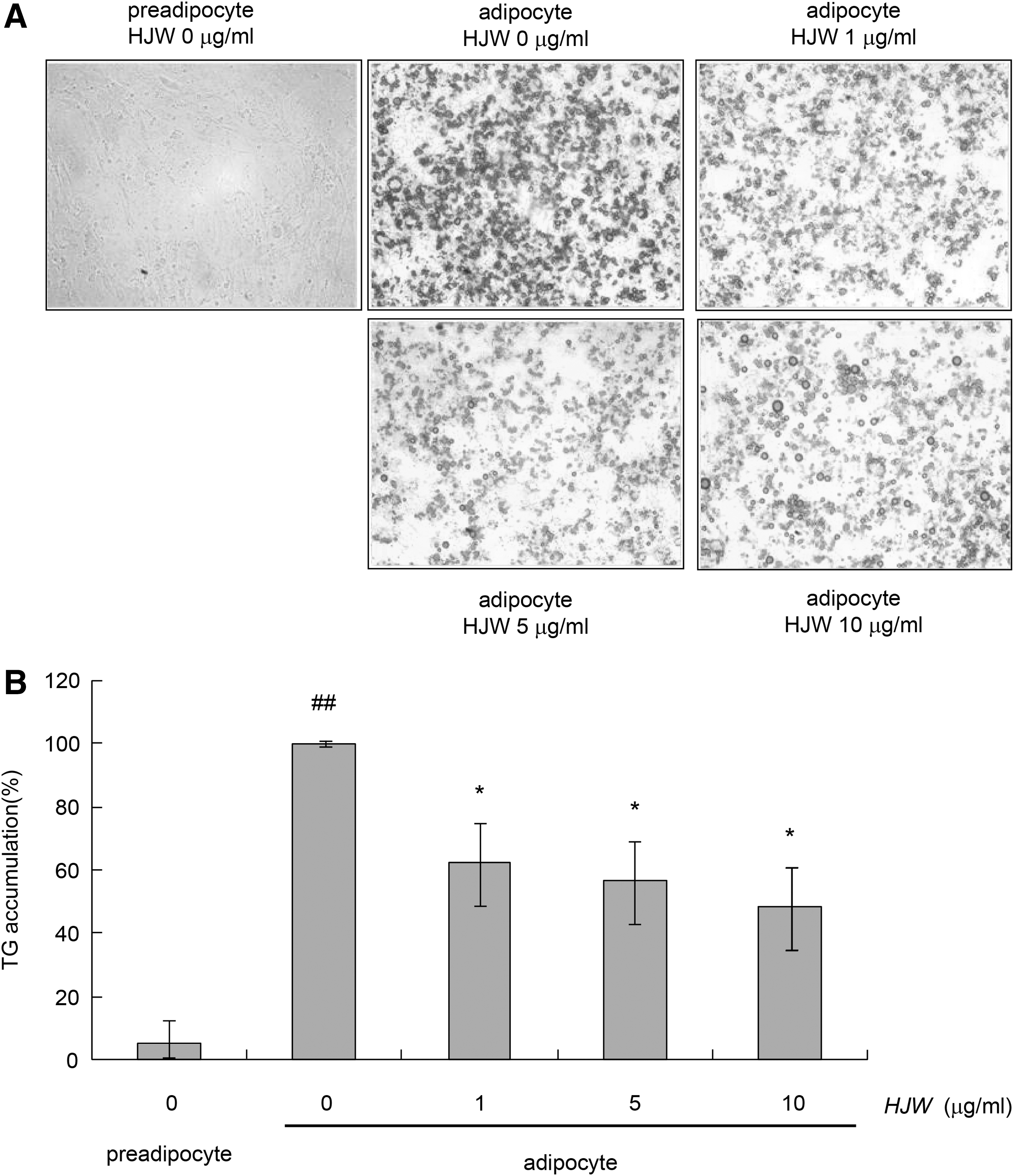
Effects of HJW on intracellular lipid accumulation in 3T3-L1 adipocytes.
Effects of HJW on intracellular lipid accumulation and FAS expression in HepG2 cells
After palmitate treatment, the degree of Oil Red O staining was increased in HepG2 cells; the extent which is reversed by HJW treatment (Fig. 9A). Treatment with HJW significantly lowered the levels of TG and FAS mRNA in a dose-dependent pattern (Fig. 9B, C).

Effects of HJW on intracellular lipid accumulation
Discussion
HJ has been used in the Orient countries as antioxidant and antihypertensive. 17 However, it was not revealed whether it could improve the HFD-induced obesity and its complications or not. In this study, we have evaluated the antiobesity activity of HJW, including its effects on several related complications. Hyperlipidemia and NAFLD-related factors were assessed to evaluate the effects of aqueous extract on several complications of HFD-induced obesity.
Excessive dietary intake, especially fat, is a major component of obesity and related complications. HFD-induced obese animal models are considered effective for assessment of obesity. 20 In this study, body weight gain was significantly increased in HFD group, the finding is attributed to increase in epididymal and mesenteric fat. HFD increased the intracellular lipid content in skeletal muscle with molecular adaptation to favor the fat storage in muscle. 3,21 Herein, there was no significant difference in food intake of HFD group, however, treatment with HJW reduced the body weight gain and accumulation of fat pad compared with HFD group. These results could suggest that HJW would increase the metabolism or fatty oxidation and inhibits cholesterol or lipid particle absorption in the gastrointestinal tract.
Whole-body lipid accumulation leads to hyperlipidemia and metabolic syndromes. 22 In this study, weight gain by HFD elevated the serum lipid profiles and atherogenic index, leading to hyperlipidemic changes. Luteolin supplement, a major component of HJ ethanolic extract, ameliorated obesity and insulin resistance in HFD mice. 23 Furthermore, dietary HJ alleviated cognitive dysfunction in type 2 diabetes and Alzheimer's disease caused by obesity. 24 In addition, luteolin lowered LDL-C and TG level, which improved heart failure in type 1 diabetic cardiomyopathy. 25 In this study, HJW (250 mg/kg) significantly decreased serum TG and atherogenic index level, while decreasing atherogenic lipoproteins such as LDL. High concentration of circulating oxidized LDL is related to metabolic syndrome and can be considered as one of the important causes of cardiovascular disease. 26 High levels of LDL-C are one of the major risk factors for atherosclerosis. 27 As TG induces lipid accumulation in liver and is associated with some metabolic diseases, such as type 2-diabetes, 28 elevated TG concentration could also be considered as a major factor for cardiovascular disease. Therefore, we expect that administration of HJW would have a beneficial effect on hyperlipidemic profiles, including reducing serum TG and LDL-C levels, and the associated complications.
NAFLD is a pathologic category of diseases ranging from isolated hepatic steatosis to nonalcoholic steatohepatitis. 29 Administration of luteolin in an animal model ameliorated ethanol-induced hepatic steatosis and injury. 30 However, in a previous study, administration of HJ has shown a protective effect against D-galactosamine-induced steatosis or alcoholic fatty liver disease, but not NAFLD. 31 In this study, HJW significantly ameliorated NAFLD as assessed by the elevation of AST and ALT and accumulated lipid in the liver. Herein, treatment with HJW significantly reduced liver-related enzyme levels compared with HFD group. The antiobesity or antihyperlipidemia effects of HJW might prevent excess lipid accumulation in the liver cells, resulting in hepatoprotective effect in NAFLD mouse induced by HFD.
Of the various genes involved in fatty acid metabolism, FAS and SCD1 are involved in the synthesis of fatty acids. 32 SCD1 is a δ-9 fatty acid desaturase necessary for the synthesis of monounsaturated fatty acids, specifically oleate and palmitoleate from stearoyl-CoA and palmitoyl-CoA, which are important substrates for TG, cholesterol esters, and phospholipids formation. SCD1 is an important regulator of lipid and glucose metabolism that could affect diabetes, insulin resistance, and hyperlipidemia. 33 In this study, the expression of SCD1 mRNA was greatly reduced by treatments with HJW in HFD group. FAS is a key enzyme involved in lipid metabolism. 34 The expression of FAS gene in mouse hepatocytes and HepG2 cells was significantly reduced by treatment with HJW. Therefore, lipid metabolism can be modulated by HJW, inhibiting hepatic fatty acid synthesis.
In conclusion, dietary HJW ameliorated HFD-induced obesity and hyperlipidemic changes through decreasing the size of adipocytes and normalizing the serum lipid parameters. Furthermore, treatment with HJW could ameliorate the HFD-induced NAFLD via reducing the levels of liver enzymes and lipid droplet accumulations. These effects are partially related to the activity of HJW, which normalize the plasma lipid profile and inhibits fat accumulation. In vitro treatment with HJW would decrease accumulated lipid in 3T3-L1 preadipocytes and HepG2 cells. Further research is needed to reveal the exact mechanisms by which HJW would alleviate obesity.
Footnotes
Acknowledgments
This study was sponsored by Basic Science Research Program through the National Research Foundation of Korea (NRF) funded by the Ministry of Education (NRF-2017R1D1A1B03028725) and the Chung-Ang University Research Scholarship Grants in 2017.
Author Disclosure Statement
No competing financial interests exist.