




Abstract
The inhibitory effects of taegeuk ginseng extract (TGE) on platelet aggregation and thrombus formation were investigated. The TGE significantly inhibited collagen- and adenosine 5′-diphosphate (ADP)-induced platelet aggregation in vitro in a dose-dependent manner. Also oral administration of TGE to rats significantly prevented ADP- and collagen-induced platelet aggregation ex vivo, but it did not affect the plasma coagulation system. The oral administration of TGE significantly delayed the occlusion of the carotid artery in ferrous chloride-treated rats in vivo. These results suggest that in vivo antithrombotic effect of TGE may be due to its inhibitory activity on platelet aggregation rather than on plasma coagulation.
Introduction
P
Ginseng, a root of Panax ginseng C. A. Meyer, has been traditionally used as a natural medicine for enhancement of human health in far-eastern Asia. 4 Ginseng possesses a lot of valuable pharmacological activities, including antiallergy, antioxidation, anticancer, antifatigue, anti-inflammatory, and antithrombotic properties. 1,5 –7 Since cardiovascular disease has been the leading cause of human death throughout the world, 8 the cardioprotective activity of ginseng has been the main subject of ginseng research. Among the pharmacologically active components of ginseng, ginsenosides Rb1, Rg1, and Rg3 have been known as main contributors to the cardiovascular beneficial effects. 8 Gisenosides Rb1 and Rg3 were reported to modulate vascular function. 9,10 Ginsenosides Rb1 and Rg1 led to relaxation of pulmonary vessels. 11 Ginsenoside Rg1 inhibited platelet aggregation in vitro. 12 Ginsenoside Rg3 and its derivatives were reported to be potent [ 3 H]-platelet activating factors. 13
According to processing conditions, ginseng products have been generally divided into four types of white, taegeuk, red, and black ginsengs. 14 White and red ginsengs are the most popular products in ginseng industry. 15 White ginseng is manufactured by hot air-drying of raw ginseng, whereas red ginseng is produced by steaming and hot air-drying. 15,16 Black ginseng is prepared by multiple steaming and drying stages. 17,18 Taegeuk ginseng has been known as an intermediate product of white and red ginsengs. 14,16 Taegeuk ginseng was reported to be prepared by steaming of raw ginseng in 60°C to 95°C for 20–25 min and then hot air-drying. 14 The content of specific ginsenosides in ginseng products varied depending on the degree of heat treatment. During steaming process, the thermally unstable malonyl ginsenosides such as Rb1, Rb2, Rc, and Rd decompose, whereas the content of ginsenosides Rb2, Rg3, and Rh2 increase. 19,20 The steaming process caused the chemical conversion in some of the ginseng components, thus leading to the change in pharmacological effects. 19,21 Red ginseng and its specific components have been the subject of extensive pharmacological research. Specifically, red ginseng and its extract possessed antithrombotic and antiplatelet activities. 1,15,22 Ginsenosides Rg1 and Rg3 were regarded as main components of red ginseng with antithrombotic activity. 23,24 Due to the difference in processing conditions, the ginsenoside content and composition of taegeuk ginseng are different from those of red ginseng. The cardiovascular beneficial effects of red ginseng have been reported as above, 1,15,22 –24 whereas those of taegeuk ginseng remain unknown. Therefore, in the present study in vitro and ex vivo antiplatelet and in vivo antithrombotic activities of taegeuk ginseng extract (TGE) were investigated to elucidate its effects on the blood circulation.
Materials and Methods
Materials
Raw ginseng (first grade, 6 years old) was purchased from Gwanghwa Nonghyup (Gwanghwa, Republic of Korea). ADP and collagen were bought from Chrono-Log (Havertown, PA, USA). Isoflurane was purchased from Piramal Healthcare (Bethlehem, PA, USA). Sodium citrate was from Sigma-Aldrich Chemical (St. Louis, MO, USA). RecombiPlasTin 2G and SynthASil were bought from Instrumentation Laboratory (Milan, Italy). Other chemicals were of analytical grade.
Preparation of TGE
Raw ginseng was washed, dried for 48 h at room temperature, and then presteamed for 25 min at room temperature and then increased to 95°C before a main steaming process for 30 min at 95°C, followed by poststeaming for 20 min at 95°C to 60°C. The steamed ginseng was dried at 60°C to 40°C in a forced-air convection oven to 15 wt.% moisture content. The processed taegeuk ginseng was extracted twice at 75°C for 10 h and then twice at 85°C with 10 volumes of distilled water. The aqueous extracts were combined, cooled at 4°C, and then filtered through a 10 μm filter. Finally TGE was prepared by vacuum concentrating at 55°C.
Chemical analyses of TGE
Moisture, crude protein, fat, and ash were analyzed by Association of Official Analytical Chemists methods. 25 For moisture determination, TGE was dried in the oven at 105°C until a constant weight, and then moisture content was calculated from the weight of dried TGE. For crude protein determination, TGE was digested by adding Kjeldahl catalyst and concentrated sulfuric acid. Nitrogen was distilled in 40% NaOH, received in 4% boric acid, and then titrated with 0.2 N HCl. For crude fat determination, the dried TGE was continuously extracted with petroleum ether for 5 h in Soxhlet apparatus and then the solvent was eliminated by evaporation at 90°C. For crude ash determination, TGE was burned in the furnace at 550°C overnight, and then the ash weight was measured.
Total carbohydrate was determined by phenol–sulfuric acid method with glucose as a standard. 26 TGE was incubated by sequentially adding 5% phenol and 96% sulfuric acid at 25°C for 20 min, and then absorbance at 490 nm was measured.
Crude saponin was determined by n-butanol extraction method. 27 TGE was mixed with distilled water and then defatted with diethyl ether. The aqueous layer was extracted with water-saturated n-butanol. The butanol-soluble fraction was dried using vacuum evaporator, and then residual solid was weighed.
The contents of ginsenosides Rb1, Rg1, and Rg3(S) in TGE were determined by high-performance liquid chromatography (HPLC) analysis. 1,15 For ginsenoside analysis, TGE was extracted with 80% methanol at 85°C for 1 h. The methanolic extract of TGE was concentrated under vacuum at 40°C, dissolved in distilled water, and then loaded into a Sep-Pak C18 HPLC column. Ginsenosides were eluted with 90% methanol.
Animals
Male Sprague–Dawley rats (Daehan Biolink, Eumsung, Republic of Korea), 5 weeks old and weighing from 130 to 160 g, were adapted to a commercial pellet diet (Samyang, Wonju, Republic of Korea), and acclimated for 1–2 weeks at a temperature of 22°C ± 2°C and humidity of 50% ± 5% before use. All animal experiments were approved by the Guide for the Care and Use of Laboratory Animals, 1,15 Chungbuk National University, Republic of Korea (CBNUR-808-15).
In vitro platelet aggregation and plasma coagulation assays
Whole blood was obtained from male Sprague–Dawley rats, 7 weeks old and weighing from 240 to 250 g, and transferred to anticoagulant acid/citrate/dextrose solution. 1,15
In vitro platelet aggregation was performed using a previously described method. 15,28 Platelet-rich plasma (PRP) was obtained by centrifuging the blood at 170 g for 7 min and PRP was again centrifuged twice at 350 g to isolate washed platelets. Tyrode's buffer solution was added to adjust the platelet concentration at 3 × 10 8 cells/mL using a hematology cell counter (Melet Schloesing Laboratories, Osny, France) to proceed for platelet aggregation assay. 1,15 Washed platelets were preincubated with 0–20 mg/mL of TGE at 37°C for 2 min along with 1 mM CaCl2. Platelet aggregation was stimulated with 2.5 μg/mL collagen and 10 μM ADP, respectively. The solutions were incubated for 5 min more with continuous stirring. The extent of aggregation was measured as the change in light transmission by using a Chrono-Log platelet aggregometer (Chrono-Log). The vehicle ratio was not more than 0.05%.
In vitro plasma coagulation was performed as previously described. 15,29 Platelet-poor plasma (PPP) was prepared by centrifugation of PRP at 2100 g for 10 min. The plasma-activated partial thromboplastin time (aPTT) and prothrombin time (PT) were measured by using an ACL 100 coagulometer (Instrumentation Laboratory). PPP was incubated with 0, 1, 5, and 10 mg/mL of TGE at 37°C for 7 min. Fifty microliters of the incubated plasma was mixed with 53 μL of aPTT reagent (SynthASil) for aPTT measurement, and mixed with 100 μL of PT reagent (RecombiPlasTin 2G) for PT measurement, respectively. In both aPTT and PT measurements, the coagulation was started by addition of 50 μL of 0.2 M CaCl2.
Ex vivo platelet aggregation and plasma coagulation assays
Male Sprague–Dawley rats, 6 weeks old and weighing from 180 to 200 g, were randomly assigned to four groups (n = 8 per group), all of which were orally administered with TGE once a day at doses of 0 (control), 10, 50, and 500 mg/kg in 1 mL of distilled water for 28 days. After 60 min postfinal administration of TGE, blood was collected from abdominal veins and treated with sodium citrate at a final concentration of 0.38%.
The ex vivo inhibitory activity on platelet aggregation was measured as described above the in vitro platelet aggregation assay. 15,28 PRP was preincubated without TGE at 37°C for 10 min. Platelet aggregation of PRP was stimulated by ADP at a final concentration of 5 μM and collagen at a final concentration of 5 μg/mL, respectively. The solutions were incubated for an additional 10 min and then platelet aggregation was measured by using the Chrono-Log platelet aggregometer (Chrono-Log). The ex vivo anticoagulation activity was investigated as described above in the in vitro plasma coagulation assay. 15,29 PPP was incubated without TGE at 37°C for 7 min. The aPTT and PT of PPP were measured by using the ACL 100 coagulometer (Instrumentation Laboratory).
In vivo thrombosis assay
Male Sprague–Dawley rats, 6 weeks old and weighing from 180 to 200 g, were randomly assigned to four groups (n = 6 per group), all of which were orally administered with TGE once a day at doses of 0 (control), 10, 50, and 500 mg/kg in 1 mL of distilled water for 21 days. The in vivo antithrombotic activity was investigated as previously described. 1,15 At 45 min after final administration of TGE, the rats were anesthetized with 2% isoflurane by inhalation, and the right carotid artery was exposed by a midline neck incision. Blood flow was measured with a laser-Doppler flowmeter (Omegawave, Tokyo, Japan). At 60 min of final administration of TGE, a 2-mm 2 Whatman no. 1 filter paper saturated with 70% ferric chloride was placed on the carotid artery to induce thrombosis. After 10 min of ferric chloride treatment, the filter paper was removed, and then blood flow was monitored for 30 min. The occlusion time meant the time point when the blood flow dropped to 10% of initial flow rate.
Statistical analysis
Data are expressed as the means ± standard errors of mean (SEM) values. Statistical analysis was performed by one-way analysis of variance (ANOVA) followed by Dunnett's t-test using an SPSS program (SPSS, Inc., Chicago, IL, USA). 1,15 P values of less than .05 were considered to be statistically significant.
Results
Chemical composition of TGE
TGE was prepared by an aqueous extraction of taegeuk ginseng, followed by concentrating to 65.8 wt.%. As shown in Table 1, the crude saponin content of TGE was 4.43%. The contents of ginsenosides Rb1, Rg1, and Rg3(S) in TGE were 4.83 ± 0.10, 1.93 ± 0.01, and 0.28 ± 0.01 mg/g, respectively (Table 1).
Chemical Compositions of Taegeuk Ginseng Extract
Data represent the mean ± standard error of mean of three independent experiments.
Inhibitory effect of TGE on platelet aggregation in vitro and ex vivo
Light transmission aggregometry was used to assess the effect of TGE on agonist-induced platelet aggregation. Different ligands such as collagen and ADP were used to induce platelet aggregation in vitro. As shown in Figure 1, TGE was found to inhibit collagen- and ADP-induced platelet aggregation in vitro in a significant and concentration-dependent manner (P < .001). Compared to the platelet aggregation ratio of the control group only treated by 2.5 μg/mL collagen, those of TGE-treated groups at concentrations of 10, 15, and 20 mg/mL with 2.5 μg/mL collagen were decreased to 72.5% ± 3.9% (P < .001), 58.3% ± 5.0% (P < .001), and 33.8% ± 6.0% (P < .001), respectively (Fig. 1A). Also, compared to the platelet aggregation ratio of control group only treated by 10 μM ADP, those of TGE-treated groups at concentrations of 10, 15, and 20 mg/mL with 10 μM ADP were 67.2% ± 5.1% (P < .001), 49.4% ± 4.8% (P < .001), and 22.7% ± 7.0% (P < .001), respectively (Fig. 1B).

In vitro effect of TGE on platelet aggregation. Washed platelets were preincubated with 0, 2, 5, 10, 15, and 20 mg/mL of TGE for 2 min.
The ex vivo effect of TGE on platelet aggregation was determined after oral administration of TGE for 28 days at doses of 10, 50, and 500 mg/kg/day. As shown in Figure 2, oral administration of TGE to rats significantly inhibited collagen- and ADP-induced platelet aggregations in a dose-dependent manner in the range from 0 to 50 mg/kg. After oral administration of TGE, the collagen-induced platelet aggregation ratios of the control group was 61.3% ± 6.0% (n = 8), whereas that of orally TGE-administered groups at doses of 10, 50, and 500 mg/kg/day were 52.8% ± 6.9% (P < .05, n = 8), 49.9% ± 7.1% (P < .01, n = 8), and 50.5% ± 4.5% (P < .01, n = 8), respectively (Fig. 2A). TGE-administration to rats significantly inhibited the collagen-induced platelet aggregation ex vivo. The ADP-induced platelet aggregation ratio of the control group was 55.5% ± 3.7% (n = 8), whereas that of orally TGE-administered groups at 10, 50, and 500 mg/kg/day were 53.9% ± 3.6% (n = 8), 51.1% ± 2.7% (P < .05, n = 8), and 51.6% ± 2.4% (n = 8), respectively (Fig. 2B). TGE-administration to rats inhibited ADP-induced platelet aggregation ex vivo.
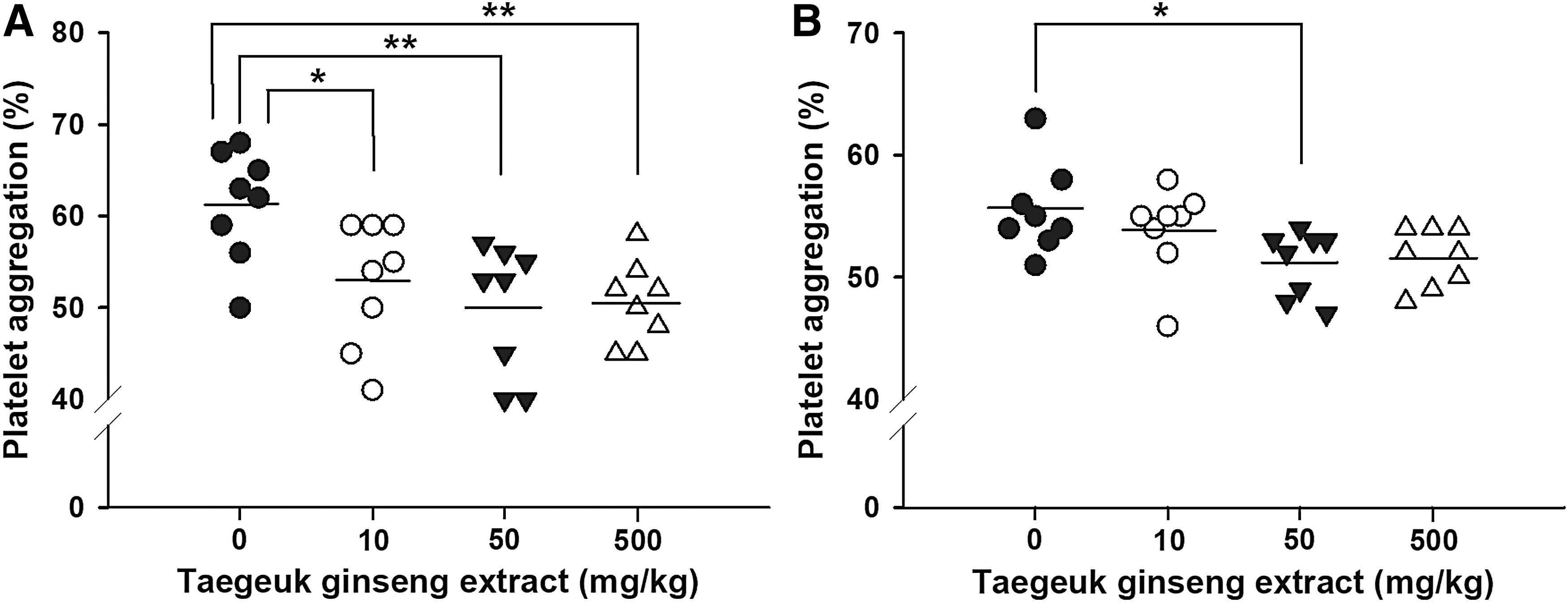
Ex vivo effect of TGE on platelet aggregation. TGE was orally administered to rats (n = 8/group) at doses of 0 (control), 10, 50, and 500 mg/kg for 28 days, respectively. Platelet-rich plasma was preincubated without TGE for 10 min at 37°C.
Effects of TGE on plasma coagulation in vitro and ex vivo
To clarify the effect of TGE on the plasma coagulation system, the coagulation times such as aPPT and PT were examined in vitro and ex vivo. After in vitro treatment of TGE to PPP, aPPT, and PT were measured (Fig. 3A). The aPPT of TGE-treated PPP at concentrations of 0, 1, 5, and 10 mg/mL were 32.7 ± 0.4 sec (n = 3), 32.6 ± 0.2 sec (n = 3), 33.0 ± 0.6 sec (n = 3), and 33.5 ± 0.9 sec (n = 3), respectively. The PT of TGE-treated PPP at concentrations of 0, 1, 5, and 10 mg/mL were 8.3 ± 0.1 sec (n = 3), 8.3 ± 0.0 sec (n = 3), 8.1 ± 0.1 sec (n = 3), and 8.1 ± 0.1 sec (n = 3), respectively. The in vitro treatment of TGE at the concentration of 10 mg/mL had no effect on aPTT and PT.

Effect of TGE on plasma coagulation. The aPTT (▪) and PT (□) were measured. The coagulation times are reported and the median calculated. Data represent the mean ± SEM (n = 8).
After oral administration of TGE for 28 days to rats, aPTT and PT of PPP were measured ex vivo (Fig. 3B). The aPPT of PPP obtained from orally TGE-administered rats at doses of 0 (control), 10, 50, and 500 mg/kg/day were 26.6 ± 1.5 sec (n = 8), 26.5 ± 0.8 sec (n = 8), 25.7 ± 0.9 (n = 8), and 26.6 ± 1.6 (n = 8), respectively. The PT of PPP obtained from orally TGE-administered rats at doses of 0 (control), 10, 50, and 500 mg/kg/day were 9.7 ± 0.7 sec (n = 8), 9.3 ± 0.4 sec (n = 8), 9.3 ± 0.3 (n = 8), and 9.4 ± 0.3 (n = 8), respectively. Compared to aPPT and TT of the control group, the oral administration of TGE did not delay the coagulation time ex vivo.
In vivo inhibitory effect of TGE on thrombus formation
The body weights of orally TGE-administered rats at doses of 0 (control), 10, 50, and 500 mg/kg/day for 28 days were 349.6 ± 13.3 g (n = 8), 348.9 ± 22.0 g (n = 8), 346.1 ± 21.3 (n = 8), and 350.6 ± 22.1 (n = 8), respectively (Fig. 4). No significant difference in body weight between control and TGE-administered groups was observed throughout the experimental period. The body weights of TGE-treated rats normally increased without any abnormal behavior and apparent signs of toxicity (Fig. 4).
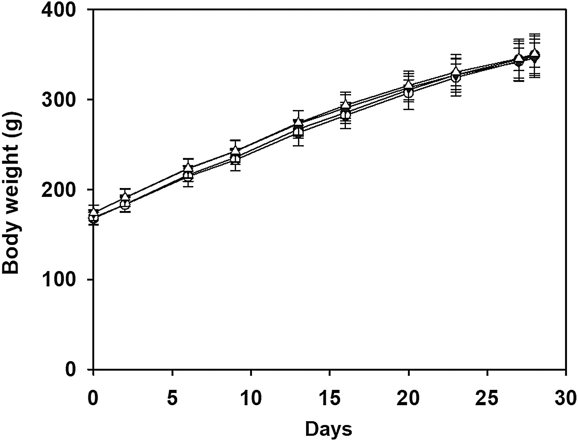
Effect of TGE on body weight of Sprague–Dawley rats. TGE was orally administered to rats (n = 8/group) at doses of 0 (control, -●-), 10 (-○-), 50 (-▾-) and 500 (-▵-) mg/kg for 28 days, respectively. Data represent the mean ± SEM (n = 8).
After ferric chloride treatment to orally TGE-administered rats, the occlusion time in the carotid artery of control rats was 9.2 ± 5.7 min (n = 6), whereas the occlusion times in that of orally TGE-administered rats at doses of 10, 50, and 500 mg/kg/day for 21 days were 37.5 ± 6.2 min (P < .01, n = 6), 32.1 ± 9.8 min (P < .01, n = 6), and 35.4 ± 7.1 min (P < .01, n = 6), respectively (Fig. 5A). The oral administration of TGE significantly delayed the occlusion of the carotid artery in ferric chloride-treated rats in vivo. After 15 min of FeCl3 treatment to orally TGE-administered rats, the blood flow in control rats decreased 92.7% (n = 6), whereas the blood flows in orally TGE-administered rats at doses of 10, 50, and 500 mg/kg/day for 21 days decreased 44.1% (P < .01, n = 6), 28.9% (P < .01, n = 6), and 25.5% (P < .01, n = 6), respectively (Fig. 5B). The blood flow of the control group rapidly decreased whereas those of TGE-administered groups decreased slowly. These results imply that there is a significant inhibition of thrombus formation in TGE-administered rats.
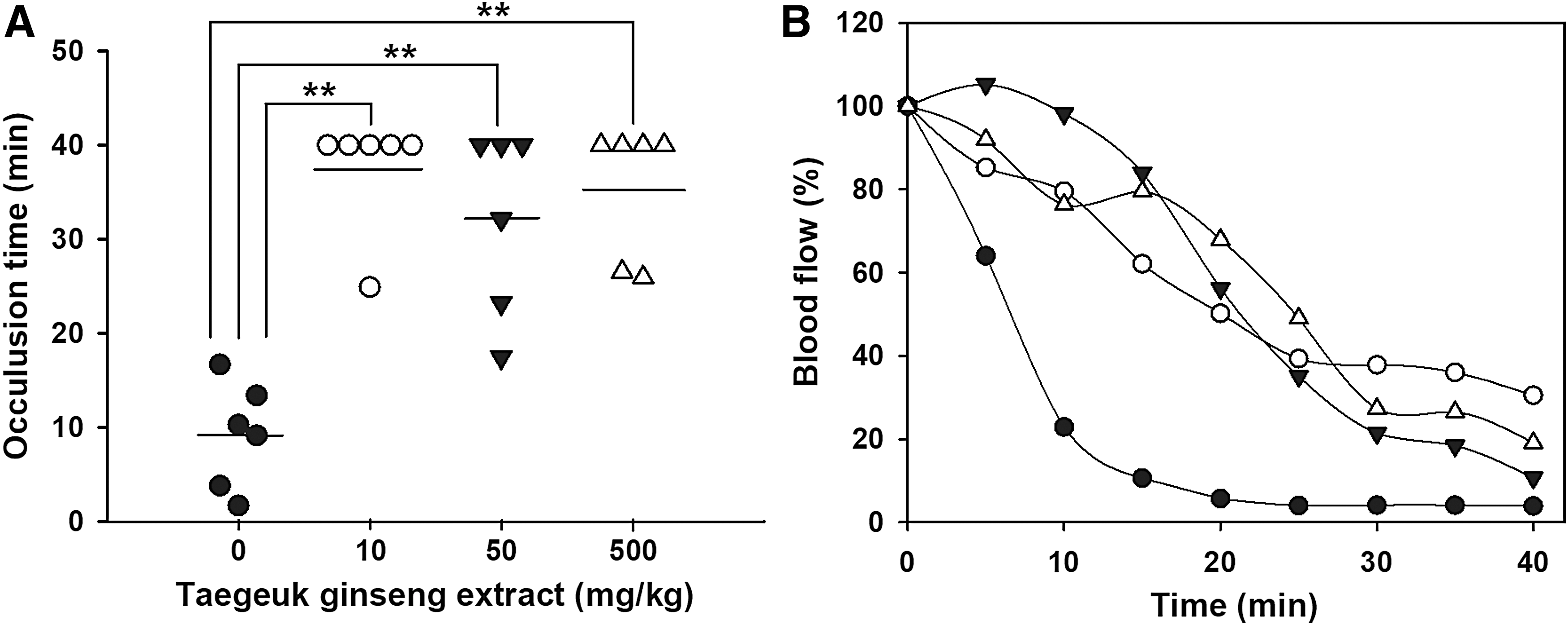
In vivo antithrombotic effect of TGE. TGE was orally administered to rats (n = 6/group) at doses of 0 (control), 10, 50, and 500 mg/kg for 21 days, respectively.
Discussion
The results of this study demonstrate that TGE significantly inhibits platelet aggregation in vitro and ex vivo, thus suppressing in vivo thrombus formation of carotid artery in ferrous chloride-treated rats.
Taegeuk ginseng is an intermediate product of fresh and red ginsengs. 16 Ginsenoside composition in ginseng products significantly varied with the degree of heat treatment. Ginsenosides Rb1, Rg1, and Rg3 were reported as antithrombotic components of ginseng products. 8 –13 Specifically ginsenosides Rg1 and Rg3 were known to inhibit platelet aggregation. 12,13 Ginsenosides Rb1 and Rg1 are major saponins of fresh ginseng, whereas ginsenoside Rg3 is a characteristic component of heat-processed ginseng such as taegeuk, red, and black ginsengs. 30,31 Heat treatment causes the decrease of ginsenosides Rb1 and Rg1, and the increase of ginsenoside Rg3. 32 It was reported that the content of ginsenoside Rg3 increased from 0.32 to 5.79 mg/g during the steaming processes of ginseng. 33 The contents of ginsenosides Rb1, Rg1, and Rg3 in red ginseng extract (RGE) were reported to be 2.66, 0.33, and 5.07 mg/g, respectively. 15 Due to the difference in heating process, the contents of ginsenosides Rb1 (4.83 mg/g) and Rg1 (1.93 mg/g) in TGE were higher than those in RGE, whereas the content of ginsenoside Rg3 (0.28 mg/g) in TGE was lower than that in RGE. The difference of ginsenoside composition and content in ginseng products has a direct influence on biological and pharmacological activities of ginseng.
Platelet aggregation occurs in an injured blood vessel via platelet-to-platelet adhesion and platelet activation by some agonists such as ADP and collagen. 34 TGE significantly inhibited ADP- and collagen-induced rat platelet aggregation (P < .001, n = 5) in vitro in a concentration-dependent manner with IC50 values of 14.2 ± 0.88 and 16.6 ± 1.16 mg/mL, respectively (Fig. 1). RGE was reported to inhibit the collagen-induced rabbit platelet aggregation in vitro with an IC50 value of 387 ± 11 μg/mL. 15 The inhibitory effect of TGE on platelet aggregation was confirmed by ex vivo experiments in TGE-administered rats. After oral administration of TGE at doses of 10, 50, and 500 mg/kg/day for 28 days, the ex vivo collagen- and ADP-induced platelet aggregations were significantly inhibited (Fig. 2). But TGE-administration had no influence on coagulation times such as aPTT and PT (Fig. 3), indicating that TGE did not affect the plasma coagulation system.
The inhibitory mechanism of taegeuk ginseng on platelet aggregation remains unknown. The antiplatelet activity of ginseng has been reported to be caused by the decrease in production and release of thromboxane A2. 1,35 It has been reported that nonsaponin fraction of red ginseng inhibited platelet aggregation by regulating the levels of cGMP and thromboxane A2. 24 Also, the crude saponin fraction of red ginseng significantly inhibited fibrinogen binding to active integrin αIIbβ3, and blocked collagen-induced phosphoinositide 3-kinase/Akt phosphorylation, which is related to signaling pathways in platelet activation and aggregation processes. 3 Further study is needed to explore the molecular mechanism underlying the antiplatelet and antithrombotic efficacy of taegeuk ginseng.
Ginseng products containing white, red, and black ginsengs are considered to be very safe and tolerable in animals. 36 In mice, LD50 values for ginseng have been reported to range from 10 to 30 g/kg. 37 In general, the oral administration of substances with LD50 values higher than 5 g/kg has been regarded as nontoxic and safe. 38 The change of body weight in experimental animals is a typical indicator of adverse effects and toxicity of test substances. 39 In this study, the normal increase of body weights was observed in orally TGE-administered rats as well as those of control rats without apparent signs of toxicity (Fig. 4). In hematological and biochemical tests of healthy human volunteers, the oral administration of ginseng extract at a dose of 2 g/day for 4 weeks has been reported to be safe and tolerable without any toxic effect. 40 Recommended intake level of ginseng in human has been known to be 1–2 g/day of ginseng products containing 4–5% ginsenoside. 37
Platelet activation eventually leads to thrombus formation. 41 The treatment with ferric chloride caused the carotid artery injury in the experimental rats, thus leading to the occlusive thrombus formation. 15 The oral administration of TGE to rats at a dose of 10 mg/kg/day for 21 days delayed occlusion time by more than fourfold of the carotid artery compared with that of the control group (Fig. 5). Oral administration of TGE was considered to significantly inhibit in vivo thrombus formation. The oral administration of RGE to rats at a dose of 500 mg/kg/day for 7 days delayed by about twofold in the occlusion time of the carotid artery compared with that of control group. 15 In vivo TGE-administration at low doses showed high antithrombotic effect. In conclusion, these results indicate that TGE significantly inhibited artery thrombosis in vivo due to the inhibitory effect on platelet aggregation rather than anticoagulation activity, and that TGE could be used as a safe agent to reduce the cardiovascular and thrombotic risks.
Footnotes
Acknowledgments
This study was supported by grants from Ministry of Agriculture, Food and Rural Affairs (Project No. 314026-3), Republic of Korea. This work was also supported by an Academic Research Fund of Chungwoon University (2015-2-09).
Author Disclosure Statement
No competing financial interests exist.