
Abstract
This study evaluated the antioxidant and protective effects of bioactive compounds isolated from pressurized steam-treated Corni Fructus (PSC). We had previously tested the protective effects of the furan fraction containing 5-hydroxymethylfurfural (5-HMF), polyphenol fraction containing gallic acid, and iridoid glycoside fraction containing morroniside and loganin. We measured the potency of antioxidant activities of the bioactive compounds isolated from PSC via oxygen radical absorbance capacity (ORAC), 2,2-diphenyl-1-picrylhydrazyl (DPPH) radical scavenging, and 2,2′-azino-bis(3-ethylbenzothiazoline-6-sulfonic acid) (ABTS) radical scavenging assays. One fraction in particular (named F-2) not only contained high amounts of phenolics but also had potent antioxidant activities. The protective effects of F-2 were evaluated by measuring the levels of the collagen-degrading enzyme, matrix metalloproteinase-1 (MMP-1), and the marker of collagen biosynthesis, procollagen type I C-peptide (PIP), in UVB-treated HS68 fibroblasts. MMP-1 levels decreased in an F-2 concentration-dependent manner, and PIP secretion from the cultured HS68 cells was significantly higher than that from the UVB-irradiated cultures alone. Further, F-2 attenuated UVB-induced MMP-1 and ameliorated UVB-downregulated collagen type I alpha 1 mRNA expression in HS68 cells. Therefore, F-2 isolated from PSC is a good candidate for the prevention of skin damage from free radicals in various skin conditions.
Introduction
R
For these reasons, fruits are important sources of compounds that protect against skin aging because of their high antioxidant levels and their abundant quantities of carotenoids, phenolics, and ascorbic acid. 6 Plant extracts and their isolated secondary metabolites have been used extensively in the cosmetic industry as, for example, skin lighteners, antioxidants, and sunscreens. 7 In vitro studies have shown that plant extracts and purified components (e.g., phenolic compounds) can diminish ROS levels and inhibit the activity of hyaluronidase, collagenase, tyrosinase, and elastase. 8,9
Corni Fructus is the fruit of Cornus officinalis Sieb. Et Zucc, which is a member of the Cornaceae family, more commonly known as dogwoods. 10 It is a popular and extensively used herbal medicine worldwide, often utilized in cosmetics, food sanitation, and medicine. 11,12 Corni Fructus has been found to contain malic acid, gallic acid, tartaric acid, morroniside, ursolic acid, sweroside, and loganin. 13 –15 Traditional uses of this herb include alleviating tinnitus, lessening excessive urination, and improving impotence. 14 Corni Fructus also has antioxidant, 16 antidiabetic, 17 and chemotherapeutic effects. 18
However, investigations on the antioxidant effects and protective effects against UVB-irradiation damage in HS68 cells by the bioactive compounds in Corni Fructus are very limited. During bioactive compound screening, treating Corni Fructus with pressurized steam significantly increased total phenol and bioactive compound (gallic acid and 5-hydroxymethylfurfural [5-HMF]) contents. 19 The aim of this study was to isolate and identify bioactive compounds from pressurized steam-treated Corni Fructus (PSC), determine their antioxidant effects via in vitro assays, and determine the protective effects of the liquid fractions against UVB irradiation by using assays in cultured HS68 cells.
Materials and Methods
Materials
Corni Fructus without seeds was purchased from a local supermarket (Uiseong, Gyeongsangbuk-do, Republic of Korea). The samples were hot-air-dried at 50°C for 24 h to adjust the moisture content to 10% and then processed by the pressurized steam treatment.
Extracts of pressurized steam-treated Corni Fructus
PSC was prepared according to our previously published method. 19 The final concentrate was suspended three times with diethyl ether: ethyl acetate (1:1, v/v), and the precipitate was concentrated and freeze dried (Free Zone 2.5; Labconco Co., Kansas, MO, USA) to yield a powder for use.
Isolation of bioactive compounds from PSC
PSC (3 g) was fractionated by silica gel 60 (70–230 mesh; Merck, Darmstadt, Germany) open-column chromatography (50 × 4 cm) with chloroform: methanol: formic acid (15:2:1, v/v). Four bioactive compounds were fractionated from the PSC. Each fraction was evaporated to dryness under vacuum to yield residues of 0.18 g for fraction 1 (F-1, nos. 14–21), 0.35 g for fraction 2 (F-2, nos. 61–68), 1.30 g for fraction 3 (F-3, nos. 81–99), and 0.82 g for fraction 4 (F-4, nos. 109–140). Total sugar content was determined by the phenol-sulfuric acid method described by Saha and Brewer, 20 and total phenol content was determined by the Folin-Denis method described by Singleton and Rossi. 21 The quantity of bioactive compounds was analyzed by high-performance liquid chromatography (HPLC, Waters 2695; Waters Co., Milford, MO, USA). HPLC consisted of an Xbridge C18 column (250 × 4.6 mm, particle size 5 μm; Waters Co.) at a column temperature of 25°C. Bioactive compounds were eluted with 0.1% acetic acid: acetonitrile: methanol (85:10:5, v/v) at a flow rate of 0.5 mL/min and were detected at 240 nm by using a photodiode detector (Waters 2996; Waters Co.).
Oxygen radical absorbance capacity assay
The peroxyl radical scavenging activity of phenolic antioxidants was assayed by the oxygen radical absorbance capacity (ORAC) method described by Ou et al.
22
The 200 μL reaction was performed in 75 mM phosphate buffer (pH 7.4). Twenty-five microliters of each sample dilution in 75 mM phosphate buffer (1 mg/mL final concentration) and 150 μL fluorescein solution (50 nM final concentration; Sigma-Aldrich Co., St. Louis, MO, USA) were added to a 96-well black plate (black, clear bottom). The mixture was preincubated for 15 min at 37°C. AAPH solution [2,2′-azobis(2-amidinopropane) dihydrochloride, 25 μL, 153 mM final concentration; Sigma-Aldrich] was added rapidly by using a multichannel pipet. The microplate was immediately placed in the Fluorescence Micro Reader (VICTOR
3
; Perkin-Elmer, Inc., Waltham, MA, USA), and the fluorescence was recorded every minute for 60 min (Excitation = 485 nm, Emission = 538 nm, Cut off = 530 nm); the microplate was automatically shaken before each reading. Trolox (0–100 μM final concentration; Sigma-Aldrich) was used as a positive control, and the sample was replaced with phosphate buffer in the blank. The antioxidant curves (fluorescence vs. time) were first normalized to the blank curve in the same assay by multiplying the original data by the factor fluorescenceblank,t = 0/fluorescencesample,t = 0. From the normalized curves, the area under the fluorescence decay curve (AUC) was calculated as:
where f0 is the initial fluorescence read at 0 min, and fi is the fluorescence read at time i. The net AUC was obtained by subtracting the AUC of the blank from that of each sample. ORAC values were obtained from the net AUC and expressed as Trolox equivalent (TE):
2,2-Diphenyl-1-picrylhydrazyl assay
The anti-radical activity was determined by the 2,2-diphenyl-1-picrylhydrazyl (DPPH) method described by Blois. 23 One hundred microliters of each sample that had been diluted in distilled water (1 mg/mL final concentration) and 900 μL DPPH ethanolic solution (400 μM final concentration; Sigma-Aldrich) were added to each well of a 24-well plate. After mixing gently and incubating for 15 min at 25°C, the absorbance was measured at 517 nm by using a Microplate Reader (UVM-340; ASYS Co., Eugendorf, Austria). Ascorbic acid (0.1 mg/mL final concentration; Sigma-Aldrich) was used as a positive control, and the blank sample was distilled water. DPPH radical scavenging activity was calculated as follows: Scavenging activity (%) = [1 − (absorbance sample/absorbance control)] × 100.
2,2′-Azino-bis(3-ethylbenzothiazoline-6-sulfonic acid) assay
The anti-radical activity was determined by the 2,2′-azino-bis(3-ethylbenzothiazoline-6-sulfonic acid) (ABTS) method described by Re et al.
24
A solution of the cation-radical, ABTS
Cell culture
The normal human dermal fibroblast, HS68 cells (ATCC CRL 1635) were obtained from the American Type Culture Collection (Rockville, MD). The complete culture medium for the HS68 cells was Dulbecco's modified Eagle's medium (Welgene, Inc., Gyeongsan, Korea) containing 10% fetal bovine serum (Gibco BRL Co., Grand Island, NY, USA) and 2% penicillin-streptomycin (Gibco BRL Co.). The cells were plated at a density of 3.0 × 10 5 cells/24-well plate and 9.0 × 10 5 cells/6-well plate, and they were maintained at 37°C in a humid incubator (MCO-18AIC; SANYO Co., Sakata, Japan) with a 5% CO2 atmosphere.
UV irradiation and sample treatment
The HS68 cells were grown in serum-free culture media overnight, followed by 24 h of sample pretreatment. The cells were washed twice with phosphate-buffered saline (PBS), and all UVB irradiations were conducted with PBS covering the cells. Non-irradiated cells were treated in parallel and maintained at room temperature. UVB irradiation was conducted by using a Radiometer (VLX-3W; Vilber Lourmat, Collégien, France), which emitted radiation in a range of 280–320 nm. The cells were exposed to a 40 mJ/cm2 dose of UVB light for 2 min.
Cytotoxicity assay
Cytotoxicity was determined by the MTT [3-(4,5-dimethylthiazol-2-yl)-2,5-diphenyltetrazolium bromide] method, as described by Denizot and Lang. 25 After the treatment of each sample and UVB irradiation, MTT solution in PBS (1 mg/mL final concentration; Sigma-Aldrich) was then added to each well at 1/10 volume of media. The cells were incubated for 4 h at 37°C, and dimethyl sulfoxide was added to dissolve the formazan crystals. The absorbance was measured at 570 nm by using a Microplate Reader (ASYS Co.). Cytotoxicity was calculated as follows: Activity (%) = (absorbance sample/absorbance control) × 100.
Matrix metalloproteinase-1 and procollagen type I C-peptide assay
After UVB irradiation, the PBS was replaced with serum-free medium and the cells were incubated for 48 h at 37°C. The following parameters were utilized to monitor the effects of PSC on the UVB-irradiated HS68 cells: matrix metalloproteinase (MMP-1) and procollagen type I C-peptide (PIP) secretion. MMP-1 was measured by using the MMP-1 human ELISA kit (ab100604; Abcam PLC., Cambridge, United Kingdom), and the absorbance was measured at 450 nm by using a Microplate Reader (ASYS Co.). MMP-1 levels were calculated as follows: Level (%) = (absorbance sample/absorbance control) × 100. The PIP release was measured by using the PIP EIA kit (MK101; Takara Bio Inc., Shiga, Japan), and the absorbance was measured at 450 nm by using a Microplate Reader (ASYS Co.). The PIP release was calculated as follows: Release (%) = (absorbance sample/absorbance control) × 100.
RT-PCR analysis of mRNA expression
To evaluate the mRNA expression levels of MMP-1 and Co1A1, total RNA (1 μg) was prepared by using the RNeasy® mini kit (Qiagen, Hilden, Germany). Total RNA was subjected to first-strand cDNA synthesis by using the RevertAid™ First-Strand cDNA Synthesis Kit (Thermo Scientific, Waltham, MA, USA). RT-PCR was performed by using the 2 × TOPsimple™ DyeMIX kit (Enzynomics Co., Ltd., Daejeon, Korea) and thermal cycler (Mastercycler® nexus; Eppendorf AG, Germany). The initial denaturation was 95°C for 2 min, followed by 30 cycles at 95°C for 30 s, annealing (MMP-1, 65°C; Col1A1, 65°C; glyceraldehyde 3-phosphate dehydrogenase [GAPDH], 55°C) for 30 s, and 72°C for 1 min, with a final extension of 72°C for 5 min. After amplification, the products were separated by electrophoresis (Mupid-ex; Advance, Tokyo, Japan) on a 1.2% agarose gel stained with Eco view (Dae-myung Science Co., Ltd., Seoul, Korea). DNA bands were visualized by UV transillumination and were documented with ImageJ software (National Institutes of health, Bethesda, MD, USA). All samples were normalized against a GAPDH control band, and the results were expressed as relative change of MMP-1/GAPDH and Col1A1/GAPDH. The sequences of primers (Bioneer, Daejeon, Korea) used in this experiment were: MMP-1, 5′-ATT CTA CTG ATA TCG GGG CTT TGA-3′ and 5′-ATG TCC TTG GGG TAT CCG TGT AG-3′; Col1A1, 5′-CTC GAG GTG GAC ACC ACC CT-3′ and 5′-CAG CTG GAT GGC CAC ATC GG-3′; GAPDH, 5′-CGG AGT CAA CGG ATT TGG TCG TAT-3′ and 5′-AGC CTT CTC CAT GGT GGT GAA GAC-3′.
Statistical analysis
All experiments were performed in triplicate, and means ± standard deviation or average values were reported. Duncan's multiple-range test was used for multiple comparisons, and null hypotheses were rejected at the 0.05 level. All data were analyzed by using SPSS/Windows software (Version 19.0; SPSS, Inc., Chicago, IL, USA).
Results and Discussion
Isolation and identification of bioactive compounds
Our previous study showed that treating Corni Fructus with pressurized steam significantly changed the total extractable phenolic and bioactive compounds (i.e., gallic acid, 5-HMF, loganic acid, morroniside, and loganin). 19 The gallic acid of the phenolic compounds and the 5-HMF of the furan compounds in PSC increased with increased treatment time. The main iridoid glycoside components of PSC were morroniside, loganin, and loganic acid, and those increased with decreased treatment time. After pressurized steam treatment at 1.2 kgf/cm2 for 2 h, the bioactive compound composition of PSC was gallic acid (6.18 mg/g), 5-HMF (20.52 mg/g), loganic acid (5.11 mg/g), morroniside (23.39 mg/g), and loganin (12.62 mg/g). 19
The biologically active compounds from PSC, 5-HMF, morroniside, gallic acid, and loganin fraction were isolated. The bioactive compound fractions that were isolated from PSC extract showed four major peaks (Fig. 1). The compounds in each fraction were identified by HPLC retention times, elution order, and their spectroscopic and spectrometric characteristics (Table 1). The main components were identified by using commercially available standards. The HPLC chromatogram recorded at 240 nm enabled the identification and quantification of four compounds: (1) gallic acid (7.718 min), (2) 5-HMF (9.744 min), (3) morroniside (13.913 min), and (4) loganin (26.869 min); 5-HMF (174.79 mg/g) and morroniside (157.72 mg/g) were the main identified components of F-1 and F-3, respectively. The predominant phenolic compound of F-2 was gallic acid (1.12 mg/g), but 5-HMF (2.50 mg/g), morroniside (0.64 mg/g), and loganin (7.59 mg/g) were also observed. The main components of F-4 were loganin (100.82 mg/g) and morroniside (20.55 mg/g). Over the past few decades, the phytochemistry of Corni Fructus has been extensively studied, and the results indicated that morroniside, gallic acid, sweroside, 5-HMF, cornuside, and loganin are its main active components. 26,27 Pharmacological studies on these components showed that they all possessed good biological activity. Gallic acid reduced intracellular production of hydrogen peroxide 28 and was anti-inflammatory 29 ; 5-HMF improved blood circulation 30 and showed hepatoprotective effects. 31 Morroniside had neuroprotective effects, 32 and loganin showed hepatoprotective effects 33 and improved learning and memory impairment. 34

Total sugar (●), and total phenol (○) of bioactive compound fractions isolated from PSC by silica gel open-column chromatography. PSC, pressurized steam-treated Corni Fructus.
Identification of Bioactive Compound Fractions Isolated from Pressurized Steam-Treated Corni Fructus by High-Performance Liquid Chromatography
Mean ± SD, n = 3.
5-HMF, 5-hydroxymethylfurfural; SD, standard deviation.
Antioxidant effects of bioactive compounds
The consumption of fruits, vegetables, and medicinal plants plays an important role in a healthy lifestyle. 35 Traditional medicinal plants are a good source of nutrients and non-nutrients, with many displaying antioxidant properties. 36 These appear to play major protective roles against cellular oxidative stress. 37 Especially, phenolic compounds have been reported to have antioxidant and anticancer activities, and they may slow the progression of aging and disease. 38 –40 Thus, our study results indicate that bioactive compound fractions might have antioxidant activities.
The antioxidant activity of the bioactive compounds was estimated by using three antioxidant assays based on radical scavenger mechanisms: ORAC, DPPH, and ABTS assays (Table 2). The ORAC value was expressed in μM/g FW of Trolox. When compared, the ORAC values were found to be in the following order: F-2 (354.28 μM/g FW) > F-4 (289.16 μM/g FW) > F-1 (236.99 μM/g FW) > F-3 (225.51 μM/g FW). The DPPH assay is widely reported for screening antioxidants and determining comparative antioxidant effectiveness.
41
The ability of the extracts to inhibit the DPPH reaction was expressed as % inhibition of DPPH radicals. Data show that F-2 (26.70%) exhibits higher DPPH radical scavenging than does F-3 (11.97%), followed by F-4 (10.36%) and F-1 (3.15%). The ABTS effect was quantified in terms of percentage inhibition of the ABTS
Antioxidant Effects of Bioactive Compound Fractions Isolated from Pressurized Steam-Treated Corni Fructus
Bioactive compound fractions were assayed at 1 mg/mL final concentration, and ascorbic acid was assayed at 0.1 mg/mL final concentration.
Mean ± SD of triplicate determinations. Means with different superscripts within a column indicate significant differences (P < .05).
ABTS, 2,2′-azino-bis(3-ethylbenzothiazoline-6-sulphonic acid); DPPH, 2,2-diphenyl-1-pycrylhydrazyl; FW, formula weight; ORAC, oxygen radical absorbance capacity.
Protective effects of bioactive compounds
Skin is an excellent model system for the study of aging. UV rays play a major role in cutaneous aging. 1 UV rays alter the cells and the extracellular structures of the skin by numerous mechanisms, such as altering structural proteins and damaging nucleic acids and membranes. 46 Specifically, UVB-induced MMPs from dermal fibroblasts increase the degradation of collagen and other extracellular matrix proteins and contribute to the photoaging of human skin. 47,48 All of these events are mediated by free radicals and are believed to be fundamental factors in photoaging. 46 In general, the skin protects itself against ROS-induced damage via enzymes such as superoxide dismutases, lactoperoxidases, glutathione peroxidases, catalases, and peroxiredoxins. 49,50 Small-molecule antioxidants, including ascorbic acid, uric acid, tocopherols, and glutathione, also play important roles as antioxidants. 51,52 F-2 exhibited high antioxidant effects, and, thus, we tried to confirm its protective effects by using the HS68 fibroblasts.
The cytotoxic effects of F-2 on HS68 cells were determined by exposing the cells to various concentrations of F-2 (0.1, 1, 10, and 100 μg/mL) for 48 h. Our results show that there were no cytotoxic effects on HS68 cells at the tested concentrations (Fig. 2). Therefore, concentrations of F-2 from 0.1 to 10 μg/mL were selected for subsequent experiments.
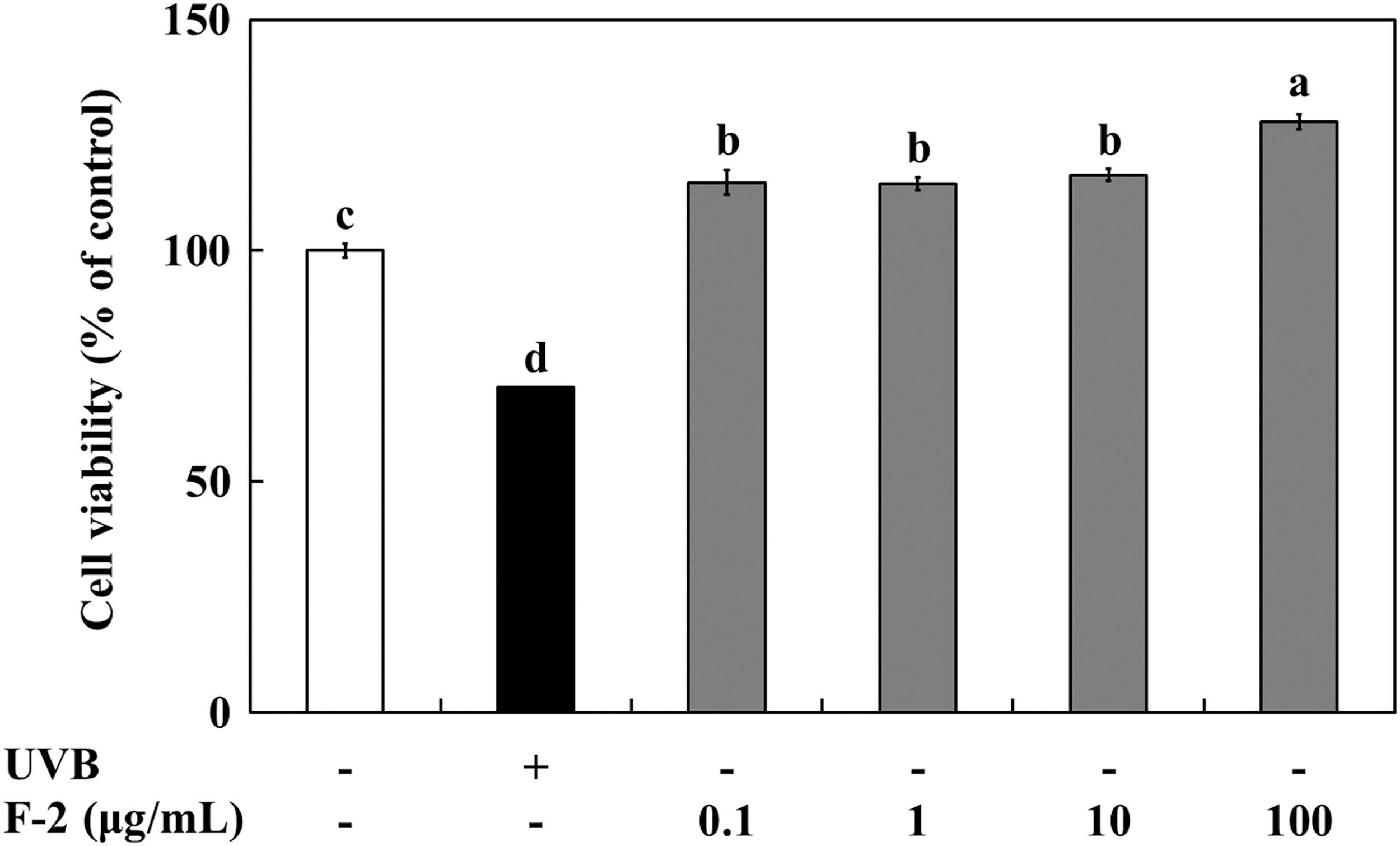
Cytotoxicity against HS68 cells of the bioactive compound fraction, F-2, isolated from PSC. Values are means ± SD of triplicate determinations. Bars with different letters indicate significant differences (P < .05). SD, standard deviation.
We confirmed the protective effects of F-2 by measuring MMP-1 levels and PIP generation after UVB exposure of HS68 cells. As shown in Fig. 3A, MMP-1 levels decreased in a concentration-dependent manner; the MMP-1 levels were 155.28%, 106.57%, and 82.97% at 0.1, 1, and 10 μg/mL of F-2, respectively. In a study by Kim, 53 plant polyphenols were shown to inhibit enzymatic activity by binding to the enzyme-substrate complex instead of directly binding to the enzyme's active site. Therefore, the reduced MMP-1 levels observed by F-2 may be due to the gallic acid in the polyphenols. As shown in Fig. 3B, PIP generation from HS68 cells was significantly higher than that from UVB-induced HS68 cells. The activity of F-2 against the UVB-induced reduction in PIP production was 125.86%, 102.47%, and 81.56% that of control cells at 10, 1, and 0.1 μg/mL, respectively.

Protective effects against UVB-irradiated HS68 cells of the bioactive compound fraction, F-2, isolated from PSC.
In addition, to determine whether F-2 affected UVB-altered MMP-1 and Col1A1 mRNA expression, we examined their mRNA levels by RT-PCR. As shown in Fig. 4, the RT-PCR results showed weak basal MMP-1 expression in the control group, which was augmented by UVB irradiation. This elevated MMP-1 gene expression was reduced by F-2 treatment in a concentration-dependent manner. F-2 treatment decreased MMP-1 gene expression by 34.8% at 1 μg/mL and 57.5% at 10 μg/mL. In contrast, UVB irradiation reduced Col1A1 mRNA expression compared with that of the control group. The Col1A1 mRNA level in cells treated with 0.1 μg/mL F-2 showed little change compared with that in cells subjected to UVB irradiation only. However, after 10 μg/mL F-2 treatment, Col1A1 mRNA level increased by 112.2% compared with that in cells subjected to UVB irradiation only.
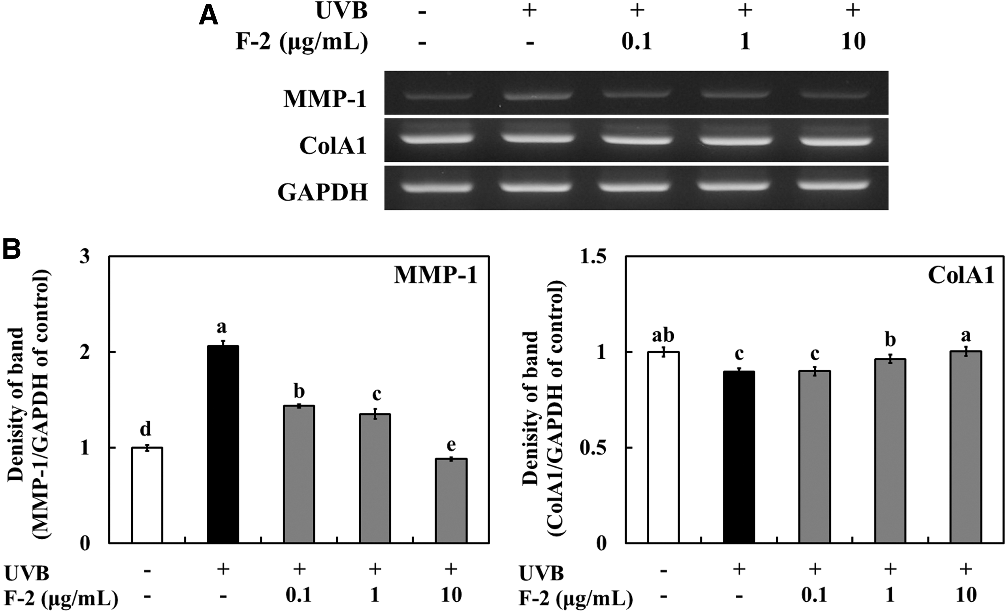
RT-PCR analysis of mRNA expression of UVB-irradiated HS68 cells treated with the bioactive compound fraction, F-2, isolated from PSC.
The dermis in normal skin comprises mostly type I collagen (85–95%) with lower amounts of type III collagen (10–15%). 54 However, fibrillar type I and type III collagens are significantly decreased in the papillary dermis, and their decrease has been shown to correlate with the clinical severity of photoaging. This decrease results from both reduced procollagen biosynthesis and increased MMP-mediated degradation. 55 In this study, the bioactive compound fractions not only activated a UVB-induced increase in type I collagen but also attenuated UVB-induced MMP-1 expression in HS68 cells. The most important skin rejuvenation process after photodamage is collagen remodeling, and the cell that is critical in this process is the dermal fibroblast.
Therefore, considering the efficacy and safety of the bioactive compound fractions in HS68 cells, these fractions hold great promise as functional food materials for protection against UVB irradiation, especially in treating photodamaged skin.
Footnotes
Author Disclosure Statement
No competing financial interests exist.