
Abstract
Despite the increasing prevalence of inflammatory bowel disease (IBD), classified as immune-mediated disorders, the exact biological mechanisms leading to its development are undetermined, and treatment strategies remain elusive. Probiotics have been proposed as potential alternatives for treating IBD. The purpose of this research was to find therapeutic candidates of probiotics for colitis. We adopted dextran sulfate sodium (DSS)-induced colitis model to demonstrate the therapeutic effects of ID-JPL934, a mixture of three live bacterial strains at a 1:1:1 ratio: Lactobacillus johnsonii IDCC9203, Lactobacillus plantarum IDCC3501, and Bifidobacterium animalis subspecies lactis IDCC4301, on IBD. The severity was scored according to the disease activity index (DAI) for colitis by observing body weight (BW) and stool status of each mouse once a day. BALB/c mice given 3.5% DSS in drinking water suffered from symptoms of colitis such as weight loss, diarrhea, and bloody excrement. In our study, administration of ID-JPL934 reduced the DAI scores in a dose-dependent manner, and treatments with ID-JPL934 108 and 109 colony-forming unit per mouse per day showed similar inhibition compared with those of sulfasalazine 500 mg per kg BW per day. Moreover, the contraction of colon length improved. ID-JPL934 also suppressed inflammatory lesions such as infiltration of immune cells in mucosa and submucosa, severe crypt damage, and loss of goblet and epithelial cells on the histological analysis. These results might be due to downregulation of the expression of proinflammatory cytokines, including tumor necrosis factor-α, interleukin (IL)-1β, and IL-6. From these results, ID-JPL934 might be an effective therapeutic candidate for IBD.
Introduction
I
Activation of immune cells triggered by inflammatory cytokines makes immunocytes easily translocate into the damaged intestinal barrier. Proinflammatory cytokines such as tumor necrosis factor (TNF)-α, interleukin (IL)-1β, and IL-6, released from the stimulated immune cells, play major roles in driving intestinal inflammation and local complications. 6 Therefore, it is a proper strategy to suppress cytokine expression to improve colitic symptoms, including weight loss, diarrhea, and bloody excrement. 7,8
To alleviate colitis, anti-inflammatory agents and immunosuppressive biologics are prescribed, but it is well known that there are undesirable side effects of these drugs such as nausea, vomiting, dyspepsia, anorexia, and infection of intracellular pathogens. 9 Various researches to find better alternatives that have less adverse effects are carried out.
Probiotics are defined as microorganisms, which confer health benefits to the host when administered in appropriate amounts. 10 The well-established safety and efficacy in benefiting human health have caused probiotics to emerge as alternatives or complements of medicinal reagents. 11 Diverse biologic advantages of probiotics, including inhibition of inflammation, regulation of metabolic diseases, and prevention of bacterial infection, have been studied. 12 –14 Especially, probiotics are promising therapeutic candidates for IBD. 15 A combination of probiotic strains is taking the spotlight to raise the beneficial efficacies in many clinical trials and experimental studies. 16 –19
The dextran sulfate sodium (DSS)-induced colitis model is well known as a standard model for IBD. This experimental model can accurately portray inflammatory changes as well as clinical symptoms of colitis such as diarrhea, occult blood, and gross rectal bleeding. 20 In this study, we aimed to determine the potential inhibitory effects of ID-JPL934, a mixture of three probiotic strains, on IBD using a murine colitis model induced by DSS and to investigate the modulation mechanisms of ID-JPL934 on the intestinal immune system.
Materials and Methods
Preparation of ID-JPL934 for in vivo experiments
ID-JPL934 consists of three live bacterial strains at a 1:1:1 ratio: Lactobacillus johnsonii IDCC9203 (isolated from infant feces), Lactobacillus plantarum IDCC3501 (isolated from Kimchi), and Bifidobacterium animalis subspecies lactis IDCC4301 (isolated from infant feces). Bacteria were cultured in growth medium at 37°C for 16 h until exponential phase and lyophilized by keeping bacterial pellets in a freeze dryer at −80°C under vacuum overnight. Each strain contained 2 × 10¹² colony-forming units (CFU)/g of viable bacteria. Lyophilized ID-JPL934 was stored at 4°C until use.
Animals
Female BALB/c mice (aged 8 weeks) were purchased from Orient Bio (Seongnam, Republic of Korea). Five animals per cage were housed in an environmentally controlled facility (temperature of 22°C ± 2°C, relative humidity of 55% ± 5%) with a 12/12 h light/dark cycle throughout the study. Animal care and treatments were carried out in accordance with the guidelines established by the National Institutes of Health Animal Research and Care and approved by the Institutional Animal Care and Use Committee of Ildong Pharmaceutical Co., Ltd (Approval No.: A1611-2).
Mouse colitis model
A mouse colitis model induced by DSS (molecular weight 36–50 kDa; MP biochemicals, Santa Ana, CA, USA) was adopted in this study. 20 After a 7 day acclimatization period, mice were divided into seven groups (n = 10 per group), adjusting average of initial body weight (BW) similarly (Table 1).
BW, body weight; CFU, colony-forming units; DSS, dextran sulfate sodium; DW, distilled water.
DSS was dissolved in drinking water to a concentration of 3.5% and supplied to all mice except the normal group from day 0 to 8. The animals had free access to water and diet. Mice in the vehicle group were orally administered 200 μL of distilled water (DW). Dose-ranging ID-JPL934 at 106–109 CFU per mouse per day and sulfasalazine (Tokyo chemical industry, Tokyo, Japan), used as a positive control, at the concentration of 500 mg per kg BW per day was suspended in DW and administered in the same volume and manner as with the vehicle group.
We observed BW and stool status of each mouse once a day to determine the disease activity index (DAI) scores, a maximum value is 12, according to the modified methods described previously (Table 2). 21 Cola from the ileocecal junction to the anus were enucleated to measure the length and washed out with phosphate buffered saline to remove remaining digesta after euthanasia with carbon dioxide gas. One-third of the distal colon was fixed with 10% formaldehyde solution to analyze pathologic histology. The remaining two-thirds of the distal colon was stored at −80°C for the protein extraction to assay proinflammatory cytokine level.
Histological analysis
The colon tissues were fixed with 10% neutral buffered formaldehyde solution and processed routinely for paraffin embedding. Paraffin-embedded sections were cut at 4 μm thickness and stained with hematoxylin and eosin (H&E). The H&E-stained colon tissues were observed using a Nikon ECLIPSE 50i optical microscope (Nikon, Tokyo, Japan) under 100 × and 200 × magnifications, and histologic assessment was performed by two pathologists in accordance with the previously described parameters (Table 3). 22 The maximum score, calculated by adding each score of three parameters, was 10.
Colonic proinflammatory cytokine level
Radioimmuno precipitation assay (RIPA) buffer (Upstate, Temesula, CA, USA) supplemented with protease inhibitor (Sigma-Aldrich, St. Louis, MO, USA) was used to extract colonic protein. After adding 300 μL of RIPA buffer, the tissue was homogenized for 1 min on ice and centrifuged at 20,000 g for 15 min at 4°C. The supernatant was collected and then quantified using bicinchoninic acid protein assay reagent kit (Pierce, Rockford, IL, USA). The levels of proinflammatory cytokines such as TNF-α, IL-1β, and IL-6 in the colon tissue were detected using commercial enzyme-linked immunosorbent assay (ELISA) kit (R&D Systems, Minneapolis, MN, USA) according to the manufacturer's protocols. The values for cytokines were revised by total quantity of each colonic protein sample.
Statistical analysis
The results presented as mean ± standard error were processed using the statistical analysis system Prism 7 (GraphPad Software, San Diego, CA, USA). All results represent averages of at least three independent experiments. Statistical comparisons between the vehicle and each treatment group were determined by one-way analysis of variance followed by Dunnett's test. A value of P < .05 was considered statistically significant.
Results
Effects of ID-JPL934 on the symptoms of colitis
To determine the severity of the colitis, the DAI scores were recorded by observing BW and fecal conditions of each mouse once a day. As the amount of DSS given to the mice increased, occurrences of bloody excrement and diarrhea became more frequent, while BW gradually declined. The vehicle group represented a steady elevation of the DAI scores from day 4 (Fig. 1A). Administrations of ID-JPL934 decreased the DAI scores dose-dependently through the prevention of weight loss, improvement of fecal appearance, and alleviation of bloody stool on day 8 (Fig. 1B). The DAI scores of ID-JPL934 108 and 109 groups were similar to that of the sulfasalazine group.
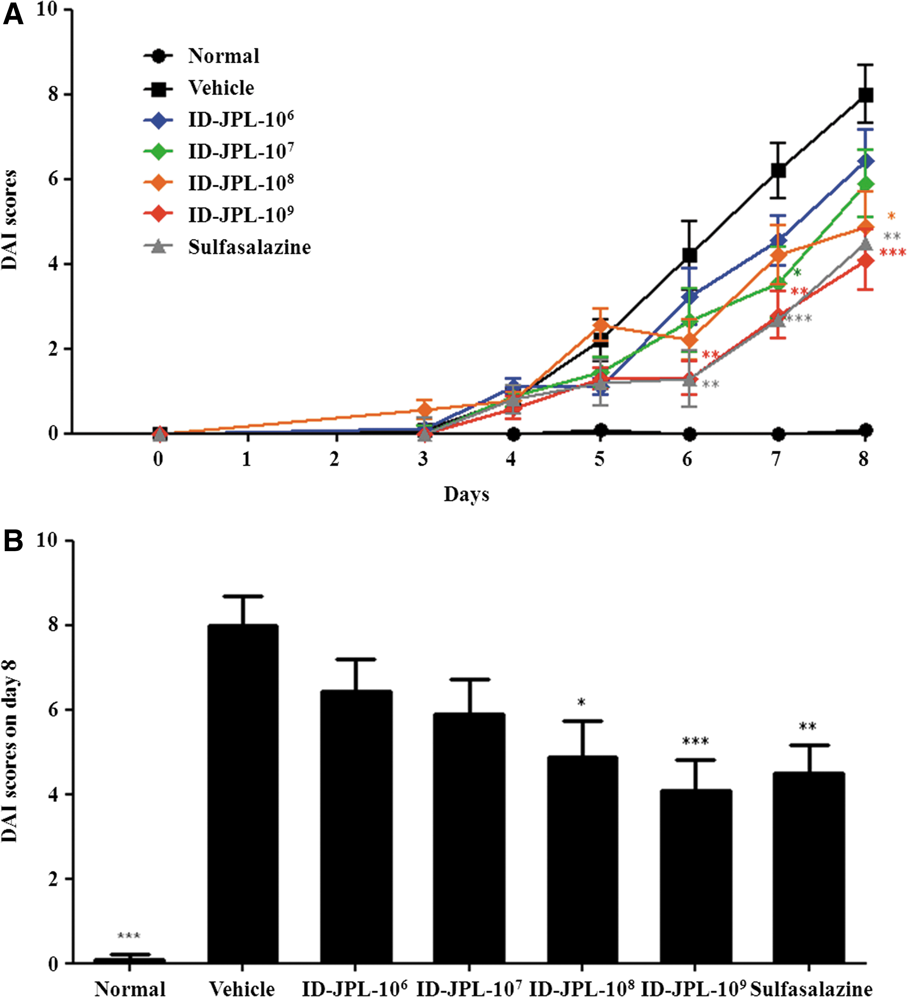
Effects of ID-JPL934 on the DAI scores.
Mice with colitis had shorter colon lengths than the normal group (Fig. 2). This shortening was prevented by ID-JPL934 in a dose-dependent manner, showing equal aspects among ID-JPL934 108, 109, and sulfasalazine groups.
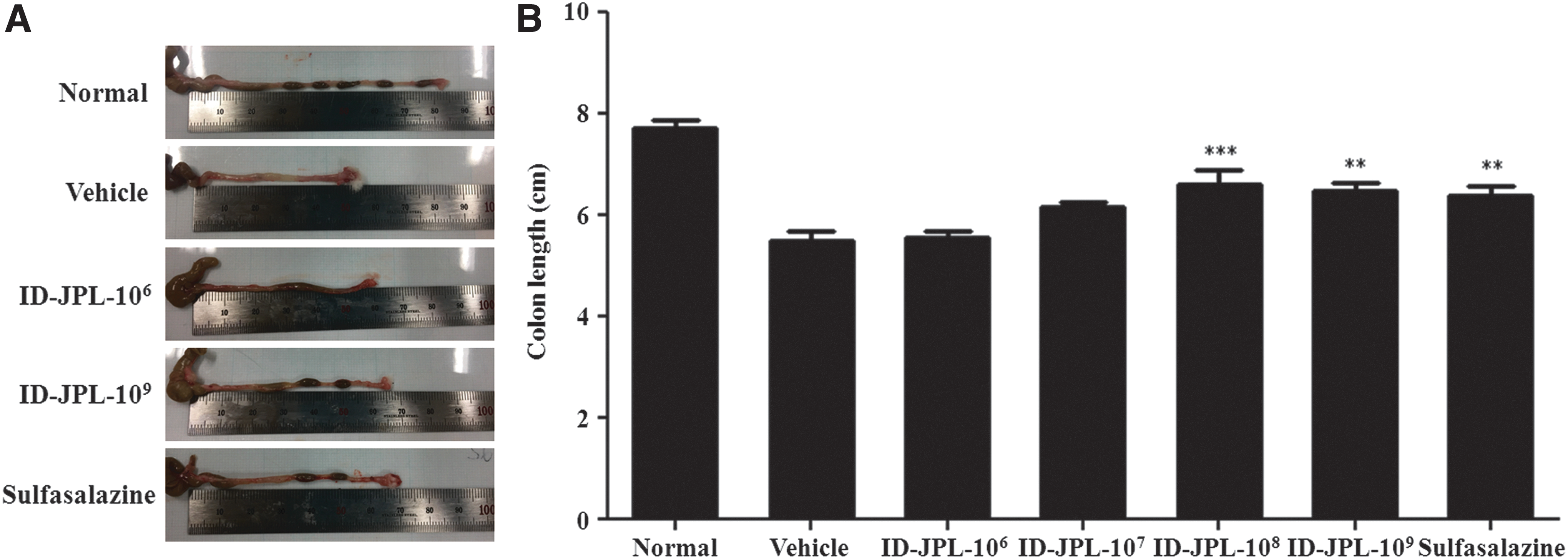
Effects of ID-JPL934 on the colon length. Cola from the ileocecal junction to the anus were enucleated to measure the length.
Effects of ID-JPL934 on the histological parameters
To assess the histologic severity of DSS-induced colitis, we observed H&E-stained colon tissues using a microscope and measured histologic scores (Fig. 3). In the vehicle group, infiltration of inflammatory cells in mucosa and submucosa, severe crypt damage, and loss of goblet and epithelial cells were observed. The histologic scores also increased to 7.2 ± 0.6, whereas that of the sulfasalazine group dramatically decreased. In ID-JPL934-treated groups, the histologic scores decreased dose-dependently. Especially, ID-JPL934 108 and 109 groups showed significant declines in the histologic scores (5.2 ± 0.5 and 4.9 ± 0.6, respectively), similar to that of sulfasalazine group (4.5 ± 0.5).

Effects of ID-JPL934 on the histological parameters.
Effects of ID-JPL934 on the expression of colonic proinflammatory cytokines
ELISA was carried out to detect the level of colonic cytokines related to inflammatory responses. Three proinflammatory cytokines, including TNF-α, IL-1β, and IL-6, were chosen because these are known to play major roles in the pathology of colitis. The expression levels of these cytokines were elevated by DSS, but were decreased by administration of ID-JPL934 in a dose-dependent manner (Fig. 4). ID-JPL934 109 group showed a similar inhibitory effect in comparison with the sulfasalazine group, used as a positive control.
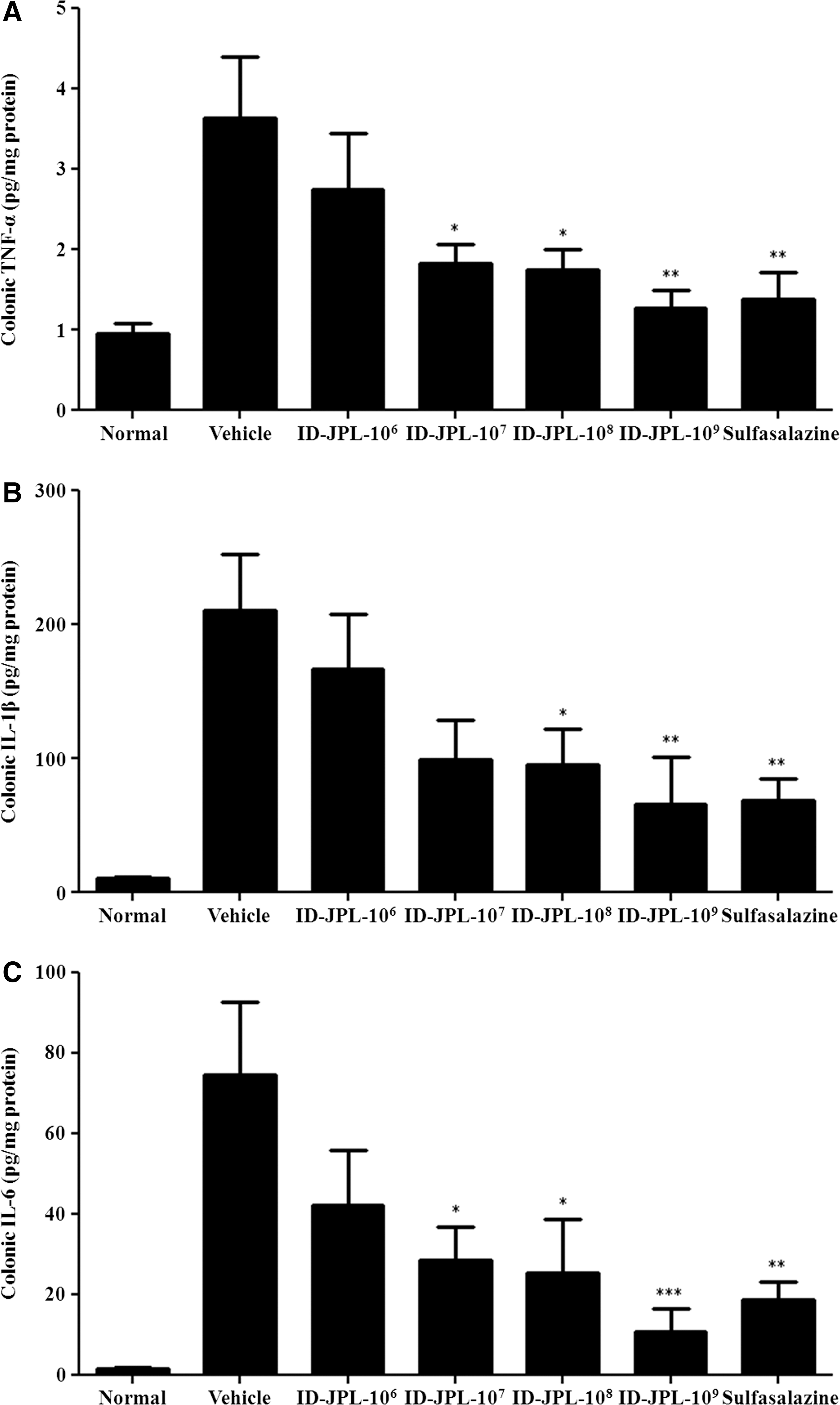
Effects of ID-JPL934 on the expression of colonic proinflammatory cytokines. After extraction of protein from colon tissues in accordance with the description in the Materials and Methods section, the expression of proinflammatory cytokines such as
Discussion
Probiotics are considered valuable sources of beneficial microorganisms and their metabolites that could potentially substitute for chemical medications. 12 –14 Indeed, respectable research groups have been working to establish the therapeutic potencies of probiotics on colitis. L. plantarum and Lactobacillus acidophilus are known to be representative bacterial strains discovered with anticolitic properties. 23 –25
Several hypotheses regarding the effects of probiotics on IBD have been suggested as follows: (1) Probiotics modulate intestinal immune system through regulation of proinflammatory cytokine expression. 23 (2) Probiotics regulate microbial balance in gastrointestinal tract through inhibition of the growth of pathogens. 25 (3) Probiotics improve the function of gut barrier through increases in the tight junction protein expression. 16 Among the three hypotheses, the anti-inflammatory effects of probiotics attracted our attention, because IBD is classified as an intestinal inflammatory disease. 1
The inhibition of inflammation has been used as a major therapeutic strategy to treat colitis. Sulfasalazine, used as a positive control in this experiment, is a representative anti-inflammatory agent. 26,27 Although physiologic benefits of a probiotic single strain are well known, efficacies of combinations of strains remain elusive. Combinations of different kinds of probiotics are a widely used method to improve their beneficial activities. Mixed probiotic strains might have additive or synergistic effects. 28 There were trials to evaluate medicinal roles of probiotic mixtures. 12,19
We designed ID-JPL934, a mixture of three live bacterial strains at a 1:1:1 ratio, and anticipating favorable alterations: L. johnsonii IDCC9203, L. plantarum IDCC3501, and B. animalis subspecies lactis IDCC4301. Three probiotics were selected from our own probiotics inventories throughout the prescreening test to demonstrate anti-inflammatory efficacies.
The effects of ID-JPL934 were investigated on colonic injury using rodent model of IBD. The DSS-induced colitis model was adopted in this study. This model is considered to be a standard model for colitis because of its characteristic similarities to human UC. 29,30 Administrations of DSS resulted in clinical colitic symptoms such as fecal bleeding, reduced BW, shortened colon, and ulcers in the mucus membrane. 20
In our results, the group treated with DSS only had the highest DAI score and developed not only intense bloody excrement, severe diarrhea, and weight loss but also significantly shorter colon lengths. These factors were dose-dependently ameliorated, when ID-JPL934 was administered. Especially, the treatment of ID-JPL934 108 and 109 dramatically improved DAI scores and colon length. DAI scores declined in accordance with recovery of weight loss and fecal conditions. These results demonstrated the therapeutic potency of ID-JPL934 to suppress acute coltis in mice.
To verify whether the intestinal damages were caused by the administrations of DSS, histopathological examination was performed. It was observed that there was serious injury to the crypt structure, intestinal goblet cells and epithelial cells, and infiltration of immune cells due to DSS. Such observations indicated that DSS triggered colitic responses by damaging the architectural organization of the colon tissues and inducing colonic inflammation. DSS-induced histopathological features of the colon were relieved dose-dependently in the ID-JPL934-treated groups. The migration of immune cell in mucosa and submucosa was restricted, and the constitution of the crypts was preserved.
Patients and mice suffering from colitis have abnormalities in the mucosal immune system influenced by various immunocytes, including effecter T cells, dendritic cells, macrophages, and natural killer cells. 31 Pathological invasion of inflammatory cells into colonic mucosa increases the concentration of inflammatory cytokines. 32 Proinflammatory cytokines such as TNF-α, IL-1β, and IL-6 play important roles to trigger colitis and lead to chronic phase. 7,8,33 The levels of these cytokines are markedly elevated in both UC and CD. 6
TNF-α, a major proinflammatory cytokine, exacerbates inflammatory reactions by increasing the expression of proinflammatory cytokines through the nuclear factor kappa-B pathway. 34 TNF-α antibodies such as infliximab and adalimumab have been approved to treat severe colitis patients because inhibition of TNF-α is considerably effective to regulate intestinal immunity by preventing the apoptosis of mucosal T lymphocytes. 35 IL-1β initiates colonic inflammation through recruiting granulocytes and activates CD4+ Th17 cells to secrete IL-17. 36 Infiltration of neutrophil is known to be essentially required for the progression of chronic inflammation. IL-6 promotes neutrophil migration into the colitic region. 37
There are studies that have proven pharmacological effects of drugs for suppressing anti-inflammatory cytokine in colitis. 38,39 Action mechanisms of sulfasalazine in UC also include the reduction of inflammation by inhibiting the expression of proinflammatory cytokines. 27 These reports convinced us that hindering the secretion of proinflammatory cytokines is a key strategy for suppressing colitis.
In this study, the expression of TNF-α, IL-1β, and IL-6 in the colon tissues is diminished dose-dependently by administrations of ID-JPL934, consistent with the reduction of DAI and histopathological scores. The ID-JPL934 109 group showed similar inhibition on the level of cytokine expression compared with the sulfasalazine group. These results suggested that ID-JPL934-induced improvement of colitic symptoms might be caused by reducing colonic inflammation, which results from blocking the expression of proinflammatory cytokines.
Downregulating the level of cytokines by administrations of ID-JPL934 reduced inflammation and suppressed progression of colitis through the inhibition of immune cell infiltration into the lesions. These anti-inflammatory effects could lead to the improvement of colitic symptoms and shortened cola through the recovery of the colon tissue. Increases in BW could be related to improvement of gastrointestinal functions.
In this study, it was demonstrated that the mixture of three strains of probiotics attenuated the clinical symptoms of IBD such as diarrhea, weight loss, shortened colon length, and fecal bleeding in a DSS-induced mouse colitis model for the first time. The results of our studies highlight that the regulation of inflammation through the inhibition of the expression of proinflammatory cytokines such as TNF-α, IL-1β, and IL-6 is a key strategy for improving IBD. Although there were difficulties in elucidating the exact underlying mechanisms of ID-JPL934 in the body as well as the specific contributions of each strain, this study might provide valuable biological evidence for the ability of ID-JPL934 to suppress the expression of proinflammatory cytokine, resulting in potential therapeutic effects on colitis.
In conclusion, we suggest that ID-JPL934 might be a pharmaceutical candidate for IBD by regulating intestinal inflammation.
Footnotes
Acknowledgments
This work was supported by Korea Institute of Planning and Evaluation for Technology in Food, Agriculture, Forestry and Fisheries (IPET) through High Value-Added Food Technology Development Program, funded by Ministry of Agriculture, Food and Rural Affairs (MAFRA) (116017032SB010). All authors are employed by Ildong Pharmaceutical Co., Ltd.
Author Disclosure Statement
No competing financial interests exist.