
Abstract
Periodontitis, an inflammatory disease of the gingival tissue, triggered by microbial-derived elements, such as lipopolysaccharide (LPS), collapses the periodontal tissues and resorbs the alveolar bone. This study evaluated the inhibitory effects of standardized Boesenbergia pandurata extract (BPE) and panduratin A (PAN) on periodontitis-induced inflammation and alveolar bone loss. Sprague-Dawley rats with LPS-induced periodontitis were orally administered BPE (50 and 200 mg/kg/day) and PAN (20 mg/kg/day) for 8 days. Histological analysis revealed that BPE- and PAN-administered groups showed decreased cell infiltration and alveolar bone resorption. Furthermore, the BPE and PAN significantly alleviated the mRNA and protein expression levels of nuclear factor kappa B (NF-κB), interleukin-1β, matrix metalloproteinase (MMP)-2, and MMP-8. BPE and PAN also inhibited the expression of nuclear factor of activated T cells, cytoplasmic 1, c-Fos, and ostoclastogenesis-related enzymes, including cathepsin K and tartrate-resistant acid phosphatase (ALP). BPE and PAN not only upregulated the osteoblastogenesis-associated markers, such as collagen type I (COL1A1) and ALP, but also increased the ratio of osteoprotegerin to receptor activator of NF-κB ligand. Collectively, BPE and PAN efficiently prevent destruction of periodontal tissues and stimulating the loss of alveolar bone tissues, strongly indicative of their potential as natural antiperiodontitis agents.
Introduction
P
Gram-negative oral pathogens, such as Porphyromonas gingivalis, are initiators of periodontitis. The host immune system is activated in response to microbial-derived elements, such as lipopolysaccharide (LPS), antigens, and virulence factors. 5 The inflammation activated by microbial-derived elements in the gingival tissues increases the expression of inflammatory factors (interleukin [IL]-1β and nuclear factor kappa B [NF-κB]). Furthermore, the expression of matrix metalloproteinases (MMPs) is upregulated to destroy components, including gelatins and collagens, which support the matrix of gingival tissues. 6
Inflammation accelerates the expression of receptor activator of NF-κB ligand (RANKL) from fibroblasts in gingival tissues, leading to osteoclast differentiation. 7 Osteoclasts, responsible for bone resorption, balance on osteoblasts, which form new bone under normal conditions. Periodontitis destroys the balance, and the prevailing osteoclasts collapse the alveolar bone. 8 The binding between RANKL and the RANK receptor triggers osteoclast differentiation by activating nuclear factor of c-Fos and activated T cells, cytoplasmic 1 (NFATc1), which work as transcription factors to regulate the genes involved in osteoclastogenesis. 9 Particularly, NFATc1 has an autoamplification system. As a unique NFAT protein, NFATc1 binds to the NFAT-binding sites, which are exited in NFATc1 promoters. 10 Once NFATc1 binds to the NFATc1 promoter, the expression of NFATc1 is upregulated. Thus, the differentiation signal is highly amplified. Then, bone resorption is increased by the upregulation of tartrate-resistant acid phosphatase (TRAP) and cathepsin K. 11
Boesenbergia pandurata Roxb., also commonly called as fingerroot, is a tropical plant belonging to the Zingiberaceae family. Panduratin A (PAN) (Fig. 1), which is a type of chalcone compound, is a major bioactive compound in B. pandurata. 12 B. pandurata and PAN are reported to possess various bioactivities, such as antibacterial, antioxidant, and antiobesity effects. 12 Previous studies on the anti-inflammatory effect of PAN were investigated by employing LPS-stimulated RAW264.7 cells. 13, 14 However, the antiperiodontitis activity of B. pandurata extract (BPE) and PAN using LPS-treated animal models has not yet been studied. In this study, we evaluated whether BPE and PAN would be effective in the inhibition and prevention of gingival inflammation and alveolar bone loss induced by periodontitis.
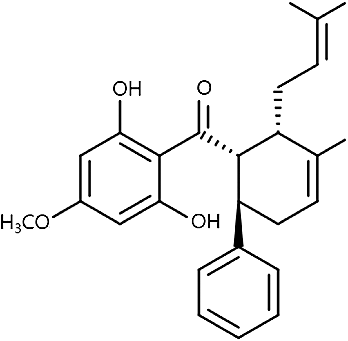
The chemical structure of PAN. PAN, panduratin A.
Materials and Methods
Preparation of standardized BPE and isolation of PAN
The dried rhizomes of B. pandurata were supplied from AAT Costech Co., Ltd. (Seoul, Korea). The ground rhizomes were extracted with 95% ethanol for 3 days at room temperature. The filtrates were subsequently evaporated by a rotary evaporator (Heidolph Instruments GmbH & Co. KG, Schwabach, Germany). The yield of BPE was 12.0% (w/w). PAN was isolated according to the previous study. 15 The purity of PAN was measured to be ≥98%. The BPE was standardized based on a bioactive compound, PAN. 12 An Agilent 1260 Infinity HPLC system (Agilent Technologies, Santa Clara, CA, USA) furnished with a LUNA C18 column (Phenomenex, Torrance, CA, USA) was used for the quantitative analysis of PAN in BPE. The mobile phase was 80% methanol; it was constantly pumped at a flow rate of 1 mL/min. The samples were detected at 290 nm. A standard curve for PAN was plotted with concentrations versus peak areas. Chromatographic peaks of BPE were identified by comparing the retention times of PAN (Fig. 2). From the standard curve, the concentration of PAN in the standardized BPE was determined to be 8% w/w.
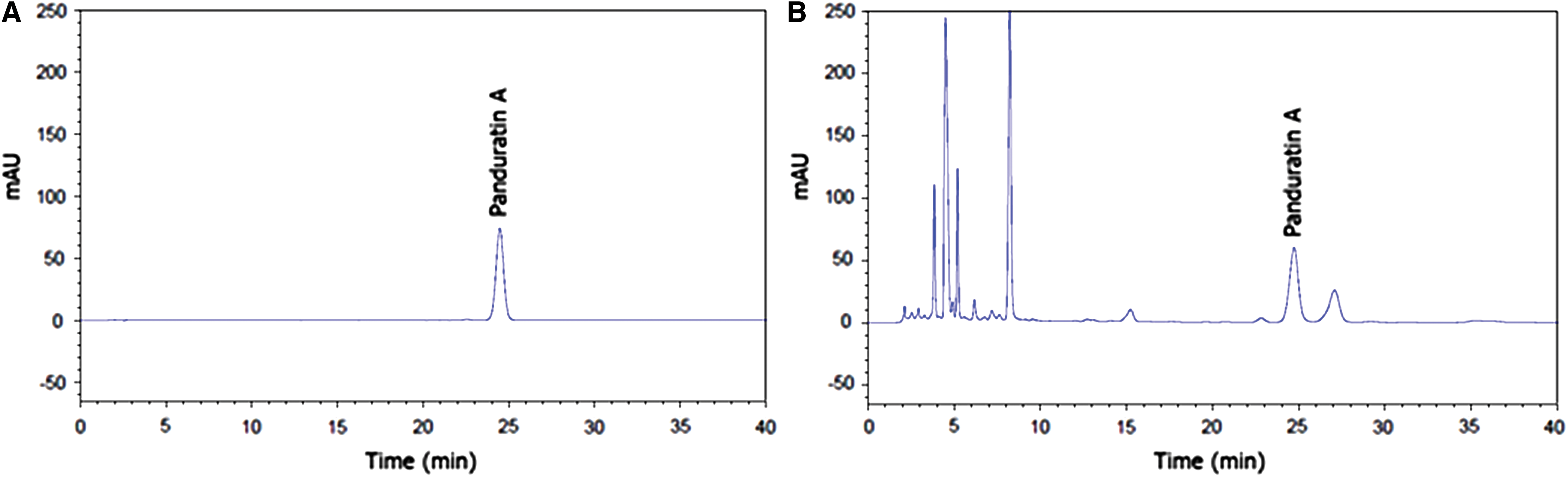
HPLC chromatograms of PAN and BPE.
Animal experiment
Animal experiments were conducted with thirty 9-week-old male Sprague-Dawley rats with an initial average weight ranging from 280 to 300 g. All animals were kept under a constant environment (24°C ± 2°C temperature, 55% ± 5% relative humidity, and 12-h light/12-h dark cycle) at the College of Pharmacy, Chung-Ang University (Seoul, Korea). Experimental groups were divided into five groups after 1 week of acclimation: (1) CON (negative control); (2) LPS; (3) BPE50 (50 mg/kg/day); (4) BPE200 (200 mg/kg/day); and (5) PAN20 (20 mg/kg/day). Periodontitis was induced in all the groups, except for the control group by injecting 10 mg/mL of Escherichia coli LPS (Sigma-Aldrich, St. Louis, MO, USA) every 2 days between the first and second mandibular molars using isoflurane (Hana Pharm. Co., Ltd., Gyeonggi-do, Korea) as an anesthetic. Following six consecutive LPS injections, BPE and PAN were administered orally for 8 days along with LPS injection. The entire experimental procedure was authorized by the Institutional Animal Care and Use Committee (IACUC) of the Chung-Ang University Laboratory Animal Research Center (Permit No. 2016-00104).
Reverse transcription-polymerase chain reaction
TRIzol reagent (TaKaRa, Shiga, Japan) was employed to isolate total RNA from gingival tissues and alveolar bone. To evaluate the quantity and quality of total RNA, NanoDrop 1000 spectrophotometer (Thermo Fisher Scientific, Inc., Waltham, MA, USA) was used. To synthesize cDNA, the RNA and the reverse transcriptase premix (Elpis Biotech, Daejeon, Korea) were mixed. Reverse transcription was performed according to the following process: 55 min at 42°C for initiation and 15 at 70°C for termination. Specific primers were designed according to our previous study (Table 1). 16 Primer pairs (Bioneer, Daejeon, Korea) were used for cDNA amplification. cDNA amplification by polymerase chain reaction was implemented according to the following process: 30 sec at 94°C for denaturation, 1 min at 55–60°C for annealing, and 1 min at 72°C for extension. The final extension was performed at 72°C for 5 min. Amplified products with loading star (Dyne Bio, Inc., Seoul, Korea) were separated using 1.5–2.0% agarose gel electrophoresis. The intensity of the bands was visualized, and they were detected with the G:BOX EF imaging system (Syngene, Cambridge, UK) and the GeneSys software version 1.3.9.0 (Syngene).
Western blotting
The lysates were extracted from the gingival tissues and alveolar bone with NP40 lysis buffer (Elpis Biotech) mixed with a protease inhibitor cocktail (Sigma-Aldrich). To determine and normalize the protein concentration of the lysate, the Bradford protein assay (Bio-Rad Laboratories, Inc., Hercules, CA, USA) was performed. After equivalent protein amounts of each sample were loaded onto 10% sodium dodecyl sulfate–polyacrylamide gel and separated by sodium dodecyl sulfate–polyacrylamide gel electrophoresis (SDS-PAGE). The separated proteins on the gels were electrophoretically transferred onto nitrocellulose membranes (Whatman GmBH, Dassel, Germany), followed by incubation with primary antibodies against IL-1β, NF-κB, MMP-2, MMP-8, NFATc1, TRAP, c-Fos, cathepsin K, alkaline phosphatase (ALP), collagen type I (COL1A1), osteoprotegerin (OPG), RANKL (1:1000 dilution; Santa Cruz Biotechnology, Inc., Santa Cruz, CA, USA), and α-tubulin (1:1000 dilution; Cell Signaling Technology, Beverly, MA, USA) for 16–18 h at 4°C. The blots were subsequently exposed to horseradish peroxidase (HRP)-conjugated antibody (1:5000 dilution; Bethyl Laboratories, Inc., Montgomery, TX, USA) for 1 h at room temperature. An enhanced chemiluminescence solution (Amersham Biosciences, Little Chalfont, UK) was used to develop the blots. The intensity of the protein bands was detected and analyzed with the G:BOX EF imaging system (Syngene) and the GeneSys software version 1.3.9.0 (Syngene).
Histological analysis
After being fixed with 10% formalin buffer, the decalcified periodontal tissues were embedded in paraffin, cut into 5-μm sections, and mounted onto slides. Hematoxylin and Eosin (H&E) was used to stain the paraffin slides. Random areas of stained tissues were analyzed using an Eclipse TE2000 U inverted microscope equipped with twin charge-coupled device (CCD) cameras ( × 200 magnification; Nikon, Tokyo, Japan) to detect cell infiltration and alveolar bone loss. To determine the expression levels of key biomarkers in the tissues, immunohistochemistry was performed using the following protocol: the paraffin slides were exposed to primary antibodies against MMP-8, MMP-2, NFATc1, TRAP, ALP, and COL1A1 (1:200 dilution; Santa Cruz Biotechnology, Inc.) at 4°C overnight. Then, the slides were exposed to HRP-conjugated antibody (1:5000 dilution; Bethyl Laboratories, Inc.) for 1 h at 4°C. The slides were analyzed using an Eclipse TE2000 U inverted microscope equipped with twin CCD cameras ( × 400 magnification; Nikon). To quantify the protein expression levels, the stained areas of the images were analyzed by the ImageJ software version 1.47 (National Institutes of Health, Bethesda, MD, USA).
Microcomputed tomography imaging
To detect the effect of BPE on alveolar bone loss, microcomputed tomography (CT) imaging was carried out using a SkyScan 1076 (SkyScan, Kontich, Belgium) under the following conditions: total rotation, 360°; rotation step, 0.5°; pixel size, 18 μm; voltage, 100 kV; current, 100 μA; and exposure time, 1475 ms. The distance between the cementoenamel junction (CEJ) and alveolar bone crest (ABC), representing the level of alveolar bone loss, was quantified using the ImageJ software version 1.47 (National Institutes of Health). The scanned images were reconstructed by NRecon (SkyScan) and CTAn (SkyScan) to obtain a 3D trabecular structure. The alveolar bone of first molar was chosen as the region of interest (ROI). The four bone structure parameters, such as bone volume per tissue volume (BV/TV), and bone mineral density (BMD), and trabecular thickness (Tb. Th) were measured and quantified within the range of the ROI: BV/TV is a value for the number of voxels classified as bone trabeculae over the total number of voxels in sample; Tb. Th is the mean trabecular bone diameter; Tb. Sp is calculated by the mean distance between the borders of the segmented trabeculae; and BMD is the amount of calcium hydroxyapatite per volume unit of bone. 17,18
Statistical analysis
All results are expressed as the mean ± standard deviation (SD) and a representative result is shown for each experiment. One-way analysis of variance (ANOVA) was carried out followed by Duncan's multiple range test to evaluate the significance of group differences using statistical package for the social sciences (SPSS) version 23.0 (SPSS, Inc., Chicago, IL, USA). P < .05 was defined as a statistically significant difference.
Results
BPE and PAN inhibit gingival inflammation in LPS-induced rats
The mRNA and protein expression levels of inflammation-related factors, NF-κB and IL-1β were upregulated in the LPS group, but significantly downregulated in the BPE- and PAN-administered groups (Fig. 3). Compared with the LPS group, the mRNA and protein expression levels of MMP-2 and MMP-8 were also suppressed by BPE and PAN administration. These results suggest that BPE and PAN exert anti-inflammatory effects by regulating the expression of inflammation-related factors and matrix degradation-related enzymes.
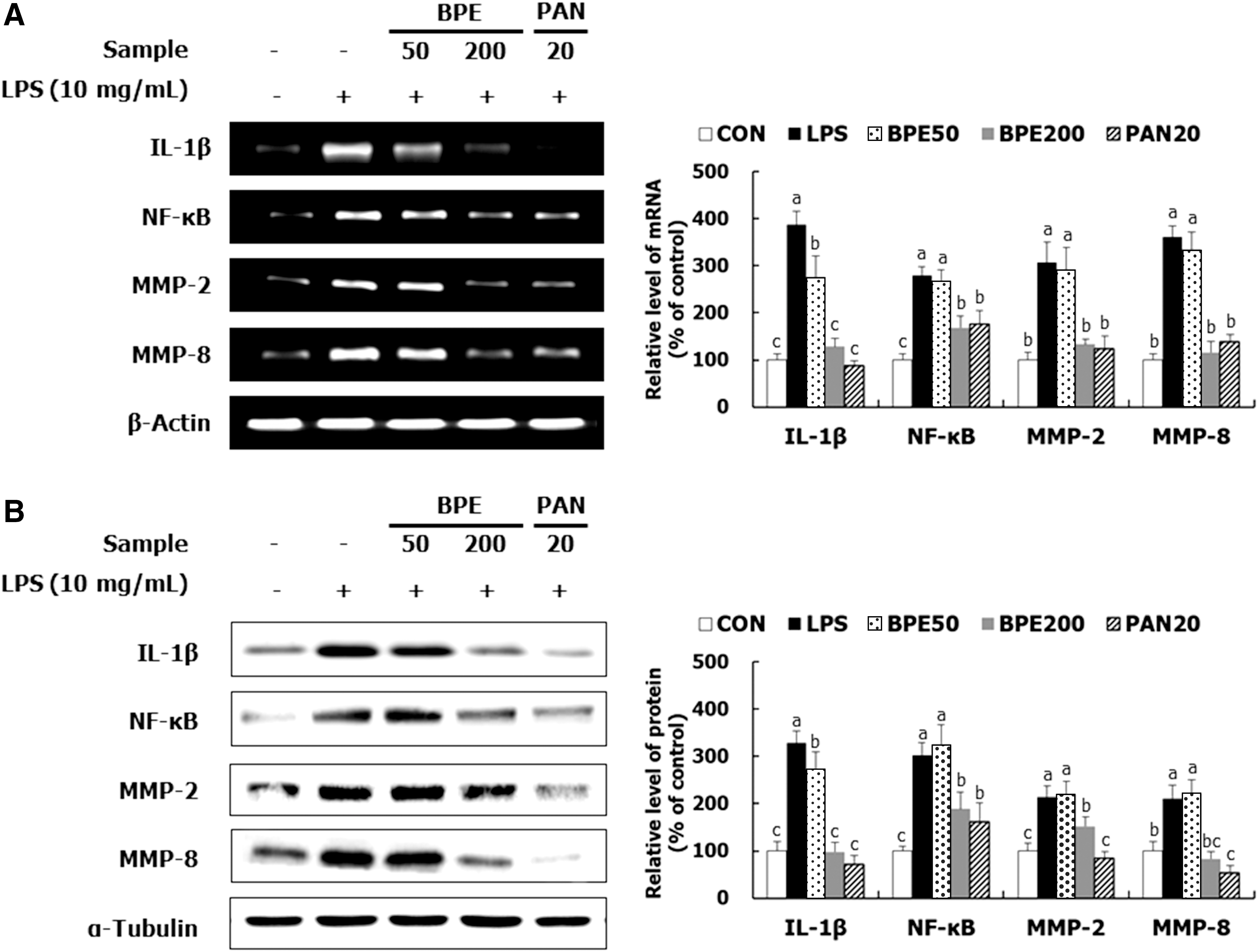
Inhibitory effects of BPE and PAN on inflammation in the gingival tissues of LPS-induced rats.
BPE and PAN inhibit osteoclastogenesis in the alveolar bone of LPS-induced rats
Compared with the LPS group, the BPE- and PAN-administered groups showed decreased mRNA and protein levels of osteoclastogenesis-related factors, such as NFATc1 and c-Fos. Additionally, the expression of bone resorption-related enzymes, cathepsin K and TRAP, was inhibited by treatment with BPE and PAN, compared with the case for LPS treatment (Fig. 4). In conclusion, BPE and PAN show antiosteoclastic effects by downregulating osteoclast differentiation-involved transcription factors and enzymes.
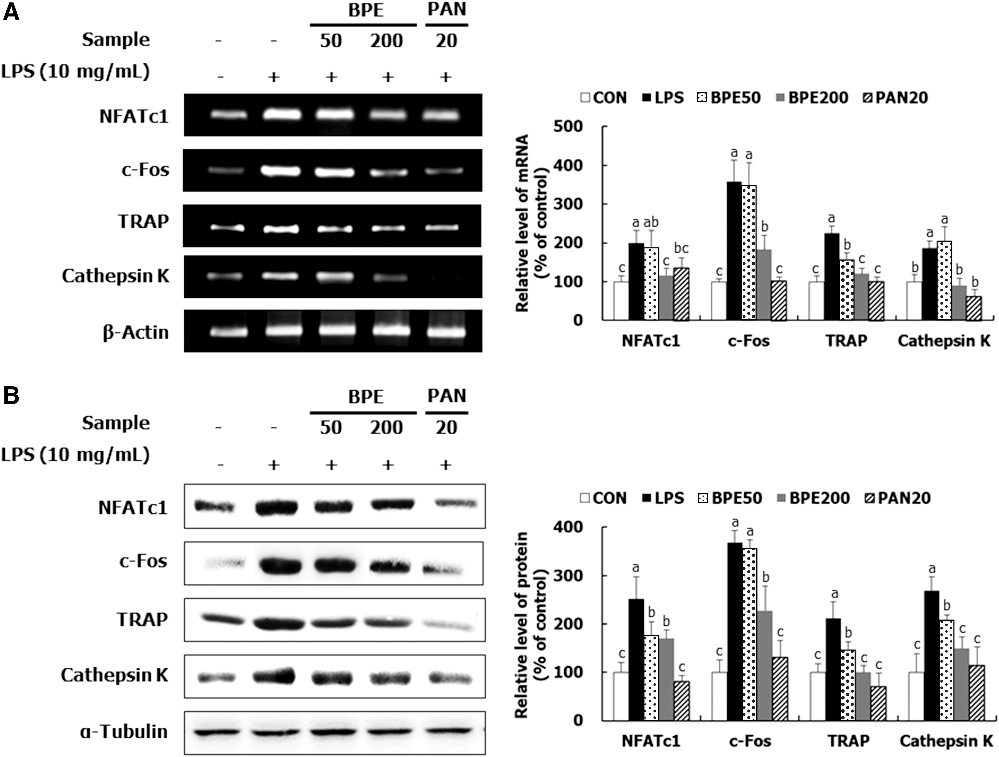
Inhibitory effects of BPE and PAN on osteoclastogenesis in the alveolar bone of LPS-induced rats.
BPE and PAN promote osteoblast differentiation in LPS-induced rats
BPE and PAN promoted the mRNA and protein expression of ALP and COL1A1 and reduced the expression of RANKL in LPS-induced rats (Fig. 5). In the BPE- and PAN-administered groups, the ratio of OPG to RANKL, which is an indicator of bone remodeling, was increased at the mRNA and protein levels, compared with the LPS group. These results suggest that BPE and PAN prevented alveolar bone resorption and contributed to bone remodeling.

Stimulatory effects of BPE and PAN on osteoblast differentiation in the alveolar bone of LPS-induced rats.
BPE and PAN improve the histological changes induced by periodontitis
H&E staining analysis revealed high levels of histological changes, such as cell infiltration and bone resorption in the LPS group (Fig. 6A). The BPE- and PAN-administered groups showed attenuation of cell infiltration and irregular surface formation caused by LPS injection. In addition, the expression of periodontitis-related proteins in the periodontal tissues was visualized by immunohistochemistry (Fig. 6B). Compared with LPS treatment, BPE and PAN treatments downregulated the protein expression of MMP-2, MMP-8, NFATc1, and TRAP, but increased the protein expression of ALP and COL1A1 in the periodontal tissues (Fig. 6C). Therefore, it can be said that BPE and PAN are effective against periodontitis at a histological level.
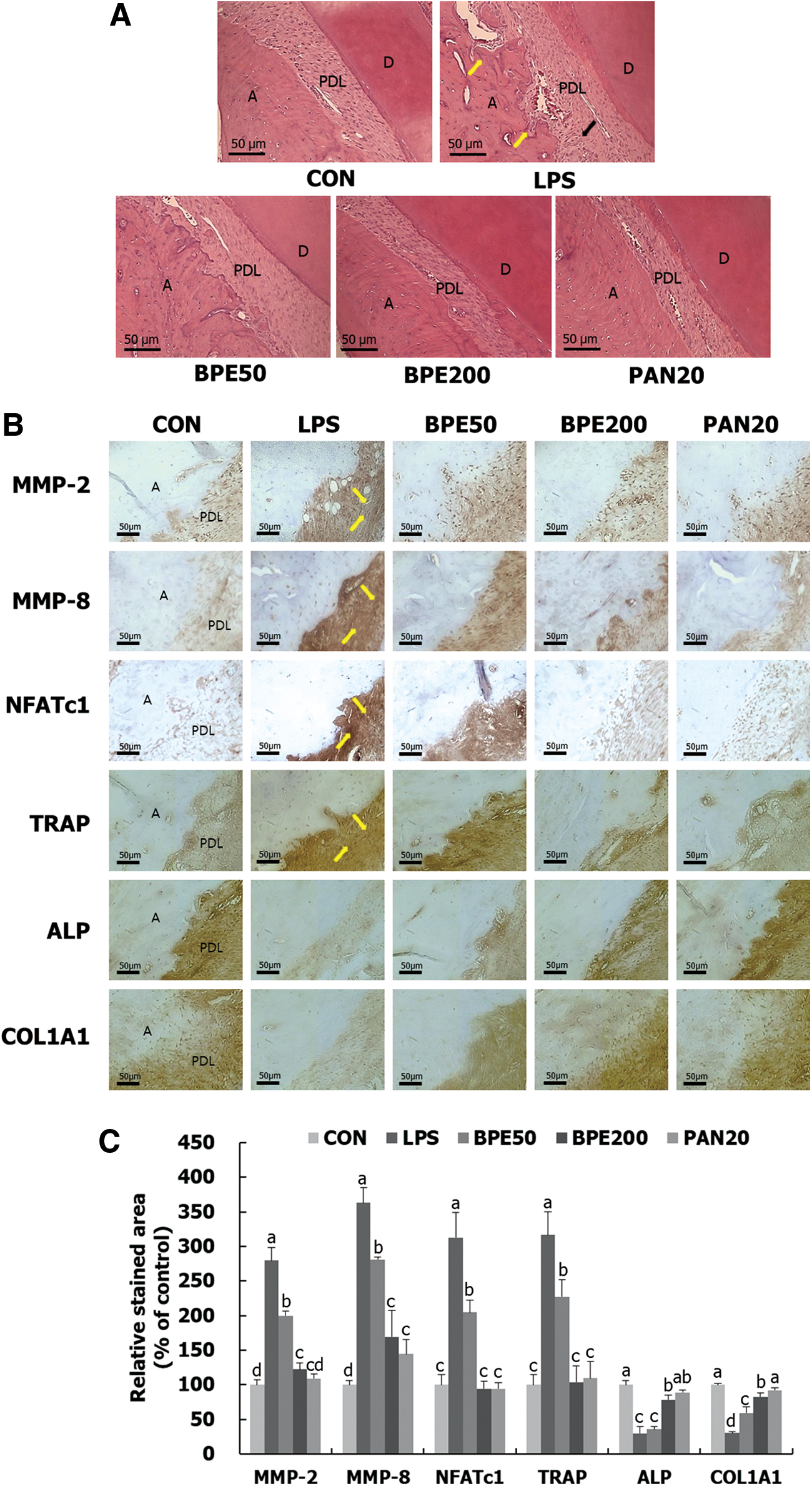
Effects of BPE and PAN on histological changes in LPS-induced rats.
BPE and PAN inhibit alveolar bone loss in LPS-induced rats
The degree of alveolar bone loss was determined using micro-CT imaging. The distance between the CEJ-ABC on the images was measured. In the LPS group, the distance of the alveolar bone loss was 1.01 ± 0.089 mm, which was 24.8% longer than that in the control group (Fig. 7). However, groups that were administered BPE (200 mg/kg/day) and PAN showed a reduction in the distance of alveolar bone loss by 19.7% and 15.4%, respectively. Moreover, the distance of alveolar bone loss in high-dose BPE group was 0.81 ± 0.037 mm, which was equivalent to that in the control group (0.81 ± 0.04 mm). Each parameter (BV/TV, Tb. Th., and BMD), which is related to bone structure, increased in the BPE- and PAN-administered groups, compared with the LPS group, as seen in the reconstructed 3D images (Fig. 8). In contrast, Tb. Sp. decreased in the BPE- and PAN-administered groups, but increased in the LPS group. Therefore, it can be said that the administration of BPE and PAN suppresses alveolar bone loss in LPS-induced rats.
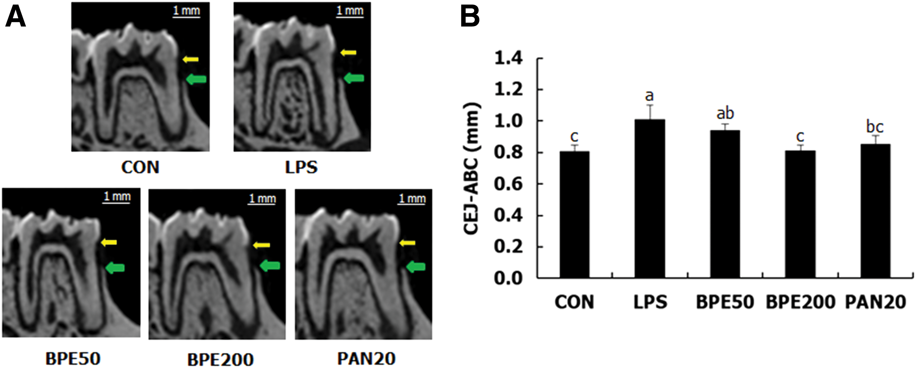
Reversible effects of BPE and PAN on alveolar bone loss in LPS-induced rats.
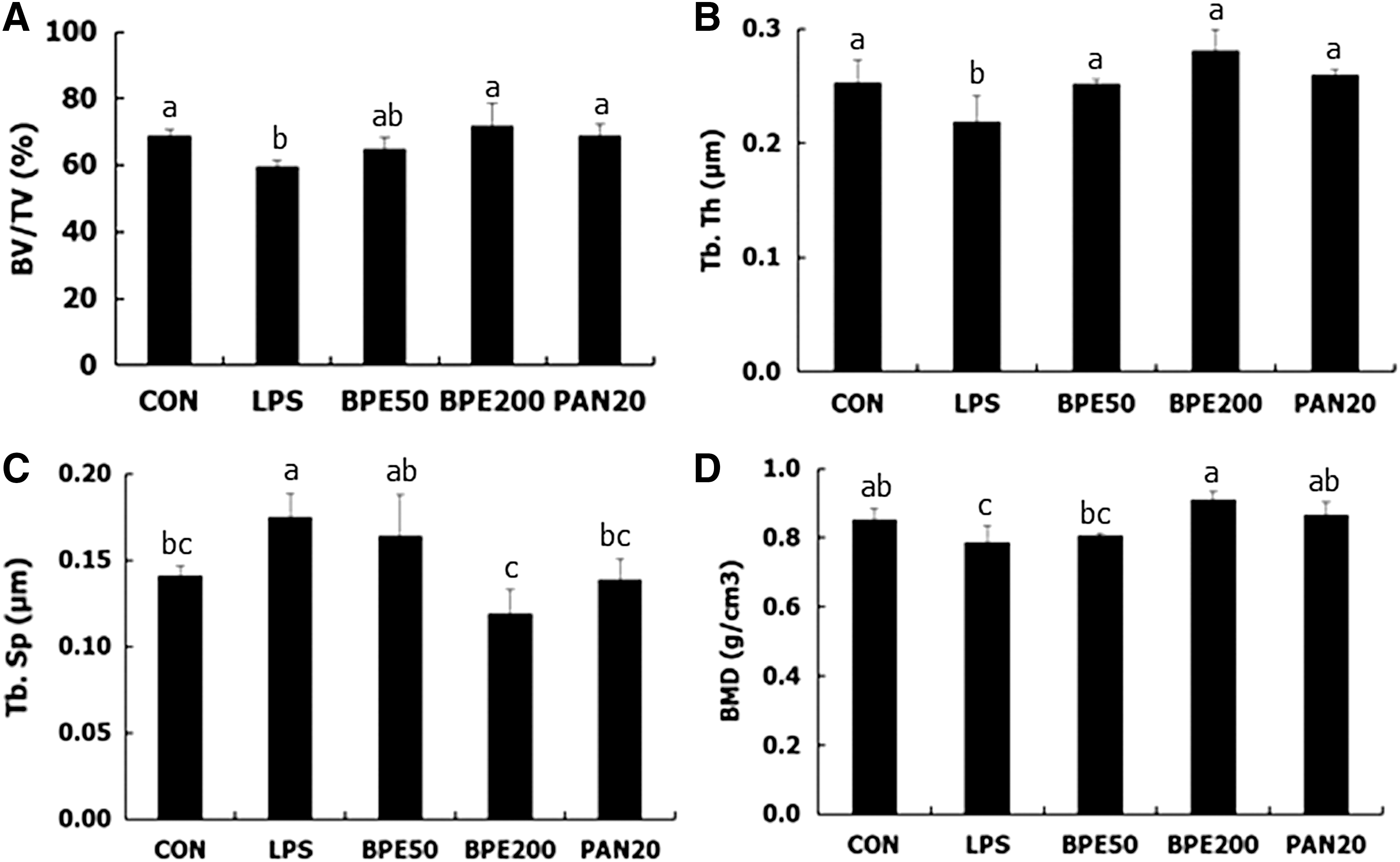
Inhibitory effects of BPE and PAN on alveolar bone destruction in LPS-induced rats. Using reconstructed 3D images,
Discussion
LPS, the main component of the Gram-negative microbial cell wall, is a stimulator of alveolar bone loss during inflammatory gingival destruction. 6 Several reports have suggested that LPS reduces osteogenic factors, including collagen synthesis and ALP activity, suggesting that oral pathogens not only stimulate bone loss, but also diminish bone formation. 19 In this study, we confirmed that BPE and PAN have the potential to prevent periodontitis, and further elucidated their effects in remodeling alveolar bone mass.
In the LPS-induced gingival tissues of rats, BPE and PAN significantly suppressed the expression of inflammatory biomarkers, MMP-2 and MMP-8 (Fig. 3). MMPs are proteolytic enzymes that destroy the extracellular matrix proteins, such as proteoglycans, collagen, and elastin in the gingival tissues. 20 Specifically, MMP-2 degrades the basilar composition of the periodontal connective tissue by dissolving collagen type IV. 21 MMP-8 is responsible for degrading collagen type I and III, which mainly support the structure of gingival tissues. 22 Accordingly, this study indicates that treatment with BPE and PAN can considerably downregulate the destruction of gingival tissues by MMPs.
In periodontitis-induced alveolar bone loss, treatment with BPE and PAN significantly reduced the expressions of TRAP and cathepsin K (Fig. 4). These results might be due to the inactivation of transcription factors, including NFATc1 and c-Fos. When RANKL binds to RANK, the target genes that are essential for osteoclastogenesis are transcribed with the entrance of NFATc1 and c-Fos into the nucleus. 23 Then, the transcription signals highly increase the expression of osteoclastogenesis-related enzymes, such as TRAP and cathepsin K. 24 However, BPE and PAN regulated the serial signal transduction by interrupting the role of NFATc1 and c-Fos. BPE and PAN not only inhibited alveolar bone resorption, but also activated bone formation by increasing the ratio of OPG/RANKL and expression of ALP and COL1A1 (Fig. 5). ALP plays a role in mineralization during bone formation in the bone matrix. 25 OPG is a powerful inhibitor of osteoclastogenesis through its binding to RANKL, which blocks the binding of RANKL to RANK, thereby affecting bone formation. 26 Several natural materials, including Salvia miltiorrhiza, Moringa oleifera, and puerarin have been shown to attenuate inflammation and alveolar bone loss triggered by LPS-induced periodontitis in rats. 27 –29 Interestingly, BPE and PAN showed that bone recovery effects as well as inhibition of bone loss in the periodontium.
H&E staining analysis revealed that the characteristic changes induced by periodontitis included cell infiltration in the gingival tissues and resorption on the surface of the alveolar bone. Cell infiltrates can induce excessive amounts of proinflammatory cytokines and osteoclastic enzymes. 30 In this study, BPE and PAN efficiently decreased cell infiltration and improved the irregular surfaces of the alveolar bone, whereas LPS injection enhanced these symptoms (Fig. 6A). Micro-CT analysis also demonstrated that BPE and PAN prevented periodontitis-induced alveolar bone loss by increasing the height of the bone crest and protecting against the deterioration of the trabecular bone structure (Figs. 7 and 8). These results suggest that BPE and PAN can prevent gingival tissue damage and alveolar bone loss induced by cell infiltration. Other phytochemicals, including quercetin, mollugin, and thymoquinone have been reported to possess antiperiodontitis activity at the histological level. Quercetin improved alveolar bone resorption induced by osteoclasts in LPS-induced periodontitis, 31 and mollugin treatment was effective against antiosteoclastogenesis in an animal model. 32 In addition, thymoquinone remarkably inhibited cell infiltration in rat models of experimental periodontitis and prevented alveolar bone loss. 33 However, the doses of these natural agents were higher than those of PAN.
The inhibitory effect of BPE on periodontitis might be mediated by the high content of PAN in addition to other phytochemicals in the extract. The PAN content (8%) of BPE mainly modulates the inflammatory responses in periodontitis based on the anti-inflammatory effects exhibited by PAN. Additionally, PAN inactivated the genes associated with bone resorption in the rat periodontium. Therefore, the PAN content of BPE could be mainly involved in delaying or inhibiting the process of periodontitis.
Collectively, the novel antiperiodontitis activities of BPE and its active compound PAN were elucidated in this study. In the LPS-induced periodontitis model, rats orally administered BPE and PAN showed histological improvements, including repression of cell infiltration and bone resorption as well as recovery of alveolar bone loss. The expression levels of inflammatory biomarkers in the gingival tissues and osteoclastic biomarkers in the alveolar bone were significantly decreased by BPE and PAN. Consequently, BPE and PAN might efficiently attenuate periodontitis induced by microbial LPS, indicating their potential role in the development of antiperiodontitis agents. Further clinical studies would provide more evidence to help confirm and enhance the antiperiodontitis activity of BPE and PAN.
Footnotes
Acknowledgments
This research was supported and funded by “The Project of Conversion by the Past R&D Results” through the Ministry of Trade, Industry, and Energy (MOTIE) (N0002221, 2016), and in part by the Yonsei University Future-leading Research Initiative of 2015 (RMS2 2015-22-0073).
Author Disclosure Statement
No competing financial interests exist.