
Abstract
Talisia esculenta (A. St.-Hil.) Radlk. is a large tree belonging to family Sapindaceae and popularly known as “pitombeira” or “pitomba.” Although species have relevant economic and medicinal uses in Brazil, no study has investigated its effectiveness as a diuretic, hypotensive, and antihypertensive agent. The aim of this study was to present a detailed anatomical and histochemical study for T. esculenta and provide important safety and efficacy parameters. After morpho-anatomical and microchemical study, a purified aqueous extract (ethanol soluble fraction obtained from T. esculenta [ESTE]) was obtained, and detailed phytochemical investigation was performed. Subsequently, acute oral toxicity test was performed in male and female rats. Moreover, diuretic, hypotensive, and antihypertensive effects on normotensive and spontaneously hypertensive rats (SHR) were investigated. Finally, the effects of prolonged treatment with ESTE on serum levels of nitrite, thiobarbituric acid reactive species, and nitrotyrosine were also measured in SHR. Oral treatment with ESTE did not induce acute toxic effects and did not affect urine production, blood pressure, or heart rate of normotensive and SHR. Prolonged treatment with ESTE was able to increase serum nitrite levels and significantly reduce oxidative and nitrosative stress markers in SHR. Data obtained showed that ESTE has a significant antioxidant activity without showing any clinical signs of acute toxicity. The use of this species as a diuretic, hypotensive, or antihypertensive agent should be carried out with caution, since administration in rodents did not produce renal and/or hemodynamic responses that justify this indication.
Introduction
T
In recent years, the phytochemical profile of T. esculenta seeds and fruits has been gradually characterized, while the chemical characteristics of its leaves remain not fully elucidated. The available data describe the presence of carbohydrate-binding proteins, 3,4 catechins, flavonoids, and organic acids, 5,6 especially in the fruits of this species.
Some preliminary biological investigations have been conducted with T. esculenta preparations, showing its insecticidal, 7,8 antifungal, 9 antibacterial, 10,11 antioxidant, antiproliferative, and antimutagenic properties. 5,6 In contrast, few toxicological studies have been conducted with this species. Recently, Riet-Correa et al. 12 reported the poisoning of sheep and cattle after the consumption of T. esculenta leaves and fruits, drawing attention due to its wide use by Brazilian populations.
Thus, considering the wide use of this species in Brazil, and especially due to the lack of data on its efficacy and safety on the cardiovascular system, a detailed ethnopharmacological investigation with a popular herbal preparation made with T. esculenta was performed. First, leaves of this species were collected, and a detailed morpho-anatomical and microchemical study was carried out. Then, a purified fraction was obtained by infusion, and its phytochemical profile was characterized through ultra-performance liquid chromatography coupled to photodiode-array detection and electrospray ionization tandem mass spectrometry (UPLC-PAD-MS/MS). Finally, a detailed acute toxicological study was carried out, and the diuretic, hypotensive, and antihypertensive properties of ethanol soluble fraction obtained from T. esculenta (ESTE) were investigated, relating its effects to a possible protective effect on lipid peroxidation and nitrosative stress.
Materials and Methods
Drugs
Hydrochlorothiazide (HCTZ), 1,1-diphenyl-2-picrylhydrazyl (DPPH), and 2,2′-Azobis(2-amidinopropane) dihydrochloride (AAPH) were obtained from Sigma-Aldrich (St. Louis, MO). Sodium chloride was obtained from Merck (Darmstadt, Germany). Acetonitrile and formic acid were purchased from J.T. Baker (Center Valley, PA) and Tedia (Fairfield, OH, EUA), respectively. All other chemicals and solvents were of analytical grade.
Plant material
T. esculenta leaves were collected in August 2015 from the preservation area near the city of Dourados, Mato Grosso do Sul (S 22°12′218″ and W 54° 45′ 05.3″). A voucher was cataloged at the UFGD Herbarium under DDMS number 5221.
Pharmacobotanical assays
T. esculenta leaves and stems (at least three samples) obtained from the sixth node and below (median, intercostal, and margin regions), as well as 5–15 cm stem fragments from shoots, were prepared for pharmacobotanical assays. The plant material was placed in a formaldehyde–acetic acid–ethanol solution and stored in 70% ethanol. For the assessment of leaf and stem material, free-hand longitudinal and cross-sections were made. The midrib, interneural regions, and lateral veins were included in leaves. Leaves and stems were stained using toluidine blue, astra blue, and basic fuchsine to obtain semipermanent slides. 13
Histochemical tests
The following standard solutions were used in histochemical tests: sulfuric acid solution for calcium oxalate crystals; Sudan III for testing lipophilic compounds; Hoepfner–Vorsatz test, modified by Reeve 14 (aqueous 10% sodium nitrate, aqueous 10% acetic acid, aqueous 10% urea, and 2N NaOH); and ferric chloride to assess phenolic substances. 15 Photomicrographs were captured using an Olympus CX 31 light microscope equipped with a C7070 digital camera.
Preparation of aqueous extracts
T. esculenta leaves were air-dried in an oven at 40°C for 5 days and then crushed and pulverized. The aqueous extract (AETE) was obtained by infusion in a similar manner to that described by Prando et al. 16 Thus, AETE was treated with three volumes of ethanol, which gave rise to a precipitate and an ethanol soluble fraction (ESTE). ESTE was filtered, concentrated, and freeze-dried (yield 6.6%). All preparations were kept in freezer until time of experiments.
Phytochemical investigation
The chemical constituents obtained from T. esculenta leaves were analyzed by ultra-high performance liquid chromatography (Acquity-UPLC™, Waters), with detection provided by photodiode array (PDA; Waters) and high-resolution mass spectrometry (HR-MS/MS). The chromatographic separation was performed in a 100 × 2.1 mm BEH-C18 column with particle size of 1.7 μm. The column temperature was 60°C, and a linear gradient of solvents was developed with a mixture of ultra-pure water (Milli-Q) and acetonitrile, both containing 0.1% of formic acid (96%), at constant flow rate of 400 μL/min. The linear acetonitrile increase was from 0% to 10% in 5 min, then to 70% in 13 min. The solvent returned to the initial condition (0% acetonitrile) at 13.5 min, held additional 2 min to re-equilibrate the system. The extract was prepared at 2 mg/mL in MeOH-H2O (1:1, v/v), with injection of 5 μL. To aid in the identification of compounds, detection was performed by PDA screening (200–400 nm) and HR-MS (m/z 150–2000).
Mass spectrometry was carried out in a LTQ Orbitrap XL (Thermo Scientific), in the negative and positive ionization modes, at atmospheric ionization pressure, with ESI source at 350°C. Desolvation was aided by nitrogen stream, with sheath gas at flow rate of 40 arbitrary units (a.u.) and auxiliary gas at 5 a.u. The energies in the positive ionization were 5 kV in the source, 30 V in the capillary, and 110 V in the tube lens; and in the negative ionization, energies were 3.5 kV, −45 V, and −200 V, respectively. Fragmentation with higher energy collisional dissociation was performed with normalized collision energy of 35%. For higher accuracy, the spectrometer was externally calibrated with Pierce™ LTQ ESI Positive Ion (Caffeine, MRFA, Ultramark-1621) and Negative Ion (SDS, sodium taurocholate, Ultramark-1621) calibration solutions (Thermo-Fisher). In LC-MS mode, resolution was set at 15,000 full width at half maximum.
Experimental approach
Animals
Male and female Wistar rats (14 weeks; 230–250 g female; 280–310 g male) were obtained from Federal University of Grande Dourados (UFGD, Brazil) and kept at controlled temperature (22°C ± 2°C) and luminosity conditions (12-h light/12-h dark cycle) with free access to filtered water and food. All procedures were previously approved by Institutional Ethics Committee of UFGD (protocol no. 16/2015) and conducted in accordance with Guidelines for the Care and Use of Laboratory Animals as adopted and promulgated by the U.S. National Health Institute.
Toxicological evaluation
Acute oral toxicity
Acute oral toxicity was determined according to procedures recommended by the Organization for Economic Co-operation and Development (OECD) protocol No. 420. 17 ESTE was administered as a single dose in male and female rats at doses of 5, 50, 300, 2000 mg/kg by oral route (gavage). Control animals received filtered water (10 mL/kg). All animals were directly monitored through the first 8 h and daily for 14 days. Behavioral parameters and clinical signs of toxicity were recorded. At the end of the experimental period, all animals were sacrificed by decapitation, and blood samples were obtained. Hematological analysis was performed in an automated hemogram analyzer (Sysmex® XN-3000). Biochemical analyses (total cholesterol, high density lipoprotein cholesterol, triglycerides, urea, creatinine, sodium, potassium, uric acid, total protein, albumin, globulin, amylase, alkaline phosphatase, aspartate aminotransferase (AST), and alanine aminotransferase [ALT]) were performed on serum samples (obtained by centrifugation; 1000 g for 10 min) in an automated biochemistry analyzer (Roche® COBAS INTEGRA 400 plus). After euthanasia, liver, heart, spleen, kidneys, ovaries, uterus, epididymis, testicles, prostate, levator ani muscle, and seminal vesicles were removed to gross pathology, and their relative weights were determined (absolute organ weight × 100/body weight of rats on the day of sacrifice). Heart, lung, spleen, kidney, and liver samples were collected for histopathological evaluation. 18
Pharmacological investigation
Diuretic activity
The diuretic activity was evaluated according to methods previously described by Gasparotto Junior et al. 19 Different single ESTE doses (30, 100, or 300 mg/kg) were orally administered in saline-loaded male rats (n = 6). Positive control animals received HCTZ at dose of 25 mg/kg. Negative controls were treated only with vehicle (filtered water; 0.2 mL/100 g). Immediately after treatment, rats were placed in metabolic cages, and urine was collected for 24 h. Urinary volume was measured and expressed as mL/100 g of body weight. At the end of experiments, under isoflurane anesthesia, blood samples were obtained by cardiac puncture. Serum was obtained by centrifugation (800 g, 10 min). Urinary and serum sodium (Na+), potassium (K+), chloride (Cl−), and calcium (Ca+2) levels were quantified in an ion selective meter (COBAS INTEGRA 400 plus; Roche®). pH was determined on fresh urine samples using digital pH meter (Q400MT; Quimis Instruments, Brazil). Density was estimated by handheld refractometer (NO107; Nova Instruments, Brazil). Excretion load of Na+, K+, Cl−, and Ca+2 was obtained by multiplying the concentration of electrolytes (mEq/L) by the urinary flow (mL/min). Results are expressed as μEq/min/100 g.
Hemodynamic measures
Hemodynamic parameters were evaluated according to methodology described by Gasparotto Junior et al. 20 For acute hypotensive activity normotensive male Wistar rats (n = 5) were anesthetized with ketamine (100 mg/kg) and xylazine (20 mg/kg) intramuscularly administered. Immediately, a bolus injection of heparin (15 IU) was subcutaneously applied. Then, the left carotid artery was isolated, cannulated, and connected to a pressure transducer coupled to a PowerLab® recording system, and an application program (Chart, v 4 .1; all from ADI Instruments; Castle Hill, Australia) recorded the systolic blood pressure (SBP) and diastolic blood pressure (DBP), mean arterial pressure (MAP), and heart rate (HR). After 15 min to stabilize cardiovascular hemodynamics, a small incision (8 mm) was performed just below the xiphoid complex, and the duodenum was isolated with the aid of surgical forceps. Then, the ESTE (30, 100, or 300 mg/kg), HCTZ (25 mg/kg), or vehicle (filtered water; 0.2 mL/100 g) was administered intraduodenally. Changes in SBP, MAP, DBP, and HR were recorded for 35 min.
For antihypertensive activity, different groups of spontaneously hypertensive rats (SHR; n = 5) received (by oral route) ESTE (30, 100, and 300 mg/kg), HCTZ (25 mg/kg), or vehicle (filtered water; 0.2 mL/100 g) once daily for 7 days. At the end of experiments, all animals were anesthetized and prepared for MAP, SBP, DBP, and HR measurements. Different cardiovascular parameters were recorded for 5 min after the hemodynamic stabilization period (15 min).
Determination of serum levels of nitrite, nitrotyrosine, and thiobarbituric acid reactive species
Five groups of SHR (n = 5) were treated by 7 days (by oral route) with ESTE (30, 100, and 300 mg/kg), HCTZ (25 mg/kg), or vehicle (filtered water; 0.2 mL/100 g). At the end of treatments, blood samples were collected by decapitation. Serum was obtained by centrifugation (800 g, 10 min). Plasma nitrite concentration was determined by enzymatically reducing nitrate according to technique described by Schmidt et al. 21 Serum nitrotyrosine (NT) was measured by enzyme-linked immunosorbent assay (BD Biosciences, CA). Thiobarbituric acid reactive species (TBARS) levels were measured using TBARS Assay Kits (Cayman Chemical, Ann Arbor, MI) according to the manufacturer's instruction.
Statistical analyses
Results were expressed as mean ± standard error of the mean (SEM). Statistical analyses were performed using one-way analysis of variance followed by Dunnett's test. P values less than .05 were considered statistically significant. Graphs were drawn, and statistical analysis was carried out using the GraphPad Prism software version 5.0 for Mac OS X (GraphPad® Software, San Diego, CA).
Results
Morpho-anatomical and microchemical study
T. esculenta leaves were composed (Fig. 1A) of the following parts: petiolule, rachis, petiole, and leaflet. The venation pattern of leaves was brochidodromous, conspicuous on both sides, and tertiary veins were reticulate (Fig. 1B). In frontal view, T. esculenta leaves showed epidermal cells with straight to slightly wavy anticlinal walls (Fig. 1C, D), which were thin on both sides.
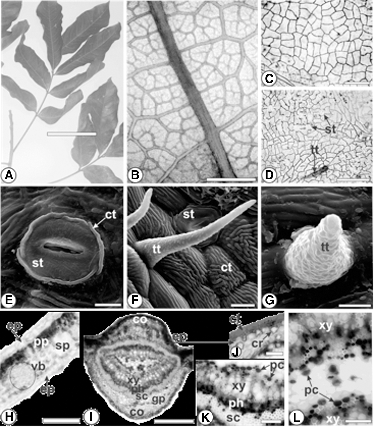
Talisia esculenta (A. St.-Hil.) Radlk. (Sapindaceae).
Leaves are hypostomatic, and anomocytic stomata are observed (Fig. 1D). Stomata measured 25 μm in length on average. The striate cuticle was located around the stomata (Fig. 1E) and on subsidiary cells (Fig. 1F). The cuticle contains cutin, which is a lipophilic polymer that is deposited on the top of the outer wall of epidermal cells. Its ornamentation is a taxonomic feature of the epidermis in leaves, appearing as striations, ridges, or papillae. 13,22
With reference to the indumentum, Talisia species can be either glabrous or have different types of trichomes (glandular and/or nonglandular). Glandular trichomes were reported for Talisia hemidasya Radlk., Talisia morii Acev.-Rodr., and Talisia setigera Radlk.; however, simple nonglandular trichomes are more common in the genus. 23 In the present study, T. esculenta showed a conic nonglandular trichome with rough cuticle (Fig. 1F, G). It is located especially on the veins on the abaxial side. In addition, glandular trichomes were not found.
In cross-section, leaves presented the uniseriate epidermis covered by a thick cuticle, and the cells were larger on the adaxial side. The mesophyll was dorsiventral and formed by a layer of palisade parenchyma and about six layers of spongy parenchyma. Small collateral vascular bundles were immersed in the mesophyll (Fig. 1H). Phenolic compounds were found in the chlorenchyma, mainly in the palisade parenchyma.
The midrib, in transection, had oval shape (Fig. 1I). The epidermis was uniseriate and covered by a striate and thick cuticle, which reacted positively with Sudan III in the histochemical test (Fig. 1J). Beneath the epidermis, on both sides, the chlorenchyma was interrupted, and about three strata of angular collenchyma were apparent. Several prismatic calcium oxalate crystals were evident in the collenchyma (Fig. 1J) and in the ground parenchyma. They reacted positively with sulfuric acid in the histochemical test.
The vascular system was represented by a medullated vascular cylinder. Phenolic compounds were found in the phloem and near the xylem. They reacted with ferric chloride (Fig. 1K) and in the Hoepfner–Vorsatz test (Fig. 1L).
In cross-section, the T. esculenta petiolule was concave–convex with two ribs on the adaxial side (Fig. 2A). The epidermis had the same characteristics as described to leaves, including the presence of conic nonglandular trichomes. The vascular system was represented by a medullated vascular cylinder as reported to leaves. Phenolic compounds were found in all parts of the petiole (Fig. 2A), especially in the phloem (Fig. 2B) and in the epidermis and collenchyma (Fig. 2C).
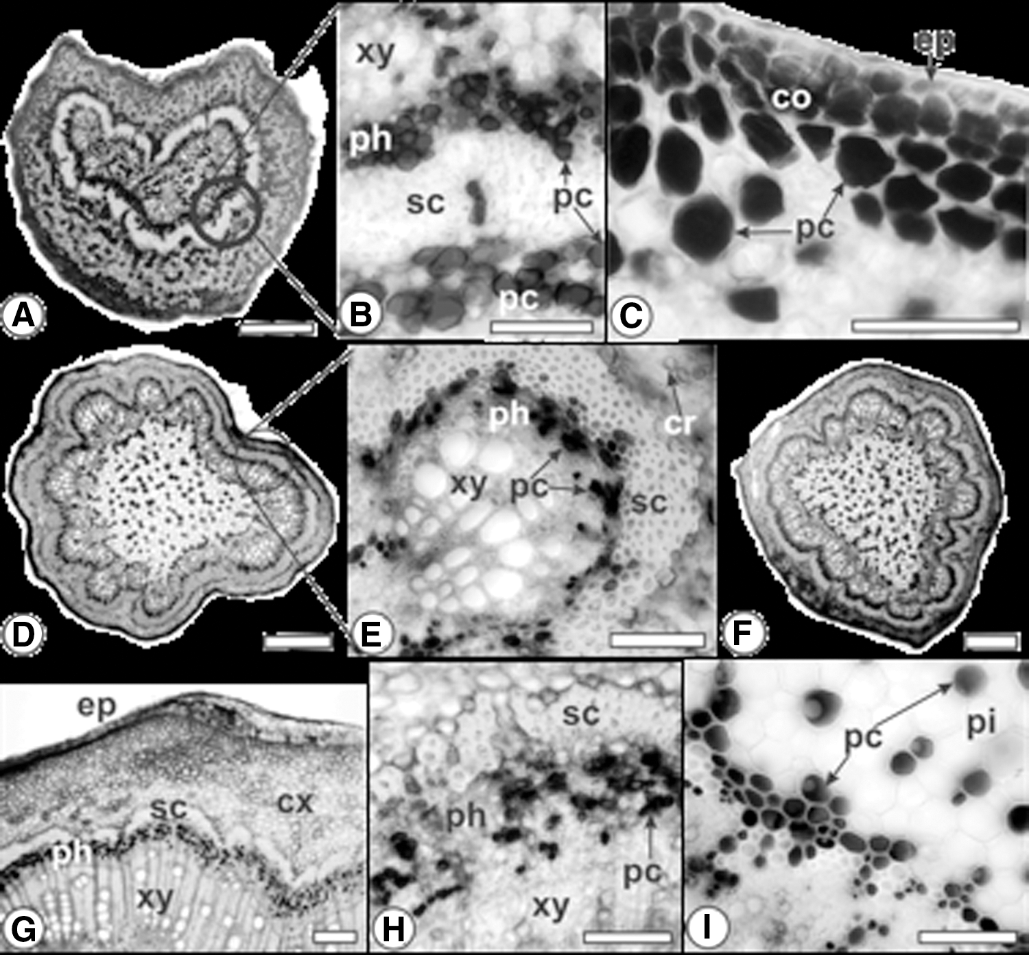
Talisia esculenta (A. St.-Hil.) Radlk. (Sapindaceae). Cross-section.
The rachis was rounded and obtusely angled (Fig. 2D). It was pubescent as the petiolule and midrib. The vascular system was represented by a collateral vascular cylinder (Fig. 2D, E). Phenolic compounds were found mainly in the phloem (Fig. 2E) and in the medullary region (Fig. 2C, D).
The petiole, in transection, had irregular shape. It had not only the same indumentum but also similar vascular cylinder as observed in the rachis and petiolule. Phenolic compounds had the same distribution as the rachis (Fig. 2F).
In transection, the stem had a rounded shape (Fig. 2G). The epidermis appeared in a single series with thickened cuticle. There were several cell layers in the cortex. The sclerenchymatous ring surrounded the vascular system. This was typical and was formed by phloem outward and xylem inward. The presence of continuous sclerenchymatous ring helped in the T. esculenta identification. Phenolic compounds were found in the phloem (Fig. 2H) and in the perimedullary region (Fig. 2I). The pith was composed of relatively small parenchymatous cells with thin walls (Fig. 2H).
Phytochemical investigation
Few compounds were identified in T. esculenta leaves. They were assigned mainly as flavonol derivatives with characteristic UV-absorbance profile with λ max at ∼255 nm (band B) and ∼360 nm (band A). Lower abundant compounds were derivatives of benzoic and cinnamic acids. HR-MS/MS allowed confirming the structures observed with UV-PDA. Thus, peak at 1.90 min which had m/z 315.072 [M − H]− was identified as dihydroxybenzoic acid-hexoside. The major peak appeared at 6.02 min (Fig. 3), producing ion at m/z 609.146 [M − H]− and 611.160 [M + H]+. Fragments obtained at m/z 611.160 (Table 1) indicated the neutral losses (NL) of 146 atomic mass units (a.m.u.), followed by 162 a.m.u., with intense aglycone ion at m/z 303.050, characteristic of the removal of rhamnosyl and hexosyl residues, respectively, as observed in the analysis of rutin. 24 Similarly, peak at 6.37 min, m/z 595.165 [M + H]+, also exhibits NL of 146 and 162 a.m.u., with aglycone fragment at m/z 287.055, characteristic of kaempferol rutinoside (or isomer). Peak at 6.46 min, at m/z 447.093 [M − H]− or 449.108 [M + H]+, was identified as quercetin-rhamnoside.
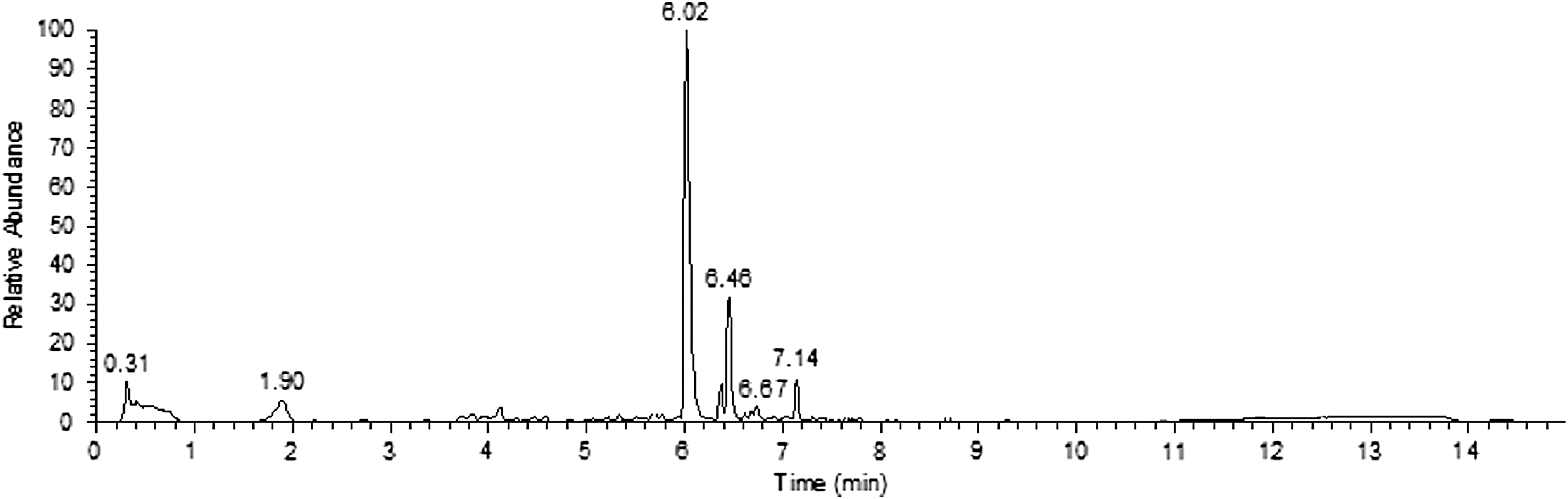
UPLC analysis of ESTE obtained from Talisia esculenta (A. St.-Hil.) Radlk. ESTE, ethanol soluble fraction obtained from T. esculenta; UPLC, ultra-performance liquid chromatography.
Phytochemical Composition of Ethanol Soluble Fraction Obtained from Talisia esculenta Obtained by Liquid Chromatography–Photodiode Array–Mass Spectrometry
First stage of mass analysis.
Second stage of mass analysis.
ESTE, ethanol soluble fraction obtained from T. esculenta; MS, mass spectrometry.
Dicaffeoylquinic acid was also identified in this extract, appearing at 6.67 min, with negative ion at m/z 515.119 and fragments at m/z 353.087, 191.055, 179.034, 173.045, and 135.045. The last peak found appeared at 7.14 min, with m/z 623.161 [M − H]− and 625.176 [M + H]+. Fragments from protonated ion appeared at m/z 479.118 (−146 a.m.u.) and 317.065 (−162 a.m.u.). This compound is 14 a.m.u. higher than rutin, and this difference was present in the aglycone moiety, consistent with methyl-quercetin-diglycoside. Similar structure has been described by Chukwumah et al. 25
Toxicological findings
ESTE obtained from T. esculenta does not induce any signs of acute toxicity in Wistar rats
All animals treated with ESTE, regardless of dose, presented a behavior pattern considered normal for the species and genus. Body weight (Table 4) and water and food intake did not show any changes throughout the experimental period (data not shown). Values obtained for blood parameters (biochemical and hematological) from ESTE-treated rats were statistically similar to vehicle-treated animals (Tables 2 and 3). In addition, all organs examined did not show any evidence of changes during macro- or microscopic evaluations (Figs. 4 and 5). Relative weight of vital or reproductive organs did not show any statistically significant changes among experimental groups (Table 4).
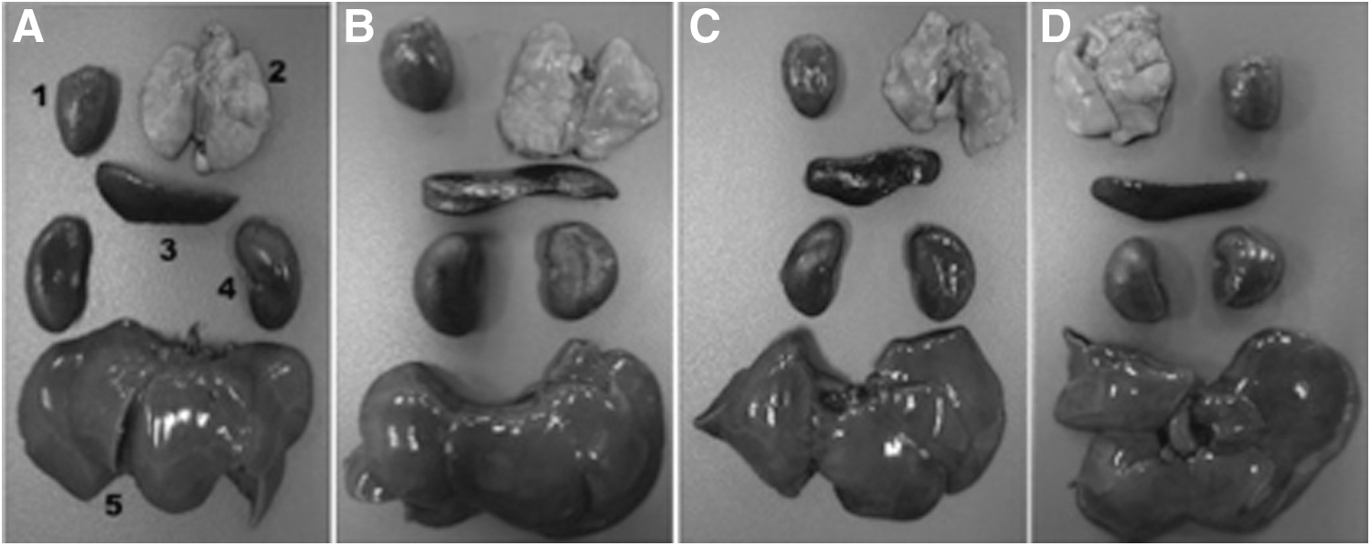
Photomacrographs of heart (1), lung (2), spleen (3), kidneys (4), and liver (5) of male
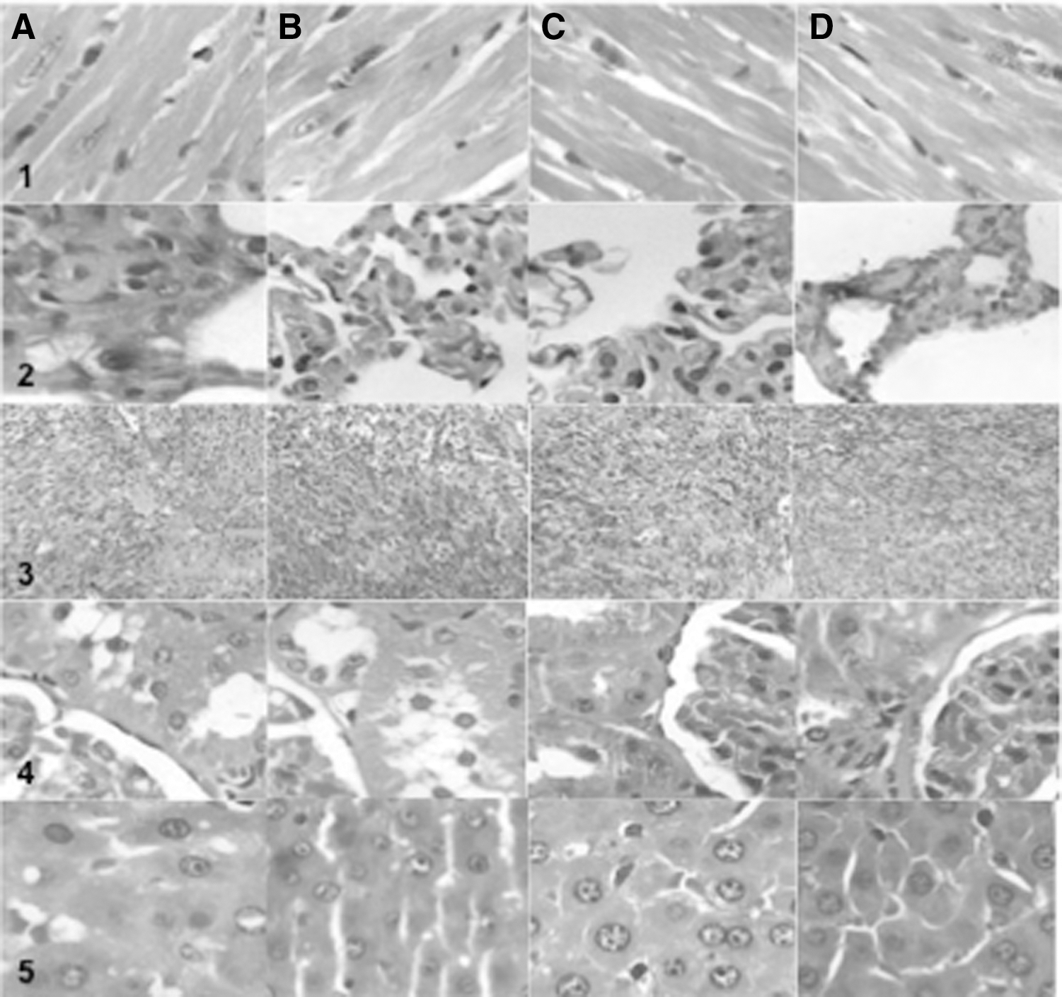
Photomicrographs of heart (1), lung (2), spleen (3), kidney (4), and liver (5) histopathology from representative male
Effect of Acute Oral Administration of Ethanol Soluble Fraction Obtained from Talisia esculenta on the Hematological Parameters of Wistar Rats After 14 Days of Exposition
Values are expressed as mean ± SEM of five rats in each group in comparison to the control using one-way ANOVA followed by Dunnett's test.
MCH, mean corpuscular hemoglobin; MCHC, mean corpuscular hemoglobin concentration; MCV, mean corpuscular volume.
Effect of Acute Oral Administration of Ethanol Soluble Fraction Obtained from Talisia esculenta on the Biochemical Parameters of Wistar Rats After 14 Days of Exposition
Values are expressed as mean ± SEM of five rats in each group in comparison to the control using one-way ANOVA followed by Dunnett's test.
ALT, alanine aminotransferase; ANOVA, analysis of variance; AST, aspartate aminotransferase; HDL, high density lipoprotein; SEM, standard error of the mean.
Effect of Acute Oral Administration of Ethanol Soluble Fraction Obtained from Talisia esculenta on the Body Weight and Relative (%) Organ Weights of Male and Female Wistar Rats Exposed for 14 Days
Values are expressed as mean ± SEM of five rats in each group in comparison to the control using one-way ANOVA followed by Dunnett's test.
Pharmacological investigation
ESTE from T. esculenta induces weak diuretic and kaliuretic response after acute treatment
The urinary volume produced by the different experimental groups after 1, 2, 8, and 24 h is shown in Table 5. ESTE (100 mg/kg) obtained from T. esculenta was able to significantly increase urinary volume only in the first 2 h after treatment. After 8 h of experiments, only HCTZ was able to increase urinary excretion significantly, returning to values similar to the control group after 24 h. The pH and urinary density values were not altered in any of the experimental groups (data not shown).
Effect of Acute Oral Administration of Ethanol Soluble Fraction Obtained from Talisia esculenta on the Urinary Volume After 1, 2, 8, and 24 H of the Treatment
Values are expressed as mean ± SEM of 10 rats in each group in comparison to the control using one-way ANOVA followed by Dunnett's test.
P ≤ .05 compared with the control.
HCTZ, hydrochlorothiazide.
The urinary Na+, K+, Cl−, and Ca+2 concentrations in the different experimental groups are presented in Tables 6 and 7. All ESTE doses tested were able to increase significantly the renal K+ elimination after 8 h from the beginning of treatments, returning to values similar to controls after 24 h. In contrast, HCTZ was able to significantly increase the urinary Na+, K+, and Cl− excretion in the first 8 h, returning to nonsignificant values at the end of the experimental period (24 h).
Effect of Acute Oral Administration of Ethanol Soluble Fraction Obtained from Talisia esculenta on Urinary Electrolyte Excretion After 8 H of the Treatment
Values are expressed as mean ± SEM of five rats in each group in comparison with the control using one-way ANOVA followed by Dunnett's test.
Saluretic index = mmol/L problem group/mmol/L control group.
P < .05.
El, excretion load
Effect of Acute Oral Administration of Ethanol Soluble Fraction Obtained from Talisia esculenta on Urinary Electrolyte Excretion After 24 H of the Treatment
Values are expressed as mean ± SEM of five rats in each group in comparison with the control using one-way ANOVA followed by Dunnett's test.
Saluretic index = mmol/L problem group/mmol/L control group.
ESTE from Talisia does not alter blood pressure or HR in normotensive or hypertensive rats
Baseline values obtained for SBP, DBP, and MAP before the administration of any substance were 165 ± 8.8, 103 ± 5.7, and 122 ± 7.9 mm Hg in SHR and 110 ± 4.6, 72 ± 4.8, and 94 ± 4.2 mm Hg in Wistar rats. In contrast, HR values were 480 ± 32.1 bpm in SHR and 282 ± 18.2 bpm in Wistar rats. The acute or prolonged administration of ESTE, in Wistar rats or SHR, respectively, regardless of dose, was not able to induce any statistically significant changes in SBP, DBP, MAP, and HR values (Figs. 6A–D and 7A–D). In contrast, HCTZ (25 mg/kg) was able to reduce SBP, DBP, and MAP values by ∼20% in SHR and Wistar rats (Figs. 6A–C and 7A–C), without affecting baseline HR (Figs. 6D and 7D).

ESTE from Talisia esculenta (A. St.-Hil.) Radlk. does not affect the values of blood pressure and the HR of normotensive Wistar rats
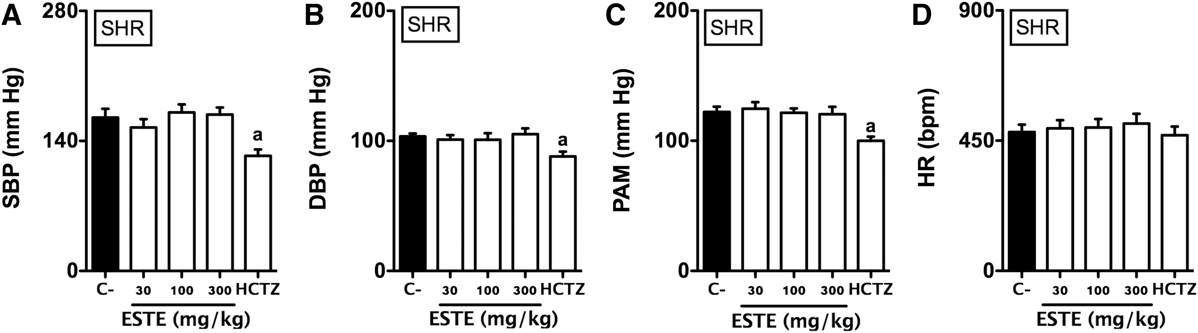
ESTE from Talisia esculenta (A. St.-Hil.) Radlk. does not affect the values of blood pressure and the HR of SHR
ESTE treatment reduces lipid peroxidation and nitrosative stress and increases nitrite levels in SHR
ESTE treatment at doses of 100 and 300 mg/kg was able to increase nitrite levels in ∼50% and 80%, respectively (Fig. 8A). Serum NT (Fig. 8B) and TBARS (Fig. 8C) levels in SHR were significantly reduced after 7 days of treatment. In fact, prolonged treatment with the ESTE at dose of 300 mg/kg was able to reduce NT and TBARS levels in ∼35%.
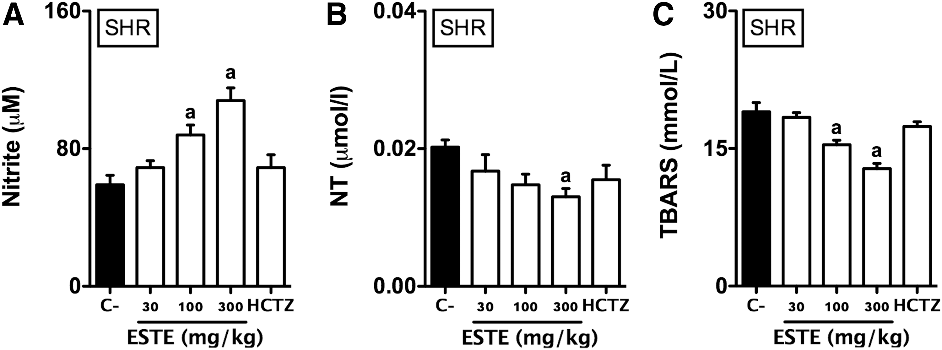
ESTE treatment increases nitrite concentration
Discussion
In this study, we performed a complete morphological and microchemical characterization of T. esculenta leaves, as well as identifying the main secondary metabolites present in a preparation commonly used in Brazil. In addition, we have shown safety aspects and evaluated the efficacy of the ESTE as a cardioprotective agent.
Initially, a detailed macro- and microanatomical study was carried out to provide important parameters for the standardization and quality control of the plant drug. Moreover, this was the first study conducted on the anatomical characteristics of this taxon. In fact, T. esculenta is not included in any pharmacopoeia, and its anatomical features provide substantial information for quality assessment in the herbal industry.
In a second stage, a semipurified aqueous extract from T. esculenta was obtained, and a detailed phytochemical profile was determined. Analyses performed by UPLC-PDA-MS/MS showed a reduced spectrum of secondary metabolites, mainly flavonol derivatives. It is now known that these compounds are among the main metabolites capable of influencing renal hemodynamics and inducing diuretic response. 26 However, many of these compounds have different toxicity degrees, 27 and safety data available for T. esculenta are very limited. Riet-Correa et al. 12 showed that the ingestion of T. esculenta leaves and fruits by cattle and sheep brought a varied spectrum of intoxication, including ruminal bloat, weakness, ataxia, and spastic paresis. Nevertheless, information on the toxicity of this species to humans and nonherbivorous animals remains unknown. This work showed that the acute administration of a wide range of ESTE doses obtained from T. esculenta leaves did not induce any signs of acute toxicity in male and female rats. It is believed that the apparent discrepancy between data obtained in herbivorous and rodents may be, in part, due to the anatomical and biochemical differences in the gastrointestinal system of these species. In addition, the fresh form of this species has different concentrations of metabolites that may not be found in significant amounts in ESTE. In fact, the results obtained in this study amplify the scientific knowledge for the species and provide important safety parameters for the rational use of T. esculenta.
The next step of this work was to investigate whether the use of this species as a probable diuretic and hypotensive agent has some scientific evidence. A positive indication is that the secondary metabolites found in this preparation, especially flavonols and chlorogenic acid derivatives, are classically recognized to have antioxidant activity, and many of them have been found to be responsible for diuretic, hypotensive, and antihypertensive effects of several natural products. 26,28 In fact, prolonged oral treatment with ESTE was able to significantly reduce the nitrosative and oxidative stress in SHR, showing superior antioxidant potential to other natural products endowed with diuretic and hypotensive activity. 16 In contrast, despite the suggestive evidence, the acute or prolonged treatment with ESTE was not able to cause any alteration in the HR or blood pressure levels of SHR or Wistar rats. In addition, the diuretic response was very limited, showing only a slight increase in urinary volume in the first 2 h after treatments and completely disappearing after 8 or 24 h. Furthermore, electrolyte excretion was concentrated only in potassium, which contributes more to adverse effects than to global saluretic response. 29
Currently, it is known that some diuretics of high efficacy, such as furosemide, have an intense and short diuretic response, which in a way compensatory to dehydration loses its effect after 8 or 24 h. However, the saluretic profile of these compounds is very different from the response obtained with ESTE and involves the extensive elimination of Na+, K+, Cl−, Ca2+, and Mg+, among others. 30 In fact, even HCTZ, a moderately effective diuretic, lost its diuretic response after 24 h, and its saluretic pattern was quite different from ESTE.
One limitation of our study was to perform pharmacological assays only with male rats. If we consider the importance of biological sex and estrogen in cardiovascular health and disease, it is likely that different responses to ESTE may occur in both male and female animals. 31 Further studies may help to explain sexual dimorphisms that exist both physiologically and in common animal models.
In conclusion, the results obtained in this study provided important anatomical and microchemical aspects of aerial vegetative organs and phytochemical data of T. esculenta leaves. In addition, it was observed that ESTE has a significant antioxidant activity without showing any clinical signs of acute toxicity. In contrast, the use of this species as a diuretic and hypotensive agent should be carried out with caution, since administration in rodents did not produce renal and/or hemodynamic responses that justify this ethnobotanical indication.
Footnotes
Acknowledgments
This work was supported by grants from the Fundação de Apoio ao Desenvolvimento do Ensino, Ciência e Tecnologia do Estado de Mato Grosso do Sul (FUNDECT, Brazil, 59/300.046/2015), and Conselho Nacional de Desenvolvimento Científico e Tecnológico (CNPq, Brazil, 449464/2014-8). The authors are grateful to the University Hospital of the Federal University of Grande Dourados for the biochemical and hematological analyses.
Author Disclosure Statement
No competing financial interests exist.