




Abstract
Morinda citrifolia (MC) and Annona muricata (AM) are popularly used for the treatment of several diseases, including cancer. Our objective was to evaluate the effects of the juice of the MC fruit, and the ethanolic extract of AM leaves on Erhlich tumor cells, in mice and in vitro fibroblastic cells. The animals were divided into G1 and G2: controls; G3: treated with AM, and G4 juice: treated with MC. The animals were subjected to intraperitoneal inoculation of 103 tumor cells and then treated with the plants. G1 and G2 received a saline solution. Cells were grown in RPMI 1640 medium, supplemented with 10% fetal bovine serum, and maintained at 37°C in 5% CO2 atmosphere. After growth, the cells were trypsinized and incubated again. Subsequently, serial dilution of extracts and juice was performed and incubated again for 48 h. We evaluated the ascites growth, the survival rate, and cytotoxicity in fibroblast cells. AM and MC did not interfere in the ascites pattern. MC, but not AM showed significance in the survival rate. Both AM and MC produced cytotoxicity activity in T3T cells. Despite advances in medicine, cancer is still one of the leading causes of death in the world and traditional medications are not always effective. The use of MC may bring beneficial effects to patients since they interfere with different mechanisms of action during carcinogenesis. However, we suggest that further investigations should be performed.
Introduction
Cancer is one of the most common causes of deaths worldwide and may be considered as a public health problem. Some authors have projected substantial augmentation in the number of cases of cancer in 2020 and 2030 possibly related to a number of reasons, such as increase in the number of adults over 65 years, modifications in the lifestyle, pollution, smoking, and genetics. 1,2 The International Agency for Research on Cancer estimated that there are about 14.1 million new cases of cancer and 8.2 million of deaths worldwide in 2012, and 21.7 million new cases of cancer and 13 millions of deaths in 2030. 3,4
A possibility to study the action of chemical, physical, and biological components on the growth, pathogenesis, immunology, cytogenetics, and tumor cell therapy is using the Ehrlich tumor. It is a transplantable malignant epithelial neoplasm, species specific, and corresponds to the mammary adenocarcinoma of the female mouse. It grows in several strains of this animal species in ascitic form when inoculated intraperitoneally, and in solid form when inoculated in the subcutaneous. 5,6
The use of conventional therapies, in addition to the side effects and the high cost, often does not allow the total eradication of neoplasias. Moreover, the available antineoplastic drugs are not specific for neoplastic cells and may affect healthy cells leading to toxicity. 7 For these reasons, there is an exponential growth in the interest and use of alternative and complementary medicines, which could give the patient an additional option for the proper treatment. 8 –10
Among several plants, we can mention Morinda citrifolia (MC), popularly known as noni and Annona muricata (AM), known as graviola. MC belongs to the Rubiaceae family and is native to South Asia. AM belongs to the Annonaceae family and originates from tropical regions of South and Central America and is vastly cultivated in the North and Northeast regions of Brazil. 11 –13
MC may be used as antiobesity, antinociceptive, anxiolytic, anti-inflammatory, antidiabetic, antioxidant, antiallergic, neuroprotective, anthelmintic, for gastrointestinal problems, nausea, and many other effects. 14,15
AM is popularly used as antiparasitic, antispasmodic, astringent, sedative, hypotensive, and anticancer. The stem, bark, and roots of the plant are commonly used as remedies for diarrhea and intestinal worms. Fruit pulp and leaves are used in the treatment of fevers, as an anti-inflammatory, gastrointestinal problems, sedative, hypotensive, hypoglycemic, kidney affections, and in the treatment of cancer. 16,17
As we observe a worldwide interest to achieve novel agents with antitumor effects, the objective of this study was to evaluate the effects of MC fruit extract and AM extract on Erhlich tumor cells in albino Swiss mice.
Methods
Group of animals
Male Swiss albino mice weighing between 20 and 30 g were used, which were kept in the laboratory of the University of Marília–UNIMAR, São Paulo (Brazil) with a 12-h light/12-h dark cycle, room temperature of 22°C ± 2°C, relative humidity of 60% ± 5%. The animals received water and were fed ad libitum and were treated according to the Guide to the Care and Use of Experimental Animals, which outlines the principles of the Canadian Council for Care of Laboratory Animals.
This work obtained approval by the Research Ethics Committee (CEUA–Comitê de Ética em Uso Animal) of UNIMAR (the University of Marília, São Paulo, Brazil) under protocol number 241.
Plant materials
The fruits of MC and the leaves of AM were purchased from the city of Pompeia, São Paulo (Brazil).
Preparation of the extract of AM
The ethanolic extract of AM was obtained by maceration of the dried and crushed leaves with ethanol (1:10) for 5 days. After this period, the filtration was carried out. The obtained extract was divided into aliquots, packed in amber glass with daily portions, and stored in the refrigerator.
Preparation of MC juice
The fruit juice of MC was prepared using water, in the proportion of 1:10 in an industrial blender. It was filtered and divided into aliquots, packed in amber glass with daily portions, and maintained in the freezer until the use. Samples were thawed daily 1 h before administration.
Obtaining and maintaining tumor cells
Erhlich tumor cells were obtained from an animal with ascites tumor, according to the method described by Brehmer. 18 A volume of 0.3 mL (containing 3 × 10 7 cells) was aspirated from the peritoneum of the tumor-bearing animal and inoculated into the peritoneal cavity of healthy animals that served as a template for obtaining the cells.
Experimental protocol
The mice were separated into four groups (n = 10). On the first day of the experiment, all animals received 103 tumor cells in the peritoneal cavity, according to the methodology described above. The group G1 received a hydroalcoholic mixture (1:10), and G2 received water throughout the experimental protocol. G3 was treated with AM ethanolic extract, and G4 treated with MC juice. The G3 and G4 groups started the treatment 7 days after inoculation (100 mg/kg of AM ethanolic extract for G3 and 100 mg/kg of MC juice for G4, administrated daily for 60 days).
The ascitic tumor growth was evaluated weekly with the aid of a manual caliper and classified (second protocol developed in the physiology laboratory of FAMEMA-Faculty of Medicine of Marília–São Paulo-Brazil) in average (0–3.5 cm), small (3.5 (4.0–5.0 cm), large (5.0–5.5 cm), very large (5.5–6.0 cm), and extra large (greater than 6.0 cm). During the experimental protocol, the weight of the animals, as well as the survival rates were evaluated weekly. The animals that survived underwent euthanasia with overdose of thiopental sodium
Ascites evaluation
The presence of ascites in the animals was evaluated weekly using a classic pachymeter; the measurements were collected and mean was performed to verify the variation of ascites size.
In vitro cytotoxicity protocol
Cell growth
Cells were cultured in RPMI 1640 medium (Cultilab Ltda., Campinas, São Paulo, Brazil) supplemented with 10% fetal bovine serum and maintained at 37°C in 5% CO2 atmosphere. After cell growth, the cells were trypsinized, and transferred to a 96-well plate, and incubated at 37°C in 5% CO2 atmosphere. After growth, a serial dilution was performed from 100 μg of the crude plant extract (respectively 100, 50, 25, 12.5, 6.25, and 3.125 μg). As well as positive and negative controls, the plate containing the cells in contact with the tested extracts were incubated for 48 h at 37°C in 5% CO2 atmosphere. We performed two controls, in the positive control was placed pure culture medium without extracts, and in the negative control restricted nutrients were placed.
Statistical analysis
The results were analyzed using the mean and standard deviation and analyzed by chi-square.
Results
Evaluation of the antitumor activity on the Ehrlich ascites tumor
The percentage of survival of the animals treated with MC juice and ethanolic extract of AM is shown in Figure 1. Our results show that G4 (a group that received noni juice) had a significant increase (P < .05) in the survival when compared with the control group (G1). Animals that received AM ethanolic extract (G3) had no significant increase in survival (P > .05) in relation to the control group (G2) (Fig. 1).
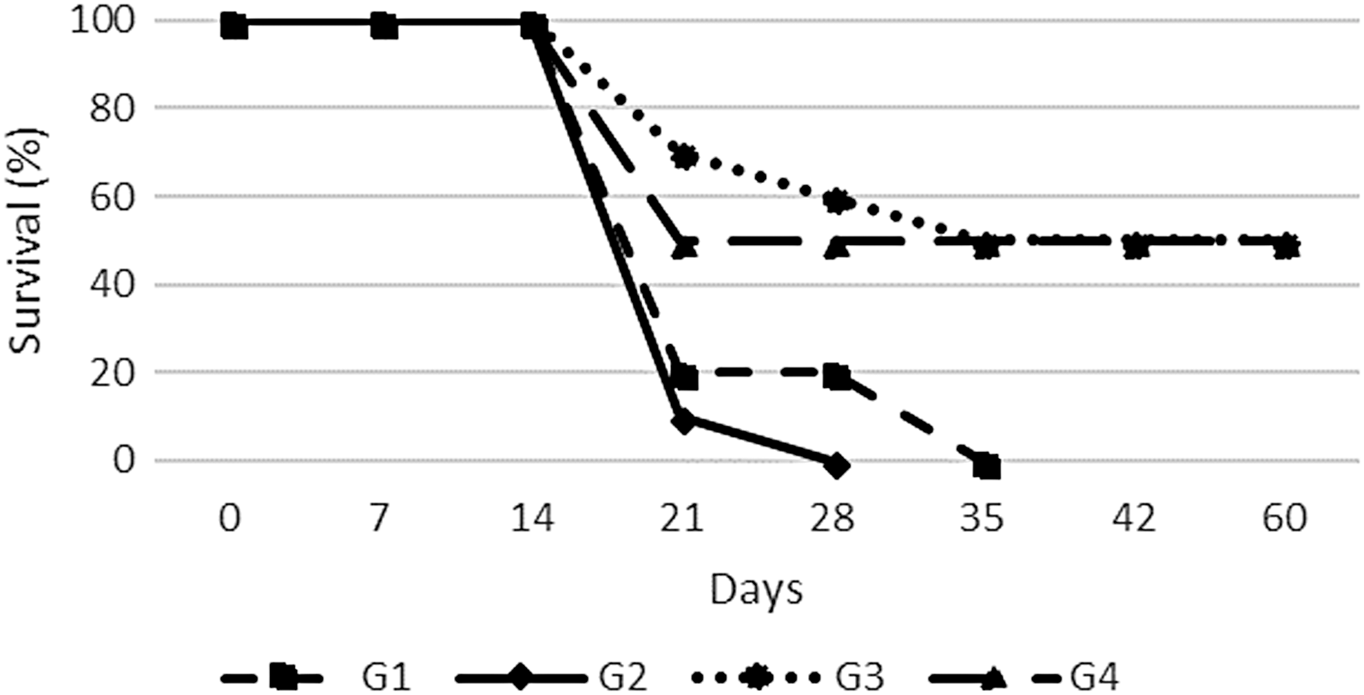
Percentage of survival of animals inoculated with Ehrlich tumor and treated with AM (100 mg/kg) (G3) and MC (100 mg/kg) (G4) juice. The animals were treated with 100 μL per gavage for 60 days. Control groups received saline (G1) and saline (G2), respectively. AM, Annona muricata; MC, Morinda citrifolia
In Figure 2 it is possible to observe that the ethanolic extracts of AM (G3) and MC juice (G4) showed no significant mean change in ascites (P > .05).
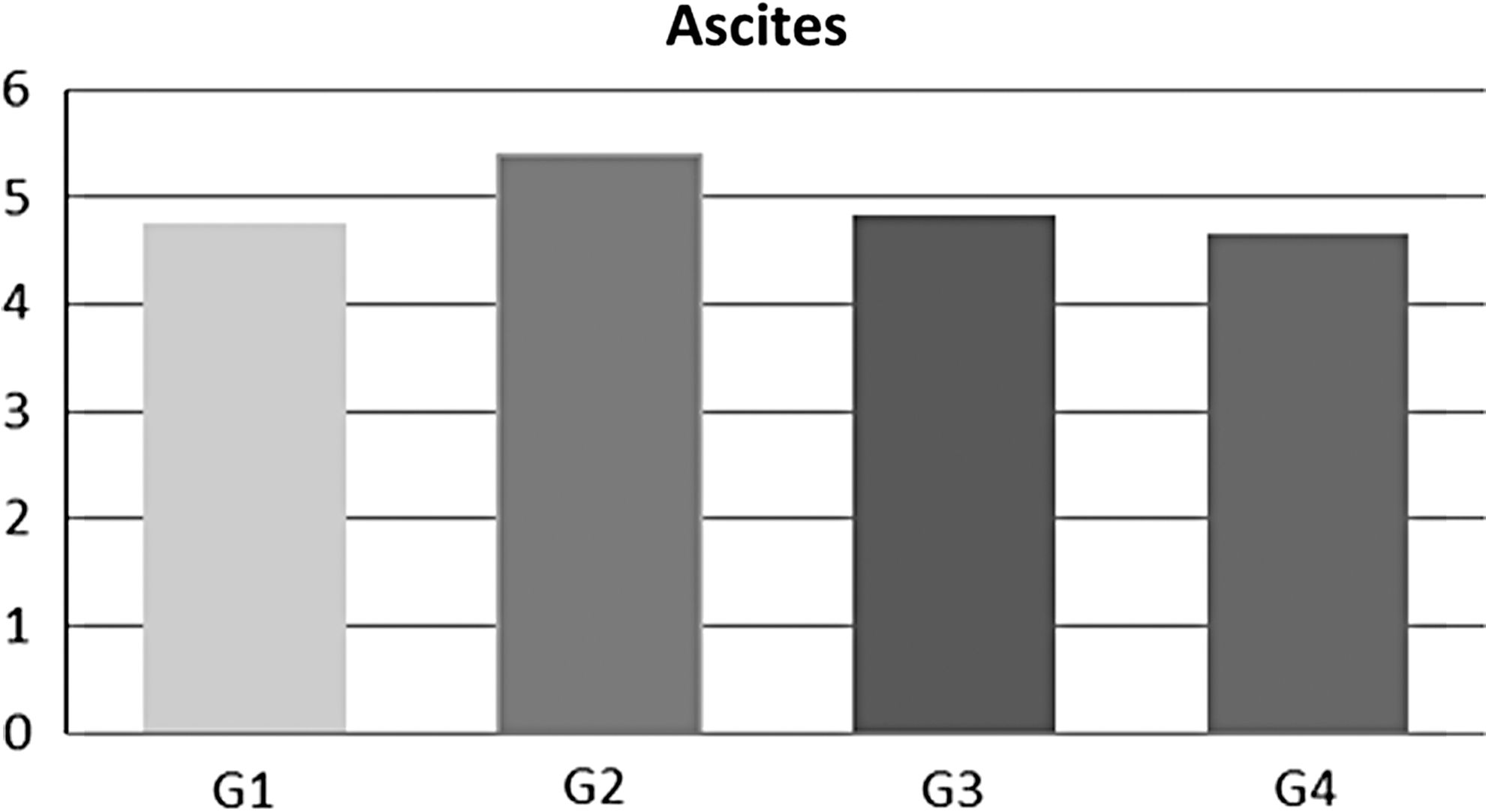
Mean of ascitic variation in the animals.
Evaluation of the cytotoxicity of extracts in fibroblast cells (T3T)
As shown in Figure 3, the ethanolic extract of AM showed significant cytotoxic activity (P = .0001) in T3T cells.
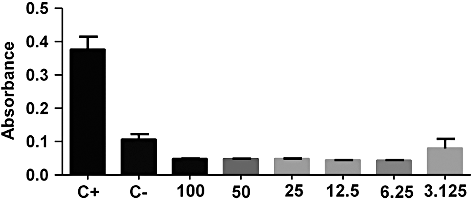
Cytotoxicity of the ethanol extracts of AM (μg/mL). T3T (fibroblastic) cells were cultured, subsequently trypsinized, and incubated again in 96-well plates with diluted ethanolic extract of AM, subsequently stained in neutral red for absorbance reading of the viable cells. C+: positive control; C−: negative control.
As shown in Figure 4 the juice of MC shows significant cytotoxic activity (P = .0012) in T3T cells.
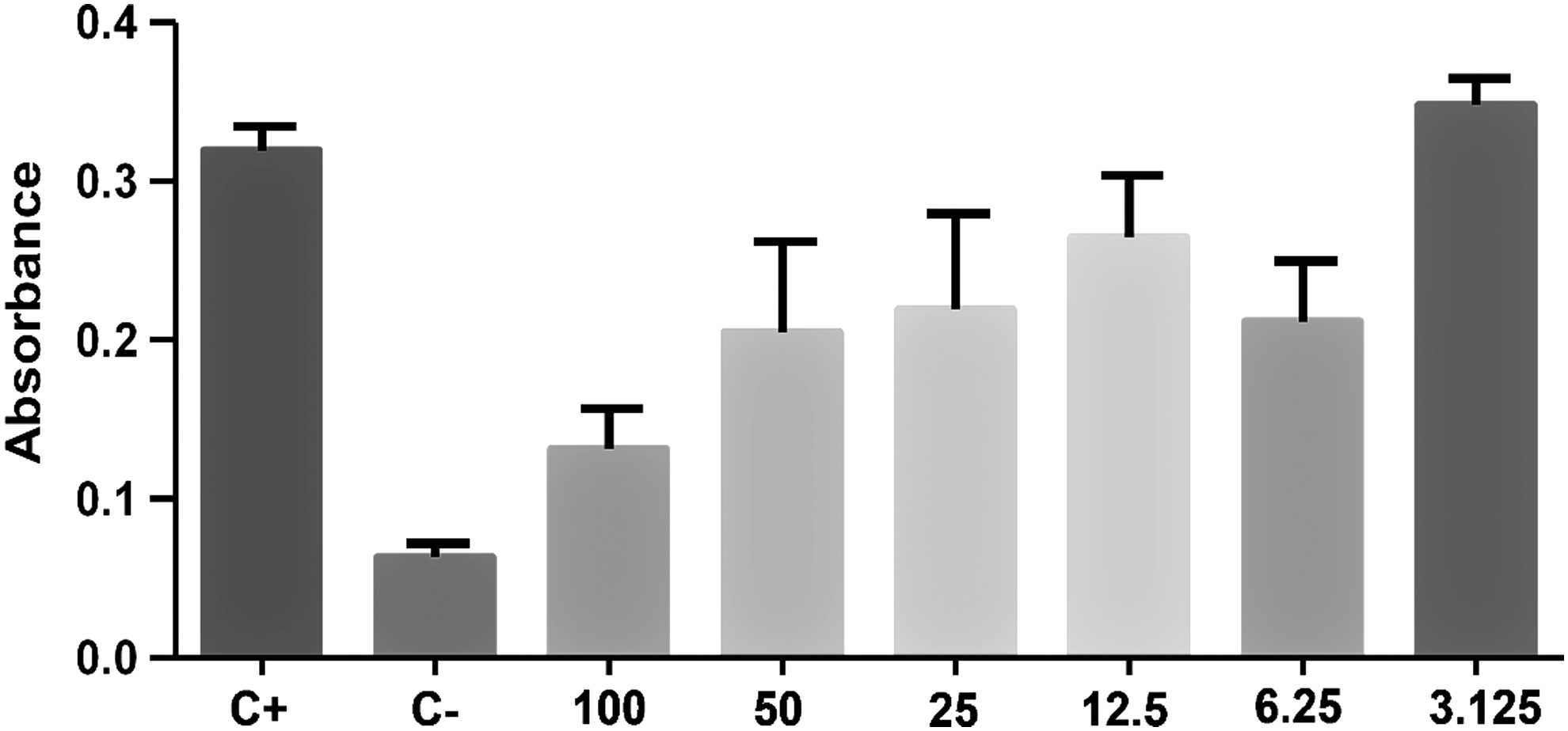
Cytotoxicity of MC juice (μg/mL). The T3T (fibroblastic) cells were cultured, subsequently trypsinized, and incubated again in 96-well plates with serial dilution of MC juice, subsequently stained in neutral red for absorbance reading of the viable cells. C+: positive control; C−: negative control.
The results of cytotoxicity demonstrate that at the concentration of 100 μg, the number of viable T3T cells was lower compared with other dilutions demonstrating that MC juice has toxic activity in healthy cells. AM extract showed high toxicity at all concentrations tested on T3T cells. The results showed that MC juice was less toxic to T3T cells than AM extracts.
Discussion
Every year it is observed that many people die due to cancer despite the existence of several drugs and different therapeutic approaches. 7 On the other side, many studies have shown that plants such as AM and MC may exhibit potential beneficial effects in this disease. 11,19
The parameters used to investigate the effects of the plants were ascites growth and survival rate, and cytotoxicity analysis in fibroblast cells (in vitro). Our results showed that the juice of MC was able to stabilize ascites growth, as well as increase the survival of the animals with Ehrlich tumor. Studies using as a model, the Ehrlich tumor in the ascitic form, has been a useful resource mainly for allowing standardization of the number of cells to be inoculated, quantification of the growth, and regression of the tumor mass. The implantation of Ehrlich's ascitic tumor induces a local inflammatory reaction, with increased vascular permeability resulting in the formation of edema, cell migration, and preprogressive formation of ascitic fluid. The formation of ascitic fluid is essential for tumor growth because its constituents serve as a nutritional source for tumor cells. Macroscopic examination of the ascitic form, after approximately 10 days of intraperitoneal tumor inoculation, shows about 90% of the peritoneal cells are tumor cells. 6,7,20,21
Murata et al. 22 investigated the effects of MC pulp on cell-mediated immunity and found that this plant could inhibit the suppression of cell-mediated immunity by compounds with immunosuppressive properties isolated from ascites of Ehrlich tumor in mice. MC also avoided decrease of interleukin-2 (IL-2) synthesis and stimulated natural killer cells (NKC) in healthy animals. Authors found the presence of deacetylasperulosidic acid and iridoid glycoside in MC extracts and used the first one in the animals. Their results showed that this compound may inhibit the suppression of IL-2 release and is also capable of activating NKC. These findings indicate that MC may promote beneficial effects in the cell-mediated immunity recover.
Taşkin et al. 23 studied the cytotoxic potential of MC on Ehrlich ascites tumor in mice and showed a reduction of 40–50% of both short or long diameters of the tumor tissues when they compared with the control group. This effect occurred due to the induction of apoptosis. They also observed a reduction in the proliferation and concluded that MC could be helpful to treat breast cancer alone or in combination with a very traditional drug for cancer treatment, doxorubicin.
We only found two studies associating Ehrlich tumor and MC, the research of Murata et al. 22 and the study of Taşkin et al., 23 which studied the cytotoxic effects of this plant. Notwithstanding, several studies are using MC in other types of cancer showing that this plant and its compounds play a potential role to be used as the antitumor agent. For example, Abu et al. 11 evaluated the effects of nordamnacanthal, which is an anthraquinone found in the MC pulp, and showed it might produce in vitro cytotoxic effects on MCF-7, MDA-MB231, and 4T1 cells. Moreover, no toxicity signs, mortality, or changes in the liver were observed in mice treated with nordamnacanthal in a subchronic toxicity model. This anthraquinone also could increase tumor immunity and delay the growth of 4T1 tumors in Balb/C mice over 28 days of treatment.
Shaghayegh et al. 19 studied the effects of nordamnacanthal and another anthraquinone, the damnacanthal compound from MC, in cytotoxicity, apoptosis, morphological changes, cell migration, and death on oral squamous cell carcinoma cells. They observed that these compounds exhibit cytotoxicity, significant apoptotic morphological modifications, inhibition of cell migration, and induction of anticipated apoptosis in the oral carcinoma. Aziz et al. 24 showed that the use of damnacanthal in combination with a common drug used to treat breast cancer, doxorubicin, improved the efficacy of the drug in inducing cell death, Annexin V, and apoptosis. Huang et al. 25 used MC pulp extracts and showed anticancer properties. They also showed that chrysin combined with apigenin promoted apoptosis due to the downregulation of S-phase kinase-associated protein-2 and the expression of LDL-c receptor-related protein in liver and human breast cancer cells. The leaves of MC were also used in lung cancer, and the effects were compared with erlotinib, an anticancer drug. The results showed that MC suppressed the growth of a lung tumor, and was more efficient than erlotinib treatment. The plant augmented spleen tissue B cells, T cells, blood lymphocyte counts, and NKC, and decreased cyclooxygenase 2, and a biomarker of the lung adenocarcinoma. The presence of phenolic compounds, such as scopoletin and epicatechin, may be involved in the anticancer properties. 26
MC extract was used in B16–F10 melanoma cells and also showed antiproliferative effects. 27 Ethanolic extract of leaves also shows anticancer properties on Lewis lung carcinoma. 28 Commercial pulp juice significantly downregulates HIF-1α protein expression in human lung cancer (A549 cell line). 29 Fresh leaf extract shows inhibitory activity on human cervical and epidermoid carcinoma, and human hepatocellular carcinoma. 30 MCF-7 and MDA-MB-23 cell proliferation related to mammary adenocarcinoma were inhibited by ethyl acetate fraction extract. 31 Ethanolic extract of the pulp decreased inflammation and oxidative stress in human colon adenocarcinoma cell (Caco-2). 32 Taskin et al. 23 showed that a commercial noni juice promoted cytotoxic effects due to apoptosis induction on the Ehrlich tumor cells (Balb/c mice), and could be employed in the treatment of breast carcinoma. The juice of MC presents polysaccharides and may play a role as antitumor agent against Lewis lung peritoneal carcinomatosis increasing the survival of the animals. 33
As mentioned for MC, there are also several studies with AM and cancer, but we did not find studies that used this plant in Ehrlich tumor. Our results showed that the animals treated with this plant showed improvement in the general state and stabilization of the ascites growth, however, the survival was not significant but 50% of the treated animals survived with progressive improvements in the tumor stability. No significant changes were observed in the weight gain and ascitic volume compared with the control group. At the treatment interruption for 10 days, the animals had a progressive increase of the ascites, demonstrating that the extract of AM influenced the stability of the tumor.
Many chemical compounds have been isolated from the roots, stem, bark, leaves, and fruits of this plant. In the leaves, annomuricins, gigantetrocin A, anonacins, goniothalamicin, muricatocins, gigantetronenin, annomutacins, and many other compounds were identified. These acetogenins exhibit cytotoxic properties against tumor cell lines 34 Acetogenins induce apoptosis by complexation with Ca2+ and Mg2+. Annonacin, for example, causes apoptosis of tumor cells, due to the induction of T24 promoting increased caspase-3 activity, whereas bullatacin (bis-THF acetogenin) induces apoptosis in Hep15 cells differently, decreasing levels of both cAMP and cGMP. 35 AM acetogenins have been reported to be selectively toxic in lung carcinoma cells, solid breast tumor cells, adenocarcinoma of the prostate, pancreatic carcinoma cells, colon adenocarcinoma cells, cell types of liver cancer, human lymphoma cells, and multiple drug-resistant breast adenocarcinoma. 36
In a review, Qazi et al. 16 show that AM may have positive effects on a plethora of cancer types. It has effects on the pancreatic adenocarcinoma due to antiproliferative effects observed in fractions of the leaves that are rich in flavonoids. 37 It may also downregulate the glycoprotein mucin, and suppress PC cell motility and invasion. 38 AM is capable of improving lung cancer because it interferes with the apoptosis, reduces NF-κB signaling, stimulates free radical production, attenuates mitochondrial membrane potential, and caspase-3/9 activation. These results are promising because most patients with this cancer do not survive due to chemotherapeutic resistance. 39 The pulp exhibits potent antiproliferative activity in PCa cell lines, and inhibition of the activity of NADPH oxidase. 40 Ko et al. 41 observed significant antitumor and antiproliferative effects of AM in breast cancer cell lines by suppressing the development of MCF-7 breast cancer cells. The extract of this plant inhibited the proliferation and growth of xenograft tumors of epidermal growth factor receptor-overexpressing MDA-MB468 cells.
Abdullah et al. 42 studied the apoptosis-inducing effect of AM leaf extract on the colorectal cancer cell line COLO-20. They observed an increase in the activities of caspase-3 and suggested that the leaf extract exerts anticancer activity in colorectal cancer. Yap et al. 43 found positive effects on endometrial cancer due to similar events on caspase-3.
For the in vitro evaluation of cytotoxicity, we used untransformed T3T fibroblasts cells, which allowed us to visualize the cytotoxic action of the juice in healthy cells. Our results showed that MC presented lower toxicity than AM.
There are only a few researchers showing the toxicity of MC in humans. A study with patients with cancer (stage 4) showed that the maximum tolerated dose was the daily ingestion of 3 g of the MC dry fruit extract. The recommendation to prevent pain, fatigue, and to keep the physical functioning was the ingestion of 1.5 to 2 g/day. Besides the benefits, some patients presented adverse effects such as abdominal discomfort and nausea. These observations indicate the need of other studies evaluating the toxicity of MC extract in humans. 14
AM is mostly used in traditional medicine and is considered useful as a therapeutic agent against certain types of cancers. Nevertheless, some authors have reported that this plant induces cytotoxicity due to the alteration of the glucose metabolism resulting in dangerous interference in the metabolism. This interference can be a target against cancer cells because of the proliferating pattern of tumor cells that requires a significant amount of energy generally provided by aerobic glycolysis and oxidative phosphorylation. 14,34 Acetogenins, such as bullatacin, are also associated with toxicity due to the interference in the mitochondrial performance and reduction in the production of energy. For these reasons, it is possible to suggest that AM inhibits proliferation, cell growth, and metabolism. 44 Our study showed that graviola presented higher toxicity than MC. On the other hand, most studies performed for dose escalation of this plant to identify the efficacy of many fractions in different diseases do not demonstrate relevant toxicity to the animal models or humans, suggesting that it is safe for the patients. 16
In conclusion, despite advances in medicine, cancer is still one of the leading causes of death in the world. Traditional medications are not always effective and are often associated with numerous adverse effects. For these reasons, the use of plants can bring numerous benefits to patients both as adjuvants and as primary therapy. The use of MC may benefit patients since they interfere with different mechanisms of action during carcinogenesis. However, we suggest that further investigations should be conducted to evaluate cell death and to standardize the dosages necessary for the effectiveness of the treatment.
Footnotes
Author Disclosure Statement
No competing financial interests exist.