
Abstract
Cardiovascular and thromboembolic disturbances are the main causes of disease-related deaths worldwide. Regardless of the etiological factors involved in thrombus formation, coagulation is mainly activated by thrombin, one of the most important blood clotting molecules. Thus, this study evaluated the Turnera subulata leaf crude extract, its ethyl acetate fraction effect on the coagulation cascade, and its possible side effects. Their phytocomposition indicated polyphenols, mainly flavonol-3-O-glycosylate and a flavone glycoside, without in vitro and in vivo toxicity. Regarding their potential anticoagulants, results displayed partial thromboplastin and prothrombin time activation, and Xa and IIa, and thrombin inhibition by heparin II cofactor, indicating significant anticoagulant activity, suggesting direct and indirect thrombin inhibition as the main mechanism of action. Therefore, T. subulata leaf active compounds exhibit therapeutic potential required to develop phytotherapeutic formulations to assist conventional anticoagulants in clinical treatments.
Introduction
Nowadays, cardiovascular and thromboembolic disorders are leading causes of death worldwide. 1,2 Arterial thrombosis is the most common cause of acute myocardial infarction, stroke, and ischemia, while deep venous thrombosis complications include pulmonary embolism and post-thrombotic syndrome. 3 Thereby, the coagulation system and its interaction with platelet aggregation are responsible for arterial and venous thrombus formation. 4
Regular indications for anticoagulant uses include prophylaxis and venous thromboembolism treatments, cardioembolic prevention in patients with cardiac arrhythmia or mechanical valve prostheses, as well as secondary prevention in patients with acute coronary syndromes or undergoing percutaneous coronary intervention. 5 At this point, nonfractionated and low molecular heparins are used as anticoagulants, even causing side effects, such as thrombocytopenia and a high risk of systemic bleeding. 6,7 This stimulates the search for new substances to aid in prolonged anticoagulant therapy. 8
Thus, plant extracts exhibit a proven ability to inhibit the blood coagulation cascade, especially regarding intrinsic and extrinsic pathway factors. 9 –12 In this context, Turnera subulata (family Passifloraceae) is widely distributed in tropical and subtropical regions and used in folk medicine 12 due to its pharmacological properties, such as anti-inflammatory, 13,14 hypoglycemic, 15 antifungal, 16 and antioxidant properties. 17 Phytochemical studies with species of this genus revealed the presence of flavonols, alkaloids, tannins, cyanogenic glycosides, fatty acids, triterpenoids, and various phenolic compounds related to bioactivities. 18
Several studies describe the use of the Turneraceae genus in inflammatory processes. 19 Thus, considering that thrombin is closely related to coagulation and inflammatory systems, this genus represents a potential reservoir for discovering compounds for clinical treatment associated with conventional anticoagulants to minimize their side effects. Therefore, this study evaluated the anticoagulant potential and toxic and hemorrhagic effects of the T. subulata leaf crude extract and its fraction of ethyl acetate.
Materials and Methods
Plant material and leaf extract preparation
T. subulata leaves, collected in Natal, Rio Grande do Norte, Brazil, were taxonomically identified by Dr. Jomar Gomes Jardim, depositing a voucher specimen (Herbarium No: 0674/08) at the Herbarium of the Department of Botany and Zoology, Federal University of Rio Grande do Norte, Natal, RN, Brazil. Leaves were air-dried at 40°C for 48 h and powdered, before preparing its T. subulata crude extract (CETS) by ethanol:water (50:50, v/v) maceration for 4 days, filtered, and then lyophilized.
To characterize CETS-active compounds, an extract portion, resuspended in methanol, was subjected to liquid–liquid partition using increasing polarity solvents: n-hexane (3 × 300 mL) and ethyl acetate (3 × 300 mL), obtaining two fractions hexanic fraction of T. subulate (HFTS) and acetate fraction of T. subulate (AFTS). Phenolic compound contents were determined in all fractions by the Folin–Ciocalteu method described 20 (data not shown). No polyphenols were detected in HFTS.
High performance liquid chromatography with diode array chromatographic profile
Chromatographic analyses in triplicates were performed on a Phenomenex C18 chromatography column (4.6 × 100 mm, particle size 2.6 μm; Torrance, CA, USA) coupled to the HPLC system (VARIAN ProStar, Walnut Creek, CA, USA) equipped with a ProStar 240 quaternary pump, autosampler (ProStar 410), and a detector (mod. 355 PDA UV/V). CETS and AFTS (5 mg/mL) were dissolved in methanol. 17 Nine microliters were injected and elution was conducted at room temperature at a flow rate of 1.3 mL/min, using 0.1% formic (A phase) and acetonitrile (B phase) in the mobile phase, under following gradient conditions: 0–3 min, 5% B; 3–7 min, 5–20% B; 7–9 min, 20% B; 9–10 min, 2–23%, B; 10–15 min, 23% B; 15–19 min, 23–50% B; and 19–20 min, 50–5% B, monitoring at 280 nm. Phenolic compounds were identified by comparison with external standards (gallic and chlorogenic acids, epigallocatechin, rutin, hyperin, quercetin, apigenin, and kaempferol). All solutions were filtered using a 0.22-μm membrane (Millipore, Billerica, MA, USA).
Animals
Three-month-old Wistar rats of both sexes, weighing 250–300 gm, were kept under standard environmental conditions with food and water ad libitum. All animal procedures were performed according to the Brazilian National Health Surveillance Agency (ANVISA), 21 Organization for Economic Cooperation and Development (OECD) 22 guidelines, and Protocol No. 035/2015, approved by the Committee on Ethics in Animal Use, Federal University of Rio Grande do Norte.
Activated partial thromboplastin time assay
This assay was performed in accordance with the activated partial thromboplastin time (aPTT) information kit (CLOT Bios Diagnostica, São Paulo, SP, Brazil), and the coagulation time was measured in triplicate using a clot timer coagulometer (Drake Electronica Commerce Ltd., São Paulo, Brazil).
Prothrombin time assay
The prothrombin time (PT) assay was performed according to the manufacturer's instructions (CLOT Bios Diagnostica, São Paulo, SP, Brazil), while the coagulation time was measured in triplicate using a clot timer coagulometer (Drake Electronica Commerce Ltd., Sao Paulo, Brazil).
Assay for anti-Xa activity
Regarding anti-Xa activity, the assay was performed on a 96-well microplate using the Biophen Heparin Anti-Xa kit (Ref: 221010; HYPHEN Biomed, Paris, France) according to the manufacturer's instructions. Absorbance was measured at 405 nm using an Epoch microplate spectrophotometer (Epoch BioTek, Winooski, VT, USA).
Direct thrombin inhibition assay (anti-IIa activity)
This assessment was conducted in a 96-well microplate using the Biophen Heparin Anti-IIa kit (ref: 221025; HYPHEN Biomed, Paris, France) according to the manufacturer's instructions, measuring absorbance at 405 nm using a microplate reader (Epoch BioTek, Winooski, VT, USA).
Indirect thrombin inhibition mediated by heparin cofactor II
This parameter was determined spectrophotometrically using standard kit assays, according to Yoon et al., 23 measuring absorbance at 405 nm using an Epoch microplate spectrophotometer (Epoch BioTek, Winooski, VT, USA), using a blank containing all the reagents without the test substance.
Residual hemorrhagic effects
The CETS and AFTS compound residual hemorrhagic effect was analyzed according to the rat topical scarification model described by Brito et al. 24 After anesthesia with ketamine and xylazine at 1:1 (v/v), an incision was performed with a surgical blade in the distal tail portion, dipping the tail in physiological saline to observe bleeding. In the following stages, the tail was dipped in CETS or AFTS or heparin solution at 100 μg/mL for 2 min and washed with saline solution before immersing in a fresh physiological saline solution for 40 min. Blood was quantified by the Drabkin test and the result expressed as the hemoglobin sum of each tube, subtracting the hemoglobin value obtained before exposure to substance test.
Cytotoxicity by 3-(4,5-dimethylthiazol-2-yl)-2,5-diphenyltetrazolium bromide assay
Mouse (3T3) and human embryonic epithelial kidney (HEK 293) cells were cultured under standard conditions in Dulbecco's modified Eagle's medium (DMEM), supplemented with 10% fetal bovine serum at 37°C, 5% CO2, and 95% humidity. Cells (1 × 10 4 cells/well) were cultured for 24 h in 96-well microplates to promote adhesion. Thereafter, cells were exposed in triplicate at different CETS and AFTS concentrations (0.1, 1, 10, 100, and 1000 μg/mL) and incubated at 37°C for 24 h. Then, 100 μL of 3-(4,5-dimethylthiazol-2-yl)-2,5-diphenyltetrazolium bromide (5 mg/mL) dissolved in DMEM was added to each well and cells incubated for 4 h. After culture medium removal, 100 μL of dimethyl sulfoxide was added to each well to assess cell viability at 570 nm using a microplate reader (Epoch BioTek, Winooski, VT, USA).
Acute oral toxicity
The acute oral toxicity was evaluated according to ANVISA 21 and OECD 22 guidelines during a period of 14 days. Animals randomized into five groups (n = 5) received, by gavage, doses of 500 and 2000 mg/kg, as recommended by OECD guidelines. 22 The control group received only distilled water. Experimental design for each group is described as follows: Group 1: normal control rats received only distilled water (vehicle); Group 2: received a single dose of 500 mg/kg of CETS; Group 3: received a single dose of 2000 mg/kg of CETS; Group 4: received a single dose of 500 mg/kg of T. subulata ethyl acetate fraction (AFTS); and Group 5: received a single dose of 2000 mg/kg of AFTS.
During the first 12 h, systematic behavioral observations were performed (e.g., vocal tremor, piloerection, hyperactivity, tremors, abdominal cramps, diarrhea, and deaths). Behavioral parameters were evaluated in all animal experimental groups. On the 14th day, animals were euthanized with thiopental sodium (100 mg/kg, i.p.), then laparotomized for cardiac puncture blood collection and evisceration. The liver, kidney, spleen, lung, heart, intestine, stomach, esophagus, and brain were removed for macroscopic and relative weight evaluations.
Biochemical and hematological parameters
Biochemical parameters (alanine aminotransferase, aspartate aminotransferase, gamma-glutamyl transferase, total protein, cholesterol, glucose, urea, creatinine, triglycerides, amylase, and bilirubin) were evaluated by commercial kits according to respective manufacturers' instructions (Labtest kits, Lagoa Santa, MG, Brazil) on the LabMax Plenno automated analyzer (Lagoa Santa, MG, Brazil). Hematological parameters were determined by ABX Micros 60 OT Equipment (ABX Diagnostics, France). Biochemical and hematological parameters were evaluated in all experimental groups of animals.
Statistical analyses
Results are expressed as mean ± SD, analyzed by a one-way analysis of variance (ANOVA) and Tukey's post hoc test, with GraphPad Prism, version 5.0. Statistical significance was considered at P < .05.
Results
CETS and AFT chromatographic analyses revealed the presence of peaks consistent with reference standards (Fig. 1), identified by comparison with ultraviolet spectra (UV) spectra and retention times (RT) of the two extracts and external patterns, as depicted in Table 1. Based on this analysis, rutin (flavonol-3-O-glycosylate) with values of 121.51 and 262.36 μg Eq/g for CETS and AFTS and values for the apigenin-like compound (flavonoglycoside) corresponding to 80.57 and 252.48 μg Eq/g for CETS and AFTS, respectively, were identified with regard to spectral similarity considering RT and UV standards.
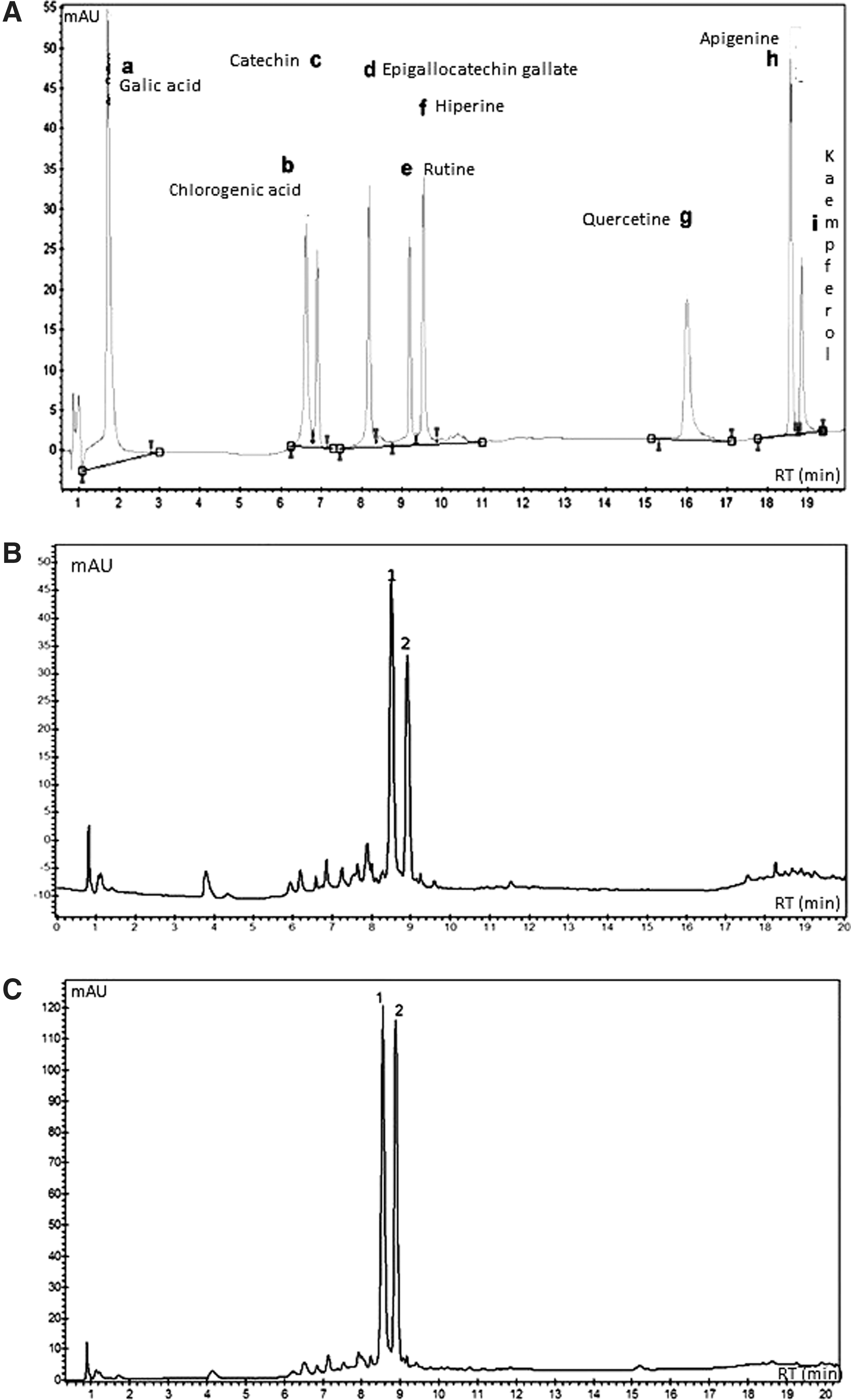
HPLC profile of phenolic compounds from Turnera subulata leaves detected at 280 nm. Profile
Quantification Parameters of Nine Phenolic Compounds Identified by Comparison Between UV Spectra and Retention Times of the Extracts and External Standards Based on the Chromatographic Method
UV, ultraviolet spectra.
CETS and AFTS anticoagulant activity was demonstrated by activated partial thromboplastin and PT evaluation, such as activated X and II factors, as well as by indirect thrombin inhibition mediated by the heparin II cofactor.
CETS, AFTS, and heparin (standard positive control) revealed a significant aPTT anticoagulant activity over 240 s (negative control: 36.05 ± 0.03 s) at 5 μg/mL, while PT >60 s (negative control: 16.65 ± 0.33 s) was revealed at 30 μg/mL, as expected and displayed in Figure 2A and B, respectively. Both CETS and AFTS inhibited clot formation through intrinsic and extrinsic pathways in concentrations >100 μg/mL.

Anticoagulant activity of CETS, AFTS, and heparin by activated partial thromboplastin time assay
Based on these data, the heparin and extract ability to directly inhibit the activity of factors Xa and IIa (thrombin) was evaluated. Figure 2C shows that heparin hindered factor Xa activity in a dose-dependent concentration, requiring 1 μg/mL for total factor inhibition, while 100 μg/mL of extracts completely hampered thrombin activity (Fig. 2D). Regarding CETS and AFTS, despite their lower activities, both extracts showed an ability to inhibit factor Xa activity in a dose-dependent concentration, reaching a value of ∼40% and 80% at ∼100 μg/mL to hinder thrombin activity.
To evaluate the extract's anticoagulant action mechanisms, an indirect inhibition assay was performed by heparin cofactor II (HCII) on thrombin. Results displayed a significant extract capacity (at 100 μg/mL) to inhibit thrombin (Fig. 2E), revealing that AFTS through HCII showed an inhibition rate of 70%, whereas CETS showed only 30%.
Heparin's adverse effects restrict its clinical use, such as thrombocytopenia and hemorrhagic complications, which interfere with the hemostatic balance. Considering the relevance of these events, CETS and AFTS effects on hemostasis were investigated.
Concerning the heparin assay (100 μg/mL), results displayed a marked hemorrhagic effect, evaluated by the high residual bleeding level determined using the rat tail scarification model after hemoglobin dosage in treating animals. Regarding CETS and AFTS treatments with 100 μg/mL, both showed anticoagulant potential with lower hemorrhagic rates, ∼50% and 10%, respectively, compared with clinical heparin (Fig. 2F).
With respect to cytotoxicity of CETS and AFTS, no cytotoxic effect was evidenced on normal fibroblast (3T3) and human embryonic kidney (HEK-293) fibroblast cells (Fig. 3A, B). No statistically significant differences were observed relative to the negative control (DMEM). During the in vivo toxicity assessment, no animal exhibited behavioral abnormalities over the trial period.
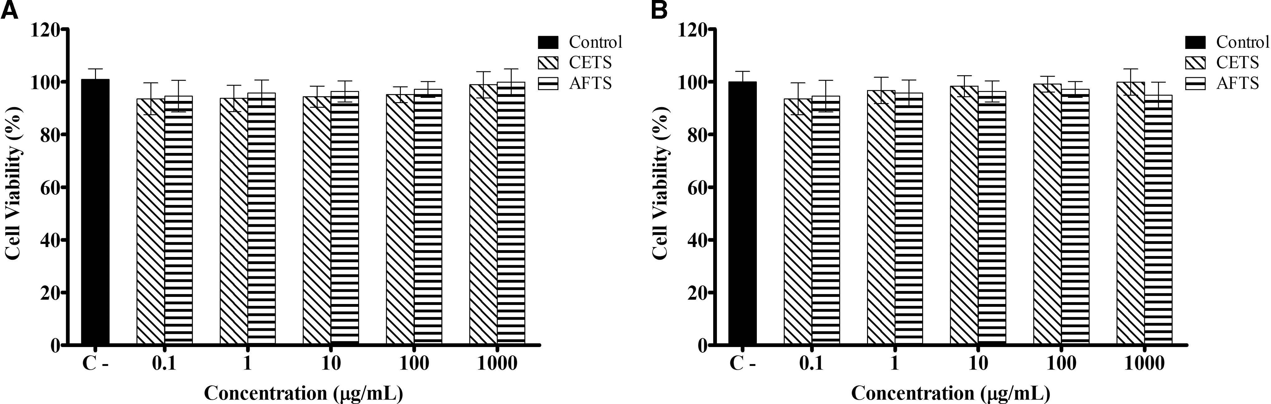
Cell viability (cytotoxicity effects) of CETS and AFTS on mouse fibroblast cells (3T3)
However, the biochemical parameter analysis in CETS- and AFTS-treated animals showed statistically significant differences with regard to glucose, triglyceride, and total cholesterol levels. These parameters exhibited a mean decrease of 50% after treatment compared with the control group (Table 2).
Biochemical Parameters of Rats After 14 Days of Treatment with CETS and AFTS
Results are expressed as mean ± SD (n = 5); control group treated with vehicle (distilled water). Comparisons between groups were analyzed with an ANOVA and Tukey's post hoc test.
P < .05 compared with the control group.
AFTS, acetate fraction of T. subulate; ALT, alanine aminotransferase enzymes; ALB, albumin; Ami, amylase; ANOVA, analysis of variance; AST, aspartate aminotransferase; CETS, T. subulata crude extract; Col, cholesterol; Cret, creatinine; DB, direct bilirubin; γ-GT, gamma-glutamyl transferase; Glo, globulin; Gli, glucose; IB, indirect bilirubin; TB, total bilirubin; TP, total proteins; Trig, triglycerides.
Regarding hematological parameters and organ weights, no significant differences were observed relative to the control, indicating no toxicity signs. Water and food intake displayed no significant difference. Data regarding hematological parameters and relative organ weights are attached as Supplementary Tables S1 and S2.
Discussion
Although anti-inflammatory, antidiabetic, and antiobesity properties have been reported for the Turnera genus, 15 –19 no data regarding its anticoagulant activity have been described so far.
Overall, plant extracts have been assessed for their ability to inhibit blood clotting factors. 25 With regard to Passifloraceae species, studies have shown their potential to treat cardiovascular diseases. The Passiflora nitida Kunth extract showed anticoagulant activity by partially activating thromboplastin, suggesting an inhibitory effect on intrinsic factors of the coagulation pathway VIII, IX, XI, and XII. 26
Concerning T. subulata, both CETS and AFTS promoted efficient inhibition of intrinsic and extrinsic pathways of the blood coagulation cascade. This was indicated by the PT test, also suggesting an anticoagulant effect due to thrombin inhibition (common pathway cascade). Therefore, extracts directly inhibited thrombin, assessed by the factor IIa inhibition assay, and indirectly by HCII as a blood coagulation action mechanism.
These parameters play a key role in coagulation inhibition. Thus, thrombin, a multifactorial enzyme, acts on the coagulation system converting soluble plasma fibrinogen into insoluble fibrin molecules and activating factor VIII. This factor binds to cross-linked fibrin polymers constituting a stable clot, amplifying the coagulation cascade with subsequent factors V, VII, VIII, and XI and platelet activation and stimulating granule release and platelet aggregation. 27
On the other hand, the long-term anticoagulant prescription such as heparin in clinical therapy causes side effects (e.g., bleeding, thrombocytopenia, hypersensitivity, skin necrosis, and intestinal toxicity). 28,29 This has encouraged the anticoagulant substance prospection to assist in clinical treatment. However, research concerning the medicinal plant anticoagulant effect is scarce, hence the relevance of this study.
Thus, the inhibitory effect of CETS and AFTS on the blood coagulation cascade may be due to the chemical composition rich in glycosylated flavonoids, mainly rutin and apigenin, besides other phenolic compounds. This phytocomposition is consistent with that previously reported for Turnera ulmifolia leaf extract, exhibiting significant polyphenolic content, such as rutin and apigenin. 18 Studies have demonstrated the significant anticoagulant effect of glycosylated flavonoids. 30
Besides anticoagulant and low hemorrhagic activities, CETS and AFTS exhibited no toxic effects in vitro using human fibroblasts and human embryonic kidney cells. An analogous result was reported with the Turnera diffusa methanolic extract, exhibiting low cytotoxicity in normal human fibroblasts. 31
Concerning the acute oral toxicity, no toxic effects were observed regarding evaluated biochemical and hematological parameters, especially those relating to hepatic and renal functions. There are no data regarding the T. subulata toxicity potential. However, the low acute oral toxicity level of T. diffusa Willd corroborates the results of the present study since no behavioral change, mortality, renal, and hepatic histopathology, as well as biochemical, parameters were observed. 32 Although AFTS displayed side effects lower than CETS due to higher polyphenol content, both extracts can be considered safe for clinical use. 33,34 Although no acute toxic effects have been observed, anatomopathological analyses of different tissues or organs such as the liver or kidney are required to corroborate the absence of damage to the liver and/or kidneys. This evaluation will be carried out in the continuation of the present study.
A relevant aspect regarding the biochemical parameters evaluated in this study was the decrease in glucose, triglyceride, and total cholesterol values after the CETS and AFTS treatments. These results suggest hypoglycemic and hypolipidemic properties, which indicate the phytotherapeutic potential of these extracts for cardiovascular disease treatment. These effects have been reported in several Passifloraceae species, especially in the Turneraceae genus, 16,35 –38 for example, in T. diffusa 35 and T. ulmifolia, 36 whose extracts were evaluated in animal models.
Overall, results regarding CETS and AFTS anticoagulant, nontoxic, and low hemorrhagic effects are due to their significant polyphenolic compound content. These compounds are associated with several pharmacological activities widely described in the literature. 39,40
In conclusion, T. subulata CETS and AFTS results displayed anticoagulant activity, inhibiting intrinsic and extrinsic pathways in the blood coagulation cascade. This suggests direct and indirect thrombin inhibition as the main action mechanism. Moreover, extracts showed low hemorrhagic and toxic effects in vitro and in vivo, while the biochemical parameters showed possible hypoglycemic and hypolipidemic activities. These biological effects can be attributed to the significant polyphenol content. Overall, experimental results are promising due to the T. subulata therapeutic potential to develop herbal formulations to assist in anticoagulant therapy, although further studies are required, considering that this is the first report evaluating the anticoagulant capacity of this plant.
Footnotes
Acknowledgments
This research was supported by the Conselho Nacional de Desenvolvimento de Científico e Tecnológico (CNPq) (Protocol No.478652/2010-0) and Banco do Nordeste (Protocol No.912011) grants. The authors would like to thank Coordenação de Aperfeiçoamento de Pessoal de Nível Superior (CAPES) and CNPq for providing a postgraduation fellowship and the Department of Biochemistry (UFRN) for cell culture technical assistance.
Author Disclosure Statement
No competing financial interests exist.
Supplementary Material
Supplementary Table S1
Supplementary Table S2
References
Supplementary Material
Please find the following supplemental material available below.
For Open Access articles published under a Creative Commons License, all supplemental material carries the same license as the article it is associated with.
For non-Open Access articles published, all supplemental material carries a non-exclusive license, and permission requests for re-use of supplemental material or any part of supplemental material shall be sent directly to the copyright owner as specified in the copyright notice associated with the article.