
Abstract
This study evaluated to determine the phenolic and flavonoids contents, and antioxidant, anti-inflammatory, and antiproliferative activity of the hydromethanolic extracts of the leaves, pulp, and seeds of Annona cacans. The isolation and structural identification of the constituent acetogenin, phenolic acid, and flavonoids were also reported. Antioxidant capacity was determined by the 2,2-diphenyl-1-picrylhydrazyl (DPPH), ethylbenzothiazoline-6-sulfonic acid (ABTS), and β-carotene/linoleic acid methods. Cell proliferation was determined by spectrophotometric quantification of the cellular protein content using a sulforhodamine B assay. Anti-inflammatory activity was evaluated in paw edema model, to myeloperoxidase (MPO) activity induced by carrageenan in mice. Fractionation resulted in the isolation of one acetogenin (annoreticuin-9-one), two flavonoids (quercetin-3-O-β-glucoside-6-O-α-rhamnoside and kaempferol-3-O-β-glucoside), and one phenolic acid (p-coumaric acid). The pulp extract presented potent antioxidant activities by the DPPH (IC50 = 44.08 μg/mL) and ABTS (IC50 = 39.32 μg/mL) methods, as well as high contents of phenols (618.95 mg GA/g) and flavonoids (477.35 mg QE/g). The bioguided fractionation demonstrated that the ethyl acetate fraction of the pulp extract and annoreticuin-9-one showed potent antiproliferative activity against ovarian cancer (GI50 = 6.4 μg/mL). The anti-inflammatory activity demonstrated significant inhibition of edema compared to the control group in 2 and 4 h; in addition, the extracts inhibited the increase in MPO activity after 6 h, when compared to the DEX and control groups. For the first time, this study demonstrated antioxidant, anti-inflammatory, and antiproliferative activity, as well as compounds isolated, suggesting that A. cacans could also be potential sources for prevention of cancer and other diseases associated with oxidative stress.
Introduction
Annona is a member of the family Annonaceae, which includes woody plants and those with arbustive or arboreal character, as well as a great variety of fruits. 1 Some species have been pharmacologically and phytochemically studied by our research group, and these studies have included the essential oil of the leaves of Annona sylvatica, and its main compounds, sesquiterpenes, presented anticancer and anti-inflammatory activities 2 : Annona dioica leaves, which exert anti-inflammatory, hypoglycemic, antiproliferative, and antioxidant effects that can be associated with the presence of flavonoids 3 ; Annona coriacea and A. sylvatica showed antioxidant activity 4 ; and Annona crassiflora showed anti-inflammatory activity and chemopreventive therapeutic potential, 5 as well as anticholinesterase and antiproliferative activities. 6 This genus is recognized as an important source of biologically active compounds and isoquinoline-derived alkaloids, 7,8 and acetogenins 9 have been reported as the main compounds. Among the popular uses of the genus, the applications of these plants as antiparasitic and anticancer therapeutics are of particular interest. 10
Annona cacans Warm. (old name Xilopiacacanes Warm.), popularly known as “araticum-cagão” or “cortição,” is used in folk medicine as purgative 11 and it is found in the states of Minas Gerais, Mato Grosso do Sul, Espírito Santo, Paraná, Rio de Janeiro, Rio Grande do Sul, Santa Catarina, and São Paulo. 12 Chemical studies of the stem of A. cacans resulted in the isolation of alkaloids (stepharine, asimilobine, michelalbine, liriodenine, and aristololactam A-11 and B-II). 13 To the best of our knowledge, there have been no chemical and biological studies reported in the literature on the fruits and leaves in this species. The maintenance of health has aroused interest of the scientific community, which they see producing many works to prove the performance of medicinal plants and their derivatives in the reduction of certain diseases. Thus, the aim of this study was the bioassay-guided fractionation, and antioxidant, anti-inflammatory, and antiproliferative investigations of A. cacans. We have also determined the contents of the constituents, isolated, and structurally identified the mono-tetrahydrofuran (THF) acetogenin (PAC-1), flavonoids (PAC-2 and PAC-3), and phenolic acid (PAC-4) (Fig. 1).
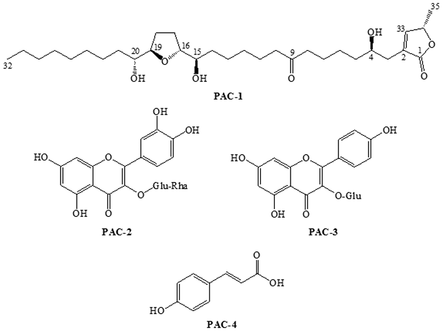
Chemical structural of the mono-THF acetogenin (PAC-1), flavonoids (PAC-2 and PAC-3), and phenolic acid (PAC-4) from Annona cacans. THF, tetrahydrofuran.
Materials and Methods
Plant material, extract preparation, isolation, and structural identification
The fruits and leaves from A. cacans were collected in December 2013, in Dourados, Mato Grosso do Sul, at an average altitude of 452 m, 23° 17′6″S latitude, and 54° 43′28″ W longitude. The plant was identified by Dr. Zefa Valdivina Pereira, teacher of the Federal University of Grande Dourados, and a sample was deposited in the Herbarium of this University (DDMS 4885). To prepare the extracts, fruit pulp, air-dried and powdered seeds, and leaves were separated, and each component was extracted for 20 days by maceration with methanol/water (MeOH: H2O, 8:2) at room temperature. The extracts were filtered, concentrated under reduced pressure, and lyophilized to obtain the hydromethanolic extract of the leaves (LHME-AC, 36.7 g), pulp (PHME-AC, 25.9 g), and seeds (SHME-AC, 10.2 g). The most potent extract evaluated by biological assays was submitted to fractionation for the isolation of the main constituents. A portion of PHME-AC (7.8 g) was dissolved in MeOH:H2O (1:1) and partitioned with n-hexane, chloroform, and ethyl acetate. The ethyl acetate fraction (PEAF-AC 3.2 g) was purified by silica gel column chromatography followed by purification on Sephadex LH-20 using H2O containing increasing amounts of MeOH to afford a known compound identified as annoreticuin-9-one (PAC-1) (163.42 mg), quercetin-3-O-β-glucoside-6-O-α-rhamnoside (PAC-2) (56.34 mg), kaempferol-3-O-β-glucoside (PAC-3) (45.60 mg), and p-coumaric acid (PAC-4) (15.60 mg).
Annoreticuin-9-one (PAC-1): 1 H NMR δ H (300 MHz, CD3OD): 2.40–2.52 (m, H-3; H-5; H-8, H-10), 3.79 (m, H-4; H-16; H-19), 1.38–1.60 (m, H-6; H-7; H-11; H-13; H-14; H-21; H-22), 1.25 (m, H-12; H-23 to H-31), 3.40 (m; H-15; H-20), 1.98–1.66 (m, H-17; H-18), 0.87 (t, J = 6.6 Hz, H-32), 7.20 (s; H-33), 5.08 (q, J = 7.5 Hz; H-34), and 1.43 (d, J = 7.0 Hz; H-35). 13 C NMR δ C (75.5 MHz, CD3OD): 174.58 (C-1), 130.4 (C-2), 33.4 (C-3), 68.4 (C-4), 35.3 (C-5), 26.4 (C-6), 23.6 (C-7), 42.6 (C-8), 208.9 (C-9), 42.5 (C-10), 23.4 (C-11), 30.3 (C-12), 25.2 (C-13), 33.3 (C-14), 75.6 (C-15/C-20), 82.4 (C-16/C-19), 29.4 (C-17), 29.7 (C-18), 32.8 (C-21), 25.6 (C-22), 29.6–29.9 (C-23, C-24, C-25, C-26, C-27, C-28, C-29), 31.6 (C-30), 22.8 (C-31), 14.1 (C-32), 153.6 (C-33), 77.4 (C-34), and 20.0 (C-35).
Quercetin-3-O-β-glucoside-6-O-α-rhamnoside (PAC-2): 1 H NMR δ H (300 MHz, CD3OD): 6.20 (d, J = 2.1 Hz, H-6), 6.39 (d, J = 2.1 Hz, H-8), 7.66 (dd, J = 8.7 e 2.4 Hz, H-2′), 6.80 (d, J = 8.7 Hz, H-3′), 7.51 (d, J = 2.4 Hz, H-6′), 5.37 (d, J = 7.8 Hz, H-1″), 5.11 (d, J = 7.8 Hz, H-1′″), and 1.17 (d, J = 6.3 Hz, H-6′″). 13 C NMR δ C (75.5 MHz, CD3OD): 157.9 (C-2), 134.2 (C-3), 177.7 (C-4), 161.9 (C-5), 98.5 (C-6), 164.4 (C-7), 93.7 (C-8), 156.5 (C-9), 104.2 (C-10), 121.1 (C-1′), 122.2 (C-2′), 115.3 (C-3′), 148.7 (C-4′), 145.1 (C-5′), 116.2 (C-6′), 102.0 (C-1″), 71.4 (C-2″), 73.4 (C-3″), 68.2 (C-4″), 76.1 (C-5″), 60.4 (C-6″), 101.1 (C-1′″), 70.8 (C-2′″), 70.7 (C-3′″), 72.5 (C-4′″), 68.7 (C-5′″), and 16.4 (C-6′″).
Kaempferol-3-O-β-glucoside (PAC-3): 1 H NMR δ H (300 MHz, CD3OD): 6.22 (d, J = 2.1 Hz, H-6), 6.42 (d, J = 2.1 Hz, H-8), 8.04 (d, J = 8.7 Hz, H-2′/H-6′), 6.77 (d, J = 8.7 Hz, H-3′/H-5′), and 5.60 (d, J = 7.8 Hz, H1″). RMN 13 C δ c (75.5 MHz, CD3OD): 159.9 (C-2), 135.8 (C-3), 177.8 (C-4), 162.7 (C-5), 100.7 (C-6), 163.7 (C-7), 95.8 (C-8), 158.1 (C-9), 107.3 (C-10), 122.5 (C-1′), 132.6 (C-2′/C-6′), 116.3 (C-3′/C-5′), 161.8 (C-4′), 105.1 (C-1″), 73.5 (C-2″), 75.4 (C-3″), 72.9 (C-4″), 75.0 (C-5″), and 67.4(C-6″).
p-Coumaric acid (PAC-4): 1 H NMR δ H (300 MHz, CD3OD): 7.08 (d, J = 15.5 Hz, H-1′), 7.04 (d, J = 8.5 Hz, H-2/H-6), 6.80 (d, J = 2.1 Hz, H-3/H-5), and 6.3 (d, J = 15.5 Hz, H-2′). RMN 13 C δ c (75.5 MHz, CD3OD): 127.3 (C-1), 131.6 (C-2/C-6), 116.5 (C-3/C-5), 161.8 (C-4), 146.6 (C-1′), 116.7 (C-2′), and 171.4 (C = 0).
The extracts were analyzed in an analytical liquid chromatography (LC) (Varian 210) system equipped with a ternary solvent delivery system, an autosampler, and a photodiode array (PDA) detector, and the eluate was monitored at λ = 200–800 nm. The LC column was a C-18 (25 cm × 4.6 mm; particle size, 5 μm; Luna, Phenomenex, Torrance, CA, USA), and a small precolumn (2.5 cm × 3 mm) containing the same packing material was used to protect the analytical column. In each analysis, the flow rate and the injection volume were 1.0 mL/min and 20 μL, respectively. All chromatographic analyses were performed at 22°C. Elution was carried out using acetic acid (6%), sodium acetate (2 mM) (solvent A), and acetonitrile (solvent B). The solvent gradient program was as follows: 0 min, 5% B; 45 min, 15% B; 55 min, 30% B; 60 min, 50% B; 65 min, 100% B; and in 75 min, a return to the initial conditions.
The contents of the components in the samples were determined by external calibration using standards [caffeic acid (98%), p-coumaric acid (98%), ferulic acid (99%), sinapic acid (98%), luteolin (98%), and kaempferol (98%)]. Aliquots of dilutions (20 μL) were analyzed by LC, and each determination was carried out five times. For each standard, a corresponding chromatogram was obtained, and a calibration graph was constructed from the mean of the peak areas plotted against the concentration of the standard (1–100 μg/mL). A linear least square regression of the peak areas as a function of the concentrations was performed to determine the correlation coefficients. The equation parameters (slope and intercept) of the standard curve were used to obtain the concentrations of the components in the samples.
Total phenols and flavonoids
Folin-Ciocalteu reagent 14 was utilized to determine the concentration of total phenol (735 nm) in each extract. Gallic acid was used as standard. The flavonoid content was evaluated at 415 nm 15 and quercetin was used as standard. The tests were performed in triplicate.
DPPH, ABTS, and β-carotene/linoleic acid—antioxidant assays
For the 2,2-diphenyl-1-picrylhydrazyl (DPPH) 16 radical, the samples were prepared in different concentrations (5, 10, 25, 50, 125, and 200 μg/mL) and read in a spectrophotometer at 515 nm. In the ethylbenzothiazoline-6-sulfonic acid (ABTS) 17 trial, the Trolox ethanolic solution was used as the standard for the preparation of the calibration curve (concentrations of 100, 500, 1000, and 2000 μg/mL were used) and the reading was performed at 734 nm. The samples of A. cacans (2 mg/mL) were evaluated in the β-carotene/linoleic acid method and read at 470 nm every 15 min to monitor oxidation (∼105 min). 18 Ascorbic acid was used as a positive control.
Antiproliferative activity assay
In vitro antiproliferative assays were conducted against human cancer cell lines provided by the Frederick National Laboratory for Cancer Research, Frederick, USA. The tested lines were U251 (glioma; CNS), MCF-7 (breast), NCI-ADR/RES (ovarian expressing the multiple drug resistance phenotype), NCI-H460 (lung and nonsmall cell), PC-3 (prostate), OVCAR-3 (ovarian), HT-29 (colon), K-562 (leukemia), 786-0 (renal), and HaCaT (human keratinocytes and immortalized nontumoral cell). Cell proliferation activities of the extracts, fractions, and individual compounds were determined by spectrophotometric quantification at 540 nm using sulforhodamine B dye. 19 Doxorubicin chloride (0.025–25 μg/mL) was used as the positive control. The activity was deduced from the concentration response, and the GI50 parameters (growth inhibitory activity) were calculated.
Animals
For the anti-inflammatory assay, 60 male Swiss mice (20–25 g, n = 6) were obtained from UFGD (Brazil). The animals were maintained under a 12-h light/12-h dark cycle, with controlled humidity (60–80%) and temperature (22°C ± 1°C). The animals were acclimatized to the experimentation room for at least 2 h before testing and were used only once throughout the experiments. All experimental procedures were carried out in accordance with U.S. National Institute of Health, and were approved by the Ethics Committee in Animal Experimentation of UFGD (005/2010).
Anti-inflammatory activity assay
Different groups of male mice were treated orally with LHME-AC (100 and 300 mg/kg), PHME-AC (100 and 300 mg/kg), SHME-AC (100 and 300 mg/kg), PEAF-AC (30 and 100 mg/kg), and saline solution (control). Another group of mice was treated with the anti-inflammatory drug dexamethasone (1 mg/kg) (0.5 h before subcutaneous injection). After 1 h of treatment, the edema was induced through 50 μL of carrageenan (Cg) injection (300 μg/paw) in the right paw and sterile saline injection (0.9%) was applied to the left paw as a control. Edema was measured with a plethysmometer at 2 and 4 h after injection and the results expressed in μL, and the difference between onset and postinjection was quantified as edema. 20
Determination of myeloperoxidase activity
To investigate whether oral treatment with extracts, fraction, or control could affect the cellular migration induced by Cg, the myeloperoxidase (MPO) activity was measured in the mouse paws. The animals were euthanized 6 h after Cg injection, as described previously. 21 For MPO activity, the tissue was homogenized in 5% (w/v) of 80 mM phosphate buffer (pH 5.4) containing 0.5% hexadecyltrimethylammonium bromide, and centrifuged at 12,000 g and 4°C for 20 min. Aliquots (30 μL) of each supernatant were mixed with 100 μL of 80 mM phosphate buffer, 85 μL of 0.22 M phosphate buffer, and 15 μL of 0.017% H2O2 on a 96-well plate. The reaction was triggered with 20 μL of 3,3,3-tetramethylbenzidine (in N,N-dimethylformamide). The plate was kept at 37°C for 3 min, after which the reaction was stopped by adding 30 μL of 1.46 M sodium acetate, pH 3.0. The enzymatic activity was determined by measuring the optical density at 630 nm and is expressed as the mOD per milligram of protein.
Results
Phytochemistry
The pulp extract (PHME-AC; 618.95 mg GA/g; 477.35 mg QE/g) and ethyl acetate fraction (PEAF-AC; 635.83 mg GA/g; 452.90 mg QE/g) exhibited the highest phenolic and flavonoid contents (Fig. 2).
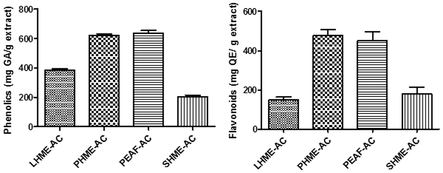
Phenolics acid and flavonoid content in samples from Annona cacans.
The PHME-AC exhibited phenolic and flavonoid contents, and an LC method was developed to identify its main components (Fig. 3). The retention times of the standards and the contents of these constituents in the samples are shown in Table 1.

Chromatogram (LC-DAD) of the PHME-AC from Annona cacans.
Retention Times of the Standards and the Contents of These Constituents in Micrograms/Gram for the PHME-AC of Annona cacans by the Liquid Chromatography Method
Fractionation and purification of the pulp extract (PHME-AC) by solvent partitioning yielded the ethyl acetate (PEAF-AC) fraction, and purification of this soluble fraction by CC with silica gel and Sephadex afforded acetogenin (PAC-1), flavonoids (PAC-2), (PAC-3), and phenolic acid (PC-4) (Fig. 1). The chemical structure was confirmed by spectral data analysis obtained through 1 H and 13 C NMR and DEPT, COSY, HMQC, and HMBC experiments, and comparison to literature data. 22,23
Antioxidant activity
The percentages of inhibition (%I) were calculated utilizing the following equation: I% = (A blank − A sample/A blank) × 100. The IC50 was calculated using the graph of %I versus concentration of the extract/fraction or compound, and ascorbic acid was used as the positive control. The results show that the three extracts (leaves, pulp, and seeds) possess free radical scavenging activity with IC50 values ranging from 89.67 to 26.25 μg/mL, and notably, the pulp extract (PHME-AC) showed an IC50value of 44.08 μg/mL (DPPH) and 39.32 μg/mL (ABTS) (Table 2). In addition, the fraction PEAF-AC showed significant activity, in both assays (DPPH, IC50 = 47.11 μg/mL and ABTS, IC50 = 26.25 μg/mL), which were comparable to that of ascorbic acid (Table 2). The PHME-AC (51.51 μg/mL) and PEAF-AC (34.34 μg/mL) also presented antioxidant activities in the lipid peroxidation assay (Table 2).
Antioxidant Activity from Annona cacans by the DPPH, β-Carotene Bleaching, and ABTS Methods
ABTS, ethylbenzothiazoline-6-sulfonic acid; DPPH, 2,2-diphenyl-1-picrylhydrazyl.
Antiproliferative activity
When extracts of the pulp and seeds were evaluated, the PHME-AC showed anticancer activity with GI50 values in the range of 50.65–6.10 μg/mL against seven of the tested human cell lines, and it was particularly potent against K-562 (GI50 = 6.10 μg/mL) and OVCAR-3 cells (GI50 = 8.8 μg/mL) (Fig. 4A). The fraction PEAF-AC showed potent activity against all of the tested cell lines with GI50 values ≤60.57 μg/mL, including OVCAR-3 (GI50 = 5.68 μg/mL), K-562 (GI50 = 7.84 μg/mL), and MCF-7 (GI50 = 11.3 μg/mL) (Fig. 4B). The acetogenin PAC-1, isolated from this fraction, was particularly effective against OVCAR-3 (GI50 = 6.4 μg/mL) (Fig. 4C) and showed GI50values ≥23.65 μg/mL against all tested cell lines (Fig. 4D).
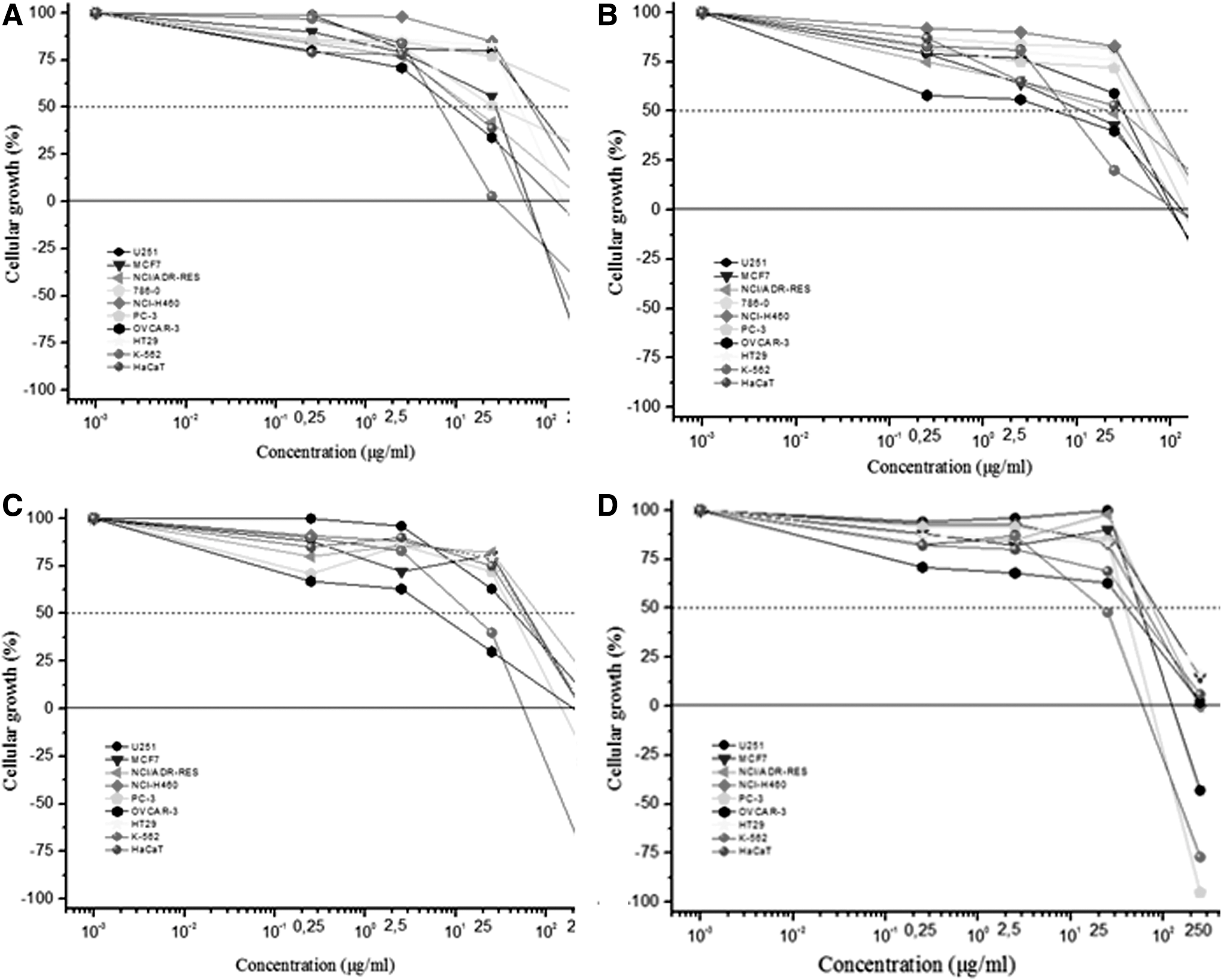
Antiproliferative activity (GI50 in μg/mL) of the pulp extract
Anti-inflammatory and MPO assay
The injection of Cg into the paw induced an edema that peaked at 2 and 4 h (Fig. 5). The results showed a significant decrease in edema compared to the control group; inhibitions for each dose were 27% ± 8% (100 mg/kg), 41% ± 6% (300 mg/kg) for LHME-AC, 50% ± 3% (100 mg/kg), 63% ± 6% (300 mg/kg) for PHME-AC, 27% ± 7% (100 mg/kg), 32% ± 5% (300 mg/kg) for SHME-AC, and 52% ± 7% (100 mg/kg), 64% ± 8% (300 mg/kg) for PEAF-AC at 2 h (Fig. 5). This reduction continued after the fourth hour of observation, 30% ± 5% (100 mg/kg), 43% ± 6% (300 mg/kg) for LME-AC, 63% ± 5% (100 mg/kg), 72% ± 5% (300 mg/kg) for PHME-AC, 37% ± 6% (100 mg/kg), 44% ± 3% (300 mg/kg) for SHME-AC, and 60% ± 6% (100 mg/kg), 67% ± 7% (300 mg/kg) for PEAF-AC (Fig. 5). In addition, the inhibition in DEX group was also significant (79% ± 3%, 2 h and 83% ± 4%, 4 h) (Fig. 5).
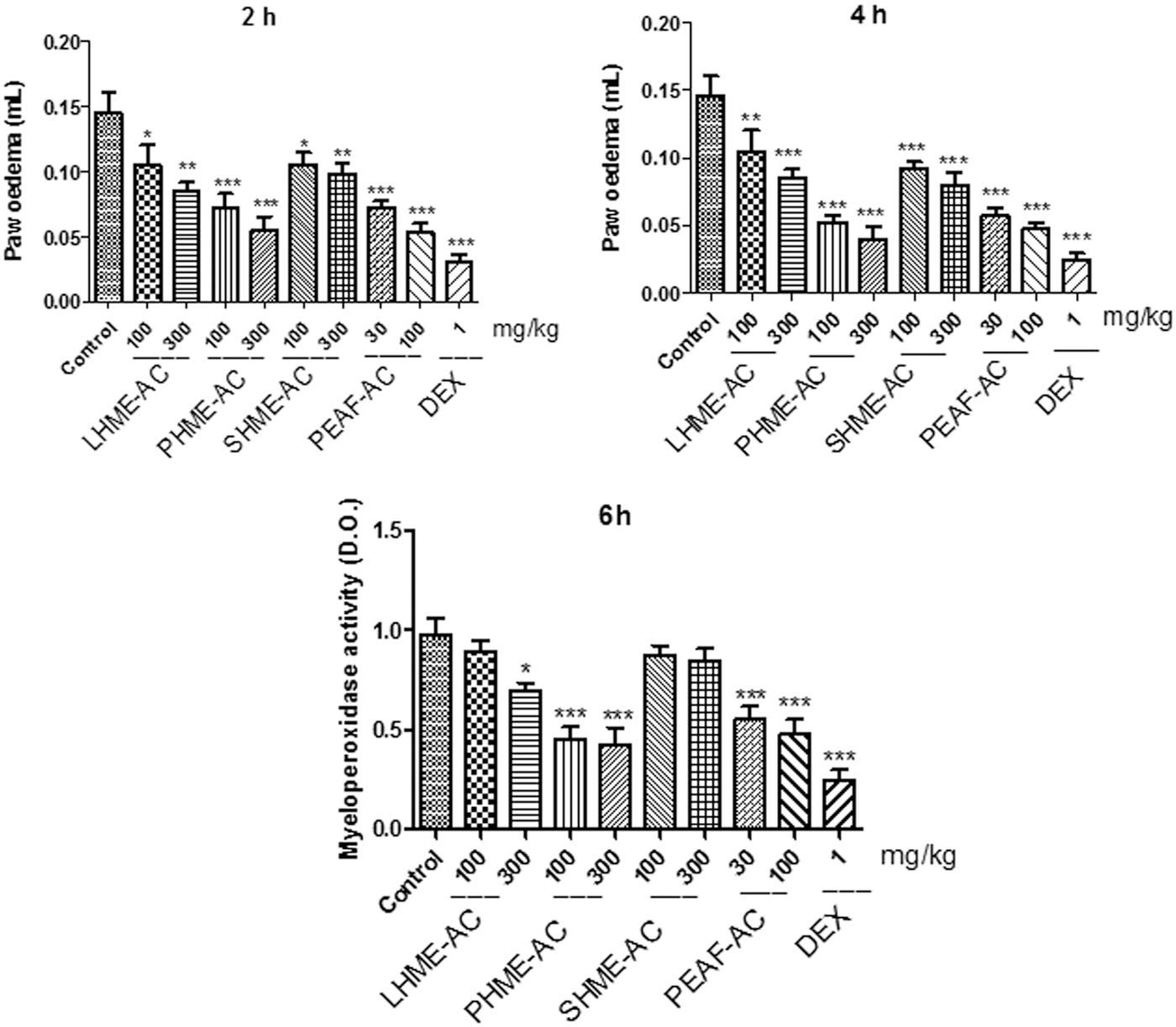
Effect of hydromethanolic extract from Annona cacans (LHME-AC) leaves, pulp (PHME-AC), seeds (SHME-AC), and ethyl acetate fraction (PEAF-AC) pulp on Cg-induced increase in MPO activity in mice. Animal received the oral treatment with LHME-AC (100 and 300 mg/kg), PHME-AC (100 and 300 mg/kg), SHME-AC (100 and 300 mg/kg), PEAF-AC (30 and 100 mg/kg), and dexamethasone (Dexa, 1 mg/kg, s.c.) or control and after 1 h, an intraplantar injection of Cg (300 μg/paw) was performed. The bars express the mean ± SEM of six animals, compared with control versus treated group. *P < .05, **P < .01, ***P < .0001, one-way ANOVA followed by Student–Newman–Keuls. Cg, carrageenan; MPO, myeloperoxidase; SEM, standard error of the mean.
The treatment with LHME-AC (300 mg/kg) and PHME-AC (100 and 300 mg/kg) extracts and PEAF-AC (30 and 100 mg/kg) ethyl acetate fraction inhibited the increase in MPO activity induced by Cg, after 6 h, with inhibition of the 28% ± 6%, 53% ± 9%, 58% ± 8%, 43% ± 5%, and 51% ± 9%, respectively (Fig. 5). The DEX was also able to inhibit MPO activity compared to the control group (74% ± 3%) (Fig. 5).
Discussion
Fractionation of the pulp extract by solvent partitioning yielded the ethyl acetate (PEAF-AC) fraction, and purification of this soluble fraction by CC with silica gel and Sephadex afforded acetogenin (PAC-1), two flavonoids (PAC-2 and PAC-3)f and one phenolic acid (PAC-4) (Fig. 1). The acetogenin PAC-1 showed the presence of an α, β-unsaturated γ-lactone moiety at δ H/C 7.20/153.6, δ H/C 5.08/77.4, δ H/C 2.40–2.52/33.4f and δ H/C 1.43/20.0, with a hydroxy group at C-4 (δ H/C 3.79/68.4) and a keto group at C-9 (δ C 208.9) in the long aliphatic chain. The oxymethine group of the mono-THF ring with two flanking hydroxyl groups was confirmed by the signals at δ H/C 3.79/82.4 and δ H/C 3.40/75.6, respectively. The keto group, which gave signals at δ C 208.9 for PAC-1, confirmed the structure of annoreticuin-9-one.
Identification of the compounds with a PDA detector scanning in the spectral range of 200–800 nm did not reveal any interference at the retention times of the samples in LC using the developed elution conditions. The standards were easily identified and quantified based on their absorption spectra in the UV region and their retention time. The standards present in the extracts were unambiguously identified by performing co-injection experiments in which aliquots of the extracts and standards were mixed, diluted to a known volume, and analyzed by LC. The calibration curves prepared using LC were determined by linear regression. The linearity of each standard was assessed for 10 concentrations. The average standard errors for the peak areas of replicated injections (n = 5) were less than 2%, which indicated good repeatability of the calibration curve. The respective coefficients of determination (r 2 values) were 0.9992 for caffeic acid, p-coumaric acid, ferulic acid, and sinapic acid, and 0.9994 for luteolin and kaempferol.
There is growing body of evidence showing that antioxidants have great therapeutic potential in the prevention and treatment of different chronic diseases, including cardiovascular and neurodegenerative diseases, as well as cancer and inflammatory diseases. 24 In vitro antioxidant assays are used as a preliminary evaluation of the beneficial effects of the samples, allowing the analysis of the inhibition of oxidative processes, to obtain exploratory or comparative information on extracts or compounds. The DPPH assay is used for methanol-soluble free radicals, whereas the ABTS assay is used to measure water-soluble free radicals. It has been found that the ABTS assay is more sensitive than the DPPH assay, since the DPPH moiety is involved only in the hydrogen transfer, while the ABTS moiety is involved in the electron transfer pathway. 25
The antioxidant activity from A. cacans was evaluated because members of this genus are used in popular medicine, and their fruits are commonly consumed. The antioxidant effects observed for the pulp extract are probably due to the presence of kaempferol and quercetin derivative. Quercetin is a natural flavonoid and has great therapeutic potential of different diseases, as well as cancer and inflammation. 26 –29 The strength of the antioxidant activity of polyphenols is dependent on the molecular structures of these compounds and is impacted by the number and position of the hydroxy groups present in the molecule, which show reducing properties. 30 The free radical quenching activities of the extracts of plants are dependent on the polarity and type of solvent used in the extraction, the activities of the pure constituents, the extraction techniques utilized, and the isolates procedure. 31
In previous studies by our research group on the constituents and antioxidant activity of Annona species, we demonstrated that pulp and seed extract of A. coriacea and A. sylvatica presented total phenolic concentrations of 147.08 ± 4.20 mg de GAE/g extract and flavonoid contents of 131.18 ± 2.31 mg QE/g extract. 4 The methanolic extract and ethyl acetate and the hydromethanolic fractions of A. dioica presented potent antioxidant activities by the DPPH method, possibly due to the presence of kaempferol, quercetin, and phenolic derivative compounds. 3 A. crassiflora has a potent antioxidant effect, since it is a natural source of quercetin. 32 Biological investigations of the others species from Annona, that is, Annona muricata, revealed antioxidant, antidiabetic, and antitumoral activity. 33 Previous phytochemical studies have attributed these activities to various compounds found in this species, such as chlorogenic and caffeic acids, procyanidins B2 and C1, epicatechin, quercetin, quercetin hexosides, and kaempferol. 34 –36 Annona glabra presents phenolic compounds and was able to inhibit the cell viability of human leukemia cells by 28%. 37 The extract and fractions from Annona hypoglauca were evaluated for cytotoxic activity in tumor cell lines and presented lethal effect on breast and colon. 38
In this context, previous studies that have confirmed antioxidant and anticancer properties in other species of Annona suggest an investigation of this activity from A. cacans. Samples from A. cacans were evaluated in vitro antiproliferative assay against HaCaT and nine human cancer cell lines (Fig. 4), highlighting the pulp and seed extract, being particularly effective against the OVCAR-3. The extracts obtained from the leaves (LHME-AC) did not show activity against the cell lines. 6
Ovarian cancer is considered the leading cause of death among gynecological cancer, and most patients are diagnosed at an advanced stage of the disease. The treatment involves chemotherapy and tumor removal through surgery. 39 Cancers are classified according to the type of cell first affected, and in the case of ovarian cancer, the FOXL2 mutation is the main cause of the disease. FOXL2 (Forkhead) is an essential transcription factor for complete ovarian formation in fetuses and is responsible for the normal progression of eyelids in newborns. 40 One in every 70 American women is diagnosed with ovarian cancer, and it mainly affects women older than 40 years. 41 Studies described in the literature with extracts of A. muricata 42 and Annona vepretorum 43 corroborate the results obtained for A. cacans, demonstrating anticancer activity against ovarian cancer.
The species of Annonaceae are widely distributed and always contain acetogenins, called annonaceous acetogenins. Such compounds are a polyester sequence containing adjacent or nonadjacent THF or tetrahydropyran (THP) moieties and an α,β-unsaturated γ-lactone ring. These compounds are derived from long-chain fatty acids (C32 or C34), and they possess potent mitochondrial inhibition (complex I) and cytoplasmic production without requiring adenosine triphosphate or oxygen. 44 Annonaceous acetogenins such as acetogenins isolated from the seeds of A. cornifolia 45 and those isolated from the leaves of A. muricata 46 have numerous biological activities, including anticancer activity, most notably against ovarian cancer. The antiproliferative activities of these compounds have also been reported against other cell lines, such the isolated of the A. muricata against prostate cancer, 47,48 Annona reticulata against four cell lineages, 49 and of the Annona squamosa against MCF-7 and A-549 lines, 50 among others described. 51 –53
The antioxidant potential and total phenolic and flavonoid content may also be useful to correlate this activity with other important ones, such as anti-inflammatory activity, that is, some plant extracts reduce inflammation by eliminating superoxide known to participate in recruitment of polymorphonuclear cells present in inflamed tissues. 54 Several studies our group has demonstrated effective anti-inflammatory activity from A. crassiflora, A. dioica, and A. sylvatica. 2,3,5 Before our work, there have been no reports available on the biological activity from A. cacans. In this way, our results showed that the samples of the A. cacans (30, 100, and 300 mg/kg) presented significant inhibition at the time after administration of Cg, highlighting the pulp extract and its fraction, suggesting that pulp A. cacans contains compounds that exert antiedematogenic properties. The inflammatory process induced in the paw of mice is accompanied by neutrophil migration (acute inflammation). The MPO is an enzyme belonging to the group of hemeperoxidases, found in the primary azurophilic granules of neutrophils (cells that play a key role in the innate immune response) and in lower concentrations in the primary lysosomes of monocytes; it is widely used as a marker of infiltration of neutrophils, because in inflammatory conditions, the MPO levels rise, inducing damage to adjacent tissues. 55,56 Our results demonstrated that extracts of A. cacans significantly reduced MPO levels in the plantar tissue of mice.
This study demonstrates, for the first time, antioxidant, anti-inflammatory, and anticancer effects, as well as acetogenin, flavonoids, and phenolic acid isolated from A. cacans. The preliminary results of the antiproliferative potential against of ovarian (OVCAR-3) cell line could be promising for the development of new cancer treatments, but future studies using other models are needed.
Footnotes
Author Disclosure Statement
No competing financial interests exist.
Funding Information
This work was supported by the FUNDECT (Foundation for Support to the Development of Education, Science and Technology of the State of Mato Grosso do Sul, Brazil, 59/300.029/2015), Capes (Coordination for Improvement of Higher Education Personnel, 2011), CNPq (National Council for Scientific and Technological Development, Brazil, 307309/2013-4), and UFGD (Federal University of Grande Dourados) for financial support and scholarships.