
Abstract
Abnormal reorganization of the dentate gyrus and neuroinflammation in the hippocampus represent characteristic phenotypes of patients suffering from temporal lobe epilepsy. Hesperetin, a flavanone abundant in citrus fruit, is known to have protective effects by preventing inflammation and oxidative stress in neuronal cultures and in the adult murine brain. However, the protective effects of hesperetin against epileptic seizures in vivo remain unclear, despite one study reporting anticonvulsant effects in vitro. In this study, we report that oral administration of hesperetin not only delays the onset of seizures triggered by kainic acid (KA) but also contributes to the attenuation of granule cell dispersion in the KA-treated hippocampus. Moreover, we observed that hesperetin administration inhibited the expression of pro-inflammatory molecules produced by activated microglia in the hippocampus. Thus, administration of hesperetin might be beneficial for preventing epileptic seizures.
T
Hesperetin (3′,5,7-trihydroxy-4′-methoxyflavanone), extracted from citrus fruit, is a flavonoid. It easily passes through the blood–brain barrier into the brain and exerts neuroprotective effects, possibly due to its radical scavenging property and by attenuating caspase-3 activity in vitro. 14,15 In addition, hesperetin could reduce neuronal cell death through antioxidant properties. 16 However, a preventive effect of hesperetin on pathological changes induced by kainic acid (KA) and epileptic seizures in vivo has not been established yet. 17 In this study, we investigated the effects of hesperetin on seizure onset, GCD formation, and neuroinflammation, in a KA-induced mouse model of epilepsy.
Mice were administered orally with hesperetin (Abcam, Cambridge, United Kingdom) (5, 10, or 20 mg/kg/day) for 8 days, starting 1 day before KA injection. 17,18 The degree of GCD was measured as the average width of the granule cell layer (GCL) in the middle and upper quadrant at top of the DG. Then, the experimental group's ipsilateral GCL was normalized to the corresponding side in the control group. 17,19 In addition, the phosphorylated form of eukaryotic translation initiation factor 4E (elF-4E)-binding protein 1 (4E-BP1) and p70 ribosomal S6 kinase (p70S6K), the markers for mTORC1 activation, and inflammatory molecules such as tumor necrosis factor-alpha (TNF-α), interleukin-1 beta (IL-1β), and inducible nitric oxide synthase (iNOS) were analyzed by western blotting and immunohistochemical staining 2 days after KA treatment. 17,18 For KA administration, C57BL/6 male mice (∼22 g, 8 weeks old), purchased from Daehan Biolink (Eumseong, Korea), were anesthetized with ketamine/xylazine solution and unilaterally injected with KA into the hippocampal CA1 region (anterior-posterior: −0.2 cm; medial-lateral: −0.12 cm; dorsal-ventral: −0.15 cm), using a stereotaxic instrument (David Kopf Instruments, Tujunga, CA, USA). 17,18 All surgical experiments were conducted according to the Animal Care Committee of the Kyungpook National University guidelines (No. KNU 2016-42). Data were presented as mean and standard error of the mean. Significance testing was performed using a Student's t-test and one-way analysis of variance followed by Tukey's post hoc between groups (SigmaPlot 12.0; Systat Software, San Leandro, CA, USA).
As shown in Figure 1, mice that had received 20 mg/kg hesperetin exhibited a significant delay in seizure onset compared to mice that received KA injections only (Fig. 1A; # P < .05 vs. KA only group). Furthermore, treatment with 20 mg/kg hesperetin significantly narrowed the width of the KA-induced GCD (Fig. 1B, C; *P < .05 and # P < .05 vs. CON and KA only group, respectively), suggesting that hesperetin might have anticonvulsant properties and that it contributes to the maintenance of a normal DG structure following epileptic insults.
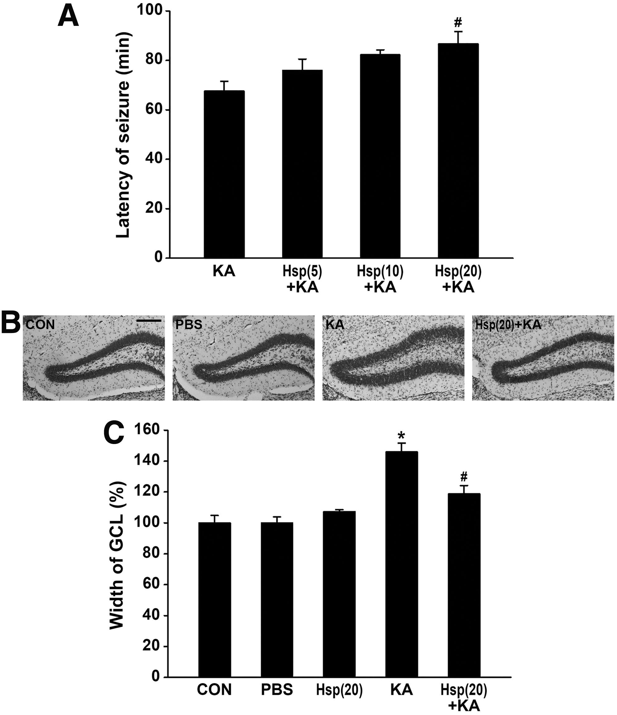
Effect of hesperetin on the seizure latency and GCD in the DG of KA-injected mice.
Next, to clarify whether 20 mg/kg hesperetin alleviates KA-induced mTORC1 activation, we sacrificed the mice 2 days after KA injection and inspected their DG. Figure 2A shows that while p-4E-BP1 expression was increased in the DG treated with KA, it was attenuated in the DG of mice that had received hesperetin before KA administration. In line with the immunostaining data, western blot results suggest that hesperetin significantly decreases both p-4E-BP1 expression (Fig. 2B; **P < .001 and ## P < .01 vs. CON and KA only group) and p-p70S6K levels (Fig. 2B; *P < .05 and # P < .05 vs. CON and KA only group).

Inhibitory effect of hesperetin on KA-induced mammalian target of rapamycin complex 1 activation in the DG.
Neuroinflammation is a natural physiological response to brain disease, and inflammatory processes strongly contribute to the pathology of chronic epilepsy, as well as to the generation of epileptic seizures. 11 Thus, in addition to investigating the beneficial effects of hesperetin on GCD and mTORC1 activity, we used immunofluorescence staining two days post-KA treatment to examine whether it could attenuate the increase of TNF-α, IL-1β, and iNOS produced by activated microglia (Fig. 3A–C). Reduced levels of pro-inflammatory cytokines and iNOS in the hesperetin receiving groups were quantitatively confirmed by western blot analysis (Fig. 3D).
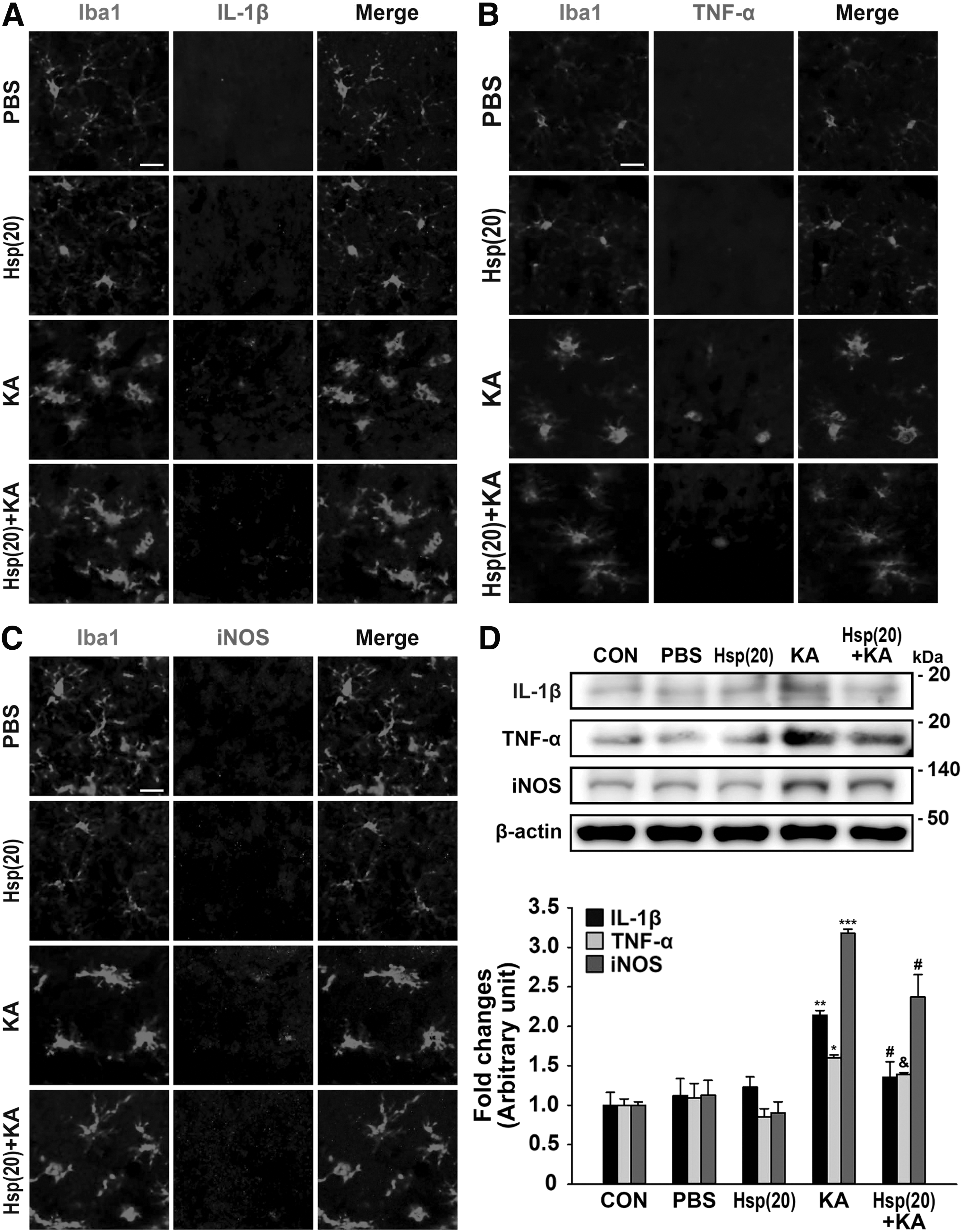
Inhibitory effect of hesperetin on KA-increased levels of pro-inflammatory cytokines and iNOS in the activated microglia..
A previous in vitro study using electrophysiology showed that hesperetin has anticonvulsant effects in convulsant-superfused hippocampal slices. 20 However, the antiepileptic effects of hesperetin had not been investigated in vivo before. In this study, we demonstrated that hesperetin attenuated epileptic progression in a KA-treated animal model of seizure and that it could suppress GCD by inhibiting mTORC1 activation. Thus, our results show that hesperetin has the potential to effectively prevent hippocampal-onset epilepsy in vivo.
Footnotes
Acknowledgments
This work was supported by grants from the Korea Healthcare Technology R&D Project, Ministry of Health and Welfare (HI16C2210), and the National Research Foundation of Korea (NRF-2017R1A2B4002675 and NRF-2017R1D1A1B03031155).
Author Disclosure Statement
No competing financial interests exist.