
Abstract
Sweet olive (Osmanthus fragrans flowers) is used to treat dysentery and reduce phlegm and stasis in traditional Chinese medicine. Recently, we found that verbascoside, the major component in the sweet olive ethanolic extract (OFE), inhibited IL-8 secretion in human colorectal adenocarcinoma WiDr cells. However, evidence-based treatment of inflammatory bowel disease (IBD) with the extract is yet to be performed. To evaluate the therapeutic effect of OFE, we measured IL-8 suppression by OFE and verbascoside in a WiDr cell culture assay. In the IL-8 secretion assay, both OFE (100 μg/mL) and verbascoside (10 μM) significantly inhibited IL-8 production in WiDr cells. Furthermore, we designed cotreated (dextran sulfate sodium [DSS]+OFE-treated) and post-treated (DSS–OFE-treated) protocols to access the therapeutic effects of OFE in vivo. Mice treated with 500 mg/kg per day OFE exhibited significant improvement in IBD symptoms, including disease activity index score, body weight, and colon length maintenance. The suppressive effects on myeloperoxidase expression and lower histopathology scores (including neutrophil infiltration) for the colon were also found. These findings suggest that OFE exerts anti-inflammatory effect on DSS-induced colitis.
Introduction
I
Despite the existence of multiple therapies, there is no effective biomedicine to improve the disease symptoms. Therefore, there is a need for continuing development of safe and effective IBD medications. 8 IL-8 is an important chemoattractant for neutrophils in IBD. 9 Neutrophil recruitment and secretion of myeloperoxidase (MPO) are crucial to understanding the effects of IL-8 on inflammation. IL-8 and MPO can be used as markers to assess neutrophil infiltration in the tissue. 10,11 In the dextran sulfate sodium (DSS)-induced model of IBD, MPO activity is used as an index of inflammation and colitis severity. 12
Sweet olive is the flower of Osmanthus fragrans (Oleaceae family), which is native to regions in Asia extending from the East Himalayas to Japan. 13 It is used as an additive in tea or other beverages and it also used to reduce phlegm production and stasis; alleviate dysentery with blood in the bowel; and treat weakened vision, halitosis, panting, asthma, cough, toothache, stomach ache, and diarrhea in traditional Chinese medicine. 14 Pharmacological studies have shown that sweet olive exhibits antioxidant properties 15 and neuroprotective effects. 16 It also inhibits nitric oxide production 17 and melanogenesis. 18,19 Our previous study showed that sweet olive ethanolic extracts (OFE) possessed significant DPPH scavenging activity and reduced IL-8 secretion in H2O2-induced human colorectal adenocarcinoma WiDr cells through the nuclear factor-κB pathway. 20 However, the therapeutic effect of OFE in IBD is yet to be clarified.
In this study, we first evaluated the IL-8 inhibitory effects of OFE and verbascoside on cotreated (H2O2 with OFE or verbascoside-treated) and post-treated (H2O2-treated, then OFE or verbascoside-treated) WiDr cells in vitro. Furthermore, since Poritz et al. have found loss of ZO-1 and increases in the colonic permeability to Evan's blue by day 3 of DSS treatment in mice, 21 we designed co-treatment (DSS with OFE-treated; DSS+OFE-treated) and post-treatment (DSS-treated, then OFE-treated; DSS–OFE-treated) C57BL/6 mice as an IBD model to evaluate the therapeutic effect of OFE. Herein, we report and compare the disease activity index (DAI) scores, MPO activity, and histopathological scores of two different treatments of OFE. In addition, the toxicity of OFE was also evaluated.
Materials and Methods
Plant materials
Dried O. fragrans flowers were supplied by Chien-Ya Hung of Chung Hwa University of Medical Technology (Tainan, Taiwan) in August 2010. O. fragrans was authenticated by Professor C. S. Kuoh (Department of Biology, National Cheng Kung University). A voucher specimen (National Formosa University, NFU no. 990801) has been deposited in the Herbarium of National Formosa University (Yunlin, Taiwan, R.O.C.).
Preparation of OFE and verbascoside
Dried O. fragrans flowers (4056.76 g) were macerated in 95% ethanol (10 L × 3) at 25°C for 24 h and then extracted with 95% ethanol (10 L × 2) under reflux for 8 h. Evaporation of the solvent under reduced pressure resulted in 1676.70 g of crude extract (OFE, 41.33%). The extract was stored at −20°C until use. OFE was found to be endotoxin-free (≤0.1 EU) in a limulus amebocyte lysate assay.
The OFE crude extract was partitioned between CHCl3–H2O and n-BuOH–H2O successively to yield CHCl3 (OFE-C; 212.10 g, 12.65%), n-BuOH (OFE-B; 898.88 g, 53.61%), and H2O (OFE-W; 565.72 g, 33.74%) layers. The OFE-B layer (708.84 g) was subjected to chromatography on Diaion HP-20 gel using mixtures of H2O and MeOH in decreasing polarity (100% H2O, 90% H2O–10% MeOH, 60% H2O–40% MeOH, 30% H2O–70% MeOH, 10% H2O–90% MeOH, and 100% MeOH, successively) to yield eight fractions, Fr. OFE-B1–B8. Fr. OFE-B3 was subjected to column chromatography over Diaion HP-20 gel to yield verbascoside (208.19 g).
Verbascoside content of OFE
OFE (10.0 mg) was dissolved in methanol (1.0 mL) and the solution was filtered using a 0.22-μm microporous membrane before analysis by high performance liquid chromatography (HPLC). The analysis was done using a Hitachi instrument equipped with an L-7100 series quaternary gradient pump (Hitachi, Tokyo, Japan) and a diode array detector (L-7455), which were linked to a Hitachi LaChrom software data handling system (D-7000 Multi-HSM-Manager). Chromatographic separation was carried out using a Lichro CART® RP-18e column (4.0 × 250 mm; i.d. 5 μm; Merck, Darmstadt, Germany). Reverse-phase HPLC was performed using 0.1% acetic acid (A) and methanol (B) as the mobile phase. The samples were separated using a linear gradient method as follows: 65% A at 0–20 min, 65–55% A at 20–25 min, 55% A at 25–30 min, 55–45% A at 30–35 min, 45–25% A at 35–55 min, 25–0% A at 35–70 min, and 100% A at 70–120 min. The flow rate of the mobile phase was set at 1.0 mL/min and the injection volume was 10 μL. The eluted components were identified by comparing their retention times with those of respective reference standards. The identity of constituents was also confirmed with a photodiode array detector by comparison with UV spectra of standards over wave length 330 nm.
Reagents preparation
A 3% DSS (molecular weight, 36,000–50,000 kDa; MP Biomedicals, USA) drinking solution was prepared daily. OFE (50 or 500 mg/kg per day) was prepared by mixing the extract with 1 × phosphate-buffered saline (1 × PBS). In the cell culture experiment, OFE and verbascoside were dissolved in dimethyl sulfoxide (DMSO; Sigma-Aldrich, USA) at different concentrations. The content of DMSO was <0.1%. In the animal model treatment, OFE (50, or 500 mg/kg per day) was prepared by mixing the extract with 1 × PBS.
Cell culture and viability assay
Human colorectal adenocarcinoma WiDr cells (ATCC® CCL-218™) were purchased from Food Industry Research and Development Institute (Hsinchu, Taiwan). WiDr cells were costimulated with 0.0015% H2O2 and different concentrations of OFE (10, 100, and 200 μg/mL; H2O2+OFE) or verbascoside (2.5, 5, and 10 μM; H2O2+VB) for 12 h. For the poststimulation assay, the cells were treated with 0.0015% H2O2 for 2 h, then different concentrations of OFE (10, 100, and 200 μg/mL; H2O2–OFE) or verbascoside (2.5, 5, and 10 μM; H2O2–VB) were added, for another 10 h incubation. After 12 h incubation period, the supernatants were collected and stored at −20°C until IL-8 secretion analysis. The control group was the cells cultured with medium only, and the positive control group was the H2O2 treatment group. Cell viability was evaluated using the 3-[4, 5-dimethylthiazol-2-yl]-2,5-diphenyl tetrazolium bromide (MTT) assay (Sigma-Aldrich). In brief, the cells were incubated in 2 mg/mL MTT solution for 4 h at 37°C. Next, the MTT solution was removed and 100 μL DMSO (Sigma-Aldrich) was added to each well. Absorbance was measured at 570 nm using an enzyme-linked immunosorbent assay (ELISA) reader (BioTek, USA).
Animals
Female C57BL/6JNarl mice (6–8 weeks old) were purchased from the National Laboratory Animal Center (Taipei, Taiwan). The animals were maintained under a 12 h/12 h light/dark cycle and at a constant temperature (23 ± 2°C) and humidity (58%). Protocols for the care and handling of the animals conformed to the NIH Guide for the Care and Use of Laboratory Animals (National Academies Press, USA; 2011). All of the experiments performed were based on the Biotechnology Department of National Formosa University affidavit of approval of animal use protocol (Animal approval number A104-01).
IBD model establishment and treatment with OFE
To investigate the therapeutic effect of OFE in IBD, we designed two different protocols in the study as shown in Figure 1. For each protocol, mice were divided into five groups (n = 5 mice per group). Mice in the normal group were given access to ddH2O as drinking water, whereas mice in the IBD group were fed 0.03 g/mL DSS in their drinking water for 10 days. In contrast, mice in the vehicle control group were administered 1 × PBS each day. To evaluate daily food uptake (50 mg/kg per day) and consider dosage for therapeutic (500 mg/kg per day) use in the future, mice in experimental groups were treated with different concentrations of OFE (50 or 500 mg/kg per day) by gavage for 10 days in the DSS+OFE-treatment experiment or for 7 days (beginning on day 3) in the DSS–OFE-treatment experiment (day 1 was the first day of treatment with DSS) according to conditions resembling colitis and colon permeability change. 21,22 During the course of the experiment, weight loss, stool consistency, and blood in stool scores for each animal were evaluated to calculate the DAI (Table 1).

Schematic diagrams showing the two treatment strategies. The schedules for the development of the IBD model (3% DSS in drinking water) and oral feeding with OFE began
Disease Activity Index Scores
Histopathological analysis of the colon
On day 10, mice were sacrificed by cervical vertebrae dislocation. In addition, their colons were removed and their lengths were measured. The livers and kidneys were also removed and weighed. The organs were fixed in 10% formalin, embedded in paraffin, cut into 3-μm sections, and stained with hematoxylin-eosin for observation under light microscopy (National Laboratory Animal Center, NARLabs, Taiwan, offered technical support for pathology analysis). The pathology of colon tissue by each section (1 cm from the anus) was scored according to the severity/extent of inflammation and characteristic epithelial lesions. Inflammatory cells infiltrate (severity) was scored as follows: 0 (normal); 1 (1–25%); 2 (26–50%); 3 (51–75%); and 4 (76–100%). The extent of inflammation was scored 0 (normal), 1 (mucosal), 2 (mucosal and submucosal), or 3 (mucosal, submucosal and transmural). Characteristic epithelial lesions were analyzed based on three parameters, which were crypt damage, erosion/ulceration, and hyperplasia, and each scored as follows: 0 (normal); 1 (1–10%); 2 (11–25%); 3 (26–50%); 4 (51–75%); or 5 (76–100%). The values obtained were added to give a maximal histologic score of 22. Moreover, each section was also scored for lesions based on severity and extent of infiltrated neutrophils as follows: severity (score 0: normal; 1: 1–10%; 2: 11–25%; 3: 26–50%; 4: 51–75%; 5: 76–100%) and extent (score 0: normal; 1: mucosal; 2: mucosal and submucosal; 3: mucosal, submucosal, and transmural).
Evaluation of IL-8 and MPO concentrations by ELISA
The concentrations of IL-8 and MPO in colonic lavage fluid were measured using ELISA kits from R&D System (Minneapolis, MN, USA). In brief, 96-well ELISA plates were coated with the capture antibody and incubated at room temperature overnight. The wells were blocked with blocking buffer and 1% bovine serum albumin in 1 × PBS to prevent nonspecific binding. The colonic lavage fluid was added to the wells that were incubated overnight at 4°C. After incubation, the plates were washed with washing buffer (0.05% Tween 20 in PBS, pH 7.2–7.4). The detection antibody was added for 2 h and the plates were washed again with washing buffer. Avidin-horseradish peroxidase was then added and the plates were incubated for 20 min at room temperature before washing. Next, a substrate solution (1:1 mixture of H2O2 and tetramethylbenzidine) was added and the plates were incubated for 20 min at room temperature. Finally, stop solution (2N H2SO4) was added to the plates and the optical density at 450 nm was measured using an ELISA plate reader.
Statistical analysis
All values in the results are expressed as mean ± standard error of the mean. Statistical evaluation of cell viability and IL-8 secretion was performed using Student's t-test with SigmaPlot software (version 10.0; Systat Software, San Jose, CA, USA). In animal experiments, differences between groups were determined using one-way analysis of variance, followed by post hoc Dunnett's multiple comparison test and Mann–Whitney U test. P values <.05 were considered statistically significant.
Results
HPLC fingerprint of verbascoside and OFE
Compound 1 was confirmed to be verbascoside, based on its retention time (37.55 min; Fig. 2A) in the HPLC analysis, as well as its UV absorption at 330 nm. The calibration curve for verbascoside was linear over a concentration range of 0.125–1.0 mg/mL (R 2 = 0.9992). The average recovery of the compound (n = 3) was 98.4%, the relative standard deviation (n = 3) was 2.80%, and the relative percentage difference was 0.10%. Based on quantitative HPLC analysis, the amount of verbascoside (compound 1) in OFE (Fig. 2B) was 22.9%.
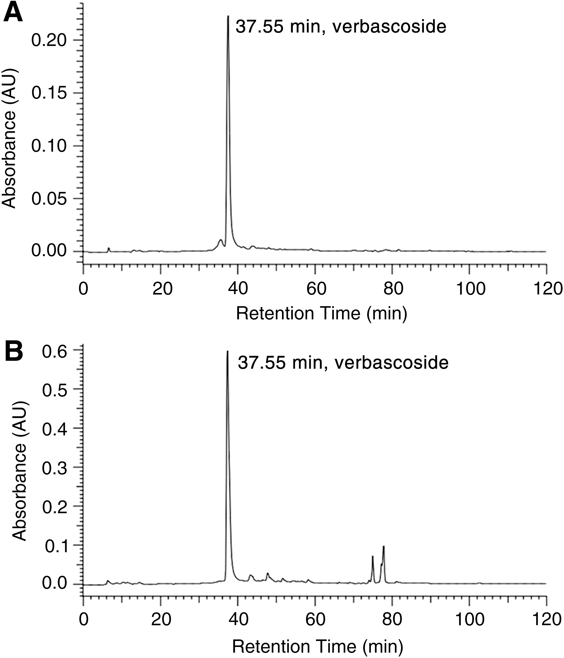
The HPLC spectrum of verbascoside standard
OFE and verbascoside inhibited IL-8 secretion by WiDr cells under oxidative stress conditions
To assess the anti-inflammatory activities of OFE and verbascoside, their effects on the suppression of IL-8 secretion under oxidative stress were assessed. Under H2O2-induced oxidative stress conditions, H2O2+OFE or H2O2–OFE (Fig. 3A, C) or H2O2+VB or H2O2–VB (Fig. 3B, D) showed IL-8 inhibition on WiDr cells at high concentrations. Cytotoxicity was observed only in H2O2+OFE at a high concentration (200 μg/mL) for 12 h (Supplementary Table S1; Supplementary Data are available online at

Inhibition of IL-8 secretion by OFE or verbascoside on WiDr cells under oxidative stress. WiDr cells were seeded in 96-well plates overnight and treated with H2O2 at different concentrations of H2O2+OFE
OFE alleviated loss of body weight and clinical symptoms of IBD in mice
In the DSS+OFE treatment study, oral administration of 500 mg/kg per day OFE to the mice significantly alleviated loss of body weight (Fig. 4A). The improvement proportion for 50 mg/kg per day OFE group was 3% and that for 500 mg/kg per day was 10%. In the DSS–OFE treatment study, 500 mg/kg per day OFE significantly relieved loss of body weight at day 10 (Fig. 4B); the improvement proportion for 500 mg/kg per day was 4%. But 50 mg/kg per day OFE with the DSS–OFE treatment protocol resulted in no significant effect on body weight loss. Both DAI scores of DSS+OFE treatment (Fig. 5A) and DSS–OFE treatment (Fig. 5B) studies were lower after treatment with 500 mg/kg per day OFE than that of 3% DSS. In the DSS+OFE group, the DAI score was improved at day 6. The DAI score was improved at day 10 from 10 of DSS group to 5 of DSS+OFE group (Fig. 5C) significantly. In contrast, the DAI score was only improved at day 7 for the DSS–OFE (500 mg/kg per day) group. The DAI score was improved at day 10 from 10 of DSS group to 6 of DSS–OFE (500 mg/kg per day) group (Fig. 5D) significantly.
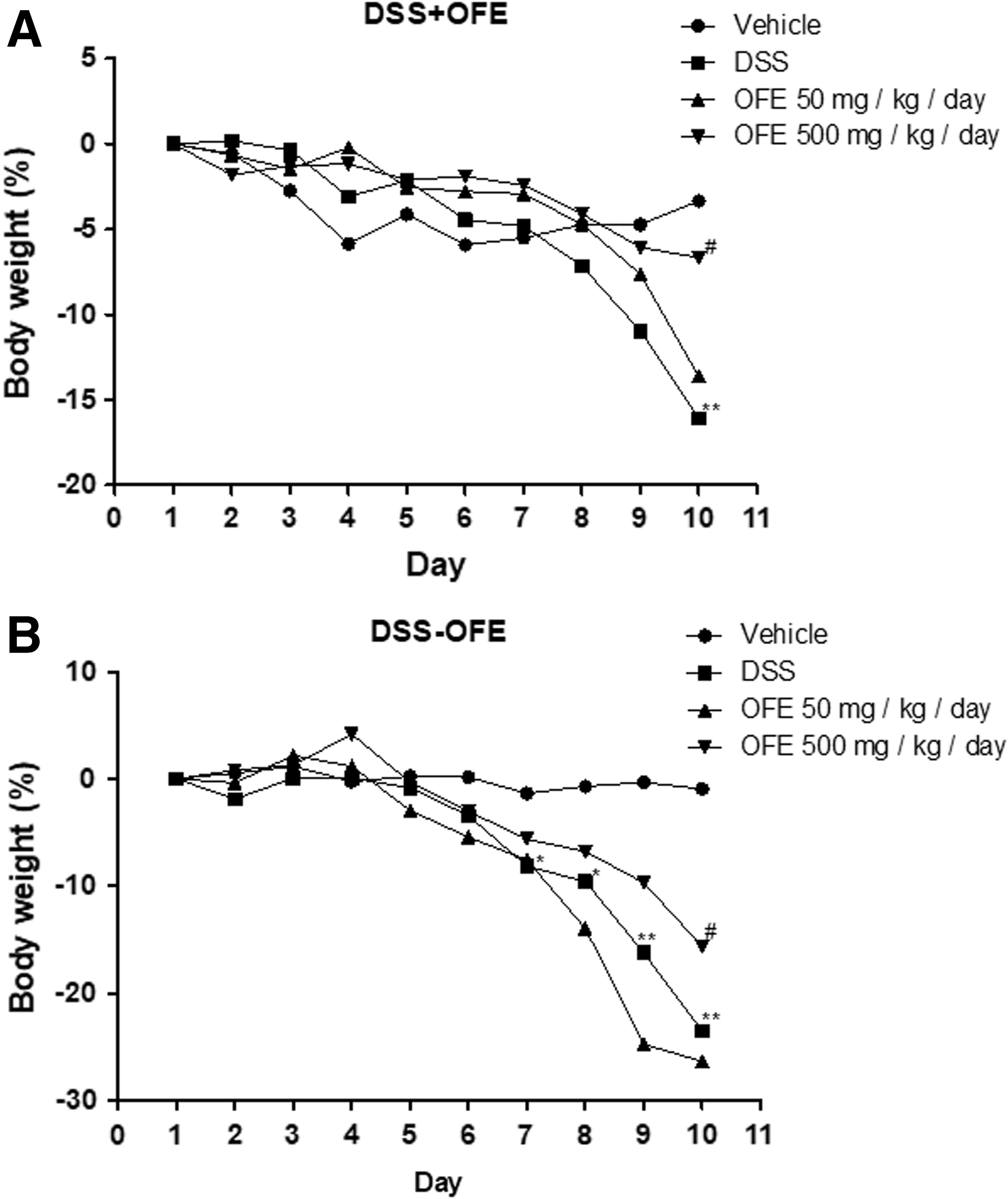
Alleviation of body weight loss by OFE in the mouse model of DSS-induced IBD. Body weight loss of mice in the DSS+OFE treatment

DAI scores after treatment with OFE in the mouse model of DSS-induced IBD. DAI was monitored daily in the DSS+OFE treatment
OFE suppressed the shortening of colon length in mice
Colon length in mice treated with DSS+OFE at 500 mg/kg per day was significantly longer those treated with DSS (Fig. 6). However, OFE did not improve colon length in the DSS–OFE treatment experiment (Fig. 7). From this result, OFE seems to improve the colon length in DSS+OFE-treated mice better than that DSS–OFE-treated mice.

Effects of DSS+OFE treatment on colon length in the IBD mouse model. The representative images of colons from each group

Effect of DSS–OFE treatment on colon length in the mouse model of IBD. The representative images of the colons from each group
OFE alleviated the pathology of IBD in mice
In the normal group, no remarkable lesions were observed in the distal colon or rectum. All the DSS-treated mice developing chronic-active colitis/rectitis showed lesions throughout the mucosa, alteration of epithelial structure, high-level of mononuclear cells, neutrophils, and fibrous tissue infiltrated into mucosa, submucosa and/or muscular area with crypt loss and damage. The incidence and severity of inflammatory and epithelial lesions were lower in DSS+OFE-treated groups at 500 mg/kg per day than in the DSS-treated group (Fig. 8A). In the DSS–OFE treatment experiment, there were no significant differences between the groups in the incidence and severity of inflammatory and epithelial lesions (Fig. 8B). After histopathology scoring by each section, the result showed that OFE at 500 mg/kg per day alleviated the severity of inflammation and inflammatory cell infiltration in the colonic mucosa, as well as reductions in erosion, ulceration, and epithelial damage in DSS+OFE treatment (Fig. 9A) and DSS–OFE treatment (Fig. 9B) groups. According to neutrophil infiltration scoring results (n-score), compared with 3% DSS-treated group in the co-treatment experiment (3% DSS n-score, 4.80 ± 0.20), the average severity scores of neutrophil infiltration revealed mild decrease in the severity in DSS+OFE (500 mg/kg per day)-treated mice (n-score, 4.00 ± 0.45) compared with DSS+OFE (50 mg/kg per day)-treated mice (n-score, 4.80 ± 0.24). In the post-treatment experiment, no alleviation of neutrophil infiltration was observed between DSS–OFE (500 mg/kg per day)-treated mice (n-score of 4.00 ± 0.55) and DSS–OFE (50 mg/kg per day)-treated mice (n-score of 4.75 ± 1.02) when compared with 3% DSS-treated group in the post-treatment experiment (3% DSS n-score, 3.80 ± 0.20). All these results suggest that 500 mg/kg per day OFE (especially in the cotreatment experiment) might attenuate DSS-induced colitis in mice through its anti-inflammatory activity.
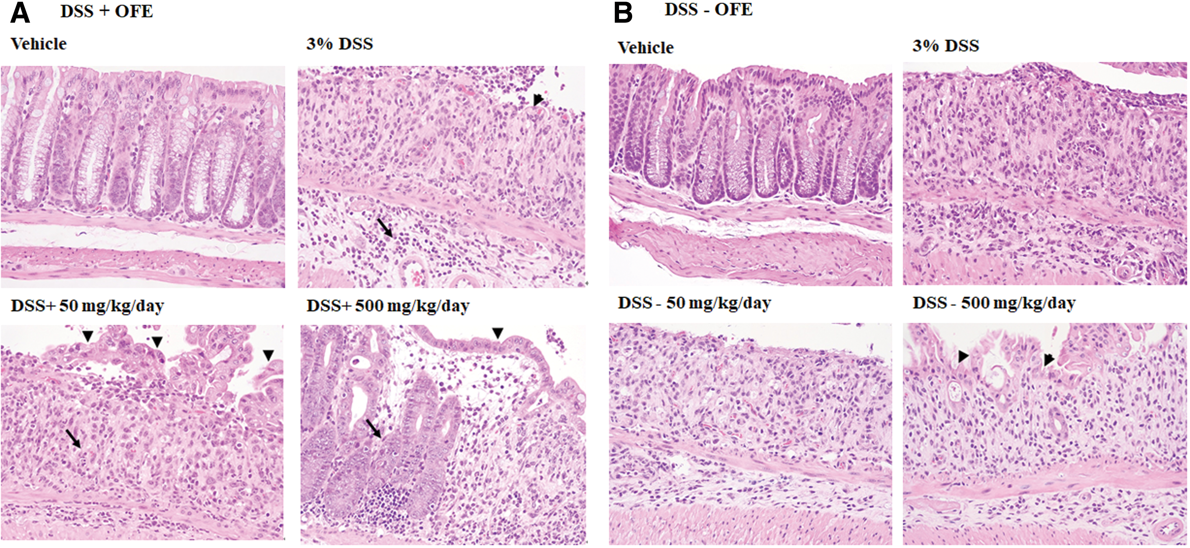
Effect of OFE on colon histopathology in the mouse model of IBD. H&E staining of colon sections from mice in different groups of DSS+OFE treatment
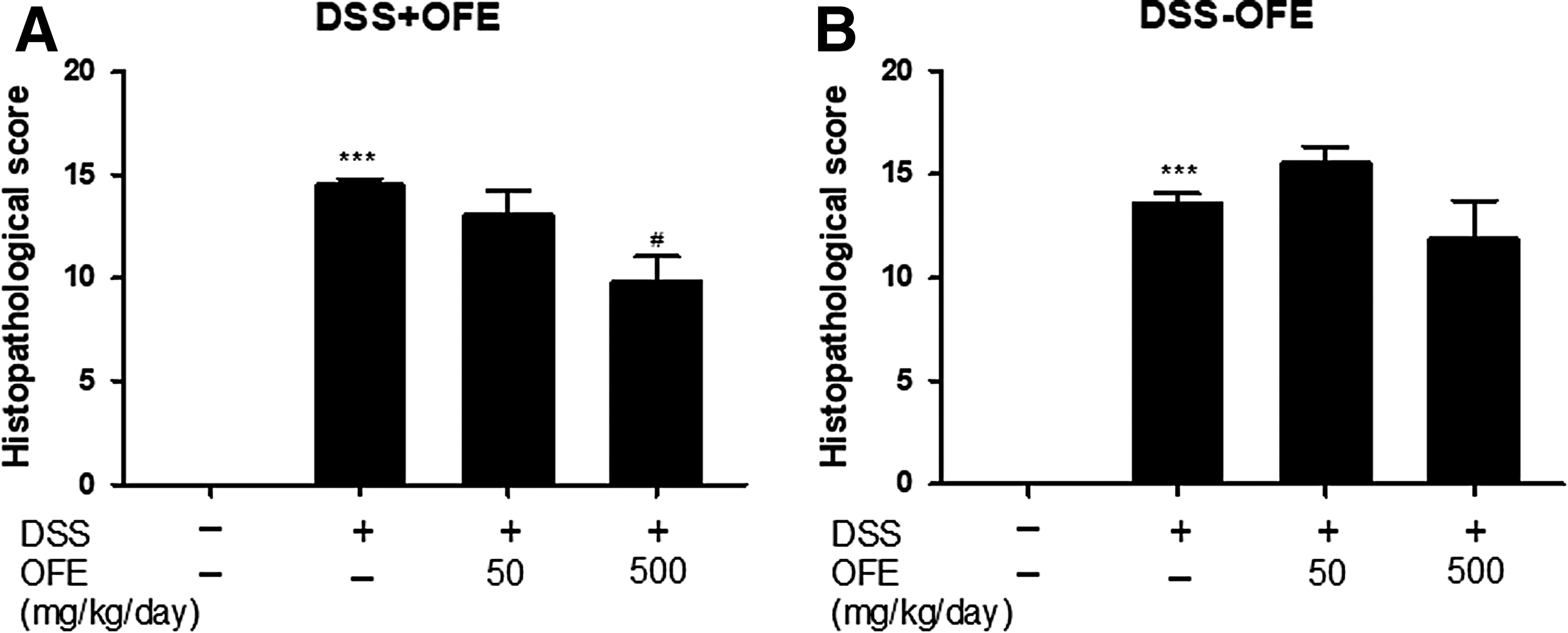
Histopathological scores after treatment with OFE in the DSS+OFE treatment
Effect of OFE on MPO activation in the colonic tissues of mice with DSS-induced colitis
Neutrophil recruitment and secretion of MPO activity are crucial to understanding IL-8 effects in inflammation. We found that MPO activity was dose dependently but not significantly reduced when mice were treated with DSS+OFE at a dose of 500 mg/kg per day (Fig. 10A). However, in the DSS–OFE treatment experiment, we observed an increase in MPO activity in the mice of DSS groups, and a significant decrease in MPO activity in DSS–OFE group at 500 mg/kg per day (Fig. 10B).
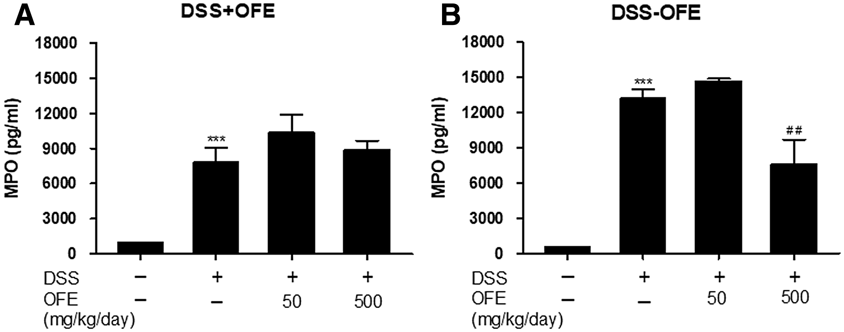
Effect of OFE on MPO activity in DSS-induced colitis. MPO activity in colonic lavage fluid was evaluated by ELISA in the DSS+OFE treatment
Effects of OFE on changes in liver and kidney weights in the mouse model of IBD
The study of Dong et al. 23 reported that DSS-induced acute colitis in mice led to colon, kidney, and liver damage; therefore, we evaluated the liver and kidney weight of mice in this study. In the DSS+OFE treatment groups, OFE at a dose of 500 mg/kg per day improved loss of liver weight significantly (Fig. 11A). In contrast, the weight of livers of DSS–OFE (500 mg/kg per day)-treated mice was heavier than those of DSS-treated mice; however, this difference was not statistically significant (Fig. 11B). In this study, we also evaluated the improvement in kidney weight loss by OFE, but no improvement has been made in co-treatment and post-treatment experiments (data not shown).
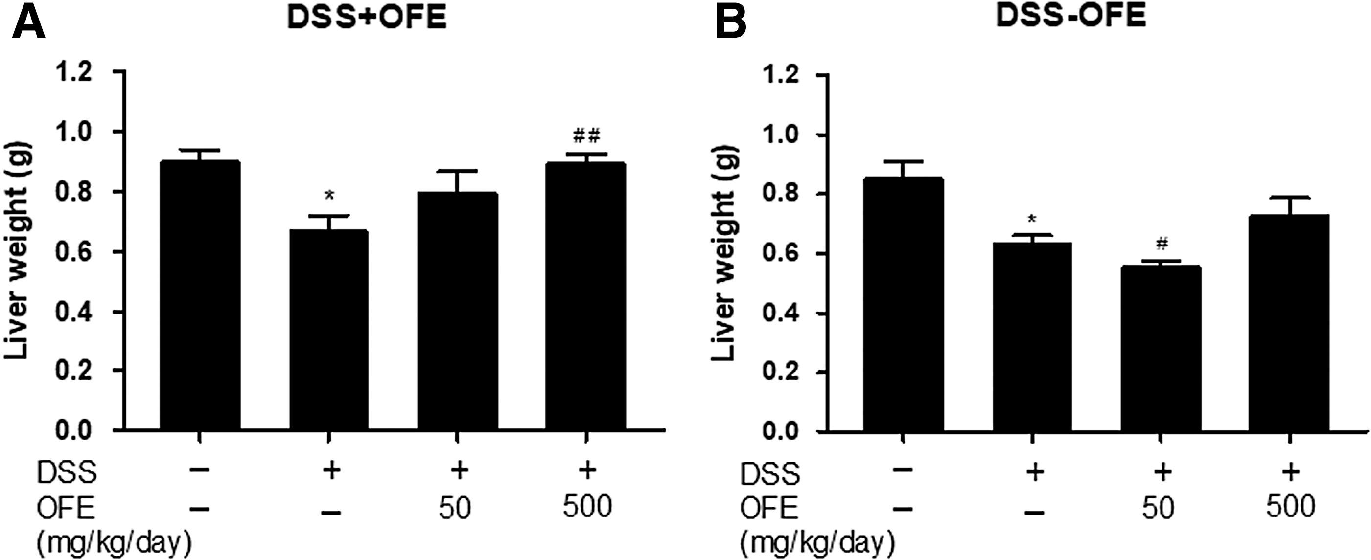
Effects of OFE on liver weight in DSS-induced colitis in the DSS+OFE treatment
Discussion
Until now, accelerating incidence of IBD in newly industrialized countries and westernized societies has been reported. 24 Also, IBD patients have a higher risk for colorectal cancer. 25 Herbal medicines for IBD therapy have been reported. 26 But the evaluation of IL-8 suppression activity for IBD therapy by herbal extracts is rare. IL-8 is an important neutrophil chemoattractant and activator for IBD patients. 9 It is also an important cytokine for angiogenesis of IBD. 27 Lee et al. reported that treatment of HT-29 cells with TNF-α significantly increased MCP-1 and IL-8, but they were significantly suppressed by treatment with Grifola frondosa (GFW). Also, oral administration with GFW for 5 days (1 g/kg per day) reversed the symptoms of colitis in 2,4,6-trinitrobenzene sulfonic acid-induced experimental rat colitis. 28 In order to mimic local damage caused by oxidative stress, we treated colonic epithelial WiDr cells with H2O2 in our study. Also, the IBD mouse model was used to evaluate the anti-inflammatory activity of OFE. The results suggest that OFE ameliorates colon inflammation by suppressing IL-8 and MPO. We designed two OFE treatment protocols for the DSS-induced colitis model. OFE prevented the loss of body weight and reduced the severity of bowel inflammation. DAI scores, MPO activity, and histopathological scores were improved in DSS-induced colitis at an optimal dose of 500 mg/kg per day in both experiments (Table 2). The therapeutic effects of OFE were better at 500 mg/kg per day than at lower doses. Moreover, OFE alleviated the severity of bowel inflammation and reduced inflammatory cell infiltration in the colonic mucosa, as well as the degree and extent of epithelial damage. The liver improvements observed in the study of Lu et al. 29 using 80% O. fragrans flower ethanolic extract (phenylethanoid glycoside-rich) were not associated with any acute lethal effect at 10 g/kg body weight in either rats or mice. Also, in the 90-day toxicity assay in rats (the highest dosage, 2.00 g/kg), the extract failed to induce any significant hematological, clinical, chemical, or histopathological organ changes (including liver). For this reason, 500 mg/kg per day, as used in our study, probably did not induce liver toxicity in mice. The preventive and therapeutic effects of OFE on the liver after long-term DSS-induced colitis should be further investigated in the future. This study is the first report to clarify the therapeutic effects of OFE in an IBD model. We provide supportive evidence for the use of OFE, which has been used in functional food as well as in medicine.
Treatment Effects
A: 500 mg/kg treatment effect better than 50 mg/kg treatment effect.
Significant improvement: * P < .05, ** P < .01, and *** P < .001 compared with DSS-treated group.
DAI, disease activity index; DSS, dextran sulfate sodium; MPO, myeloperoxidase; OFE, sweet olive ethanolic extract.
Footnotes
Acknowledgments
Financial support for this study was provided by the Ministry of Science and Technology of Taiwan, R.O.C. (grant number Most 103-2320-B-150-001). The funding source had no role in the study design; in the collection, analysis, and interpretation of data; in the writing of the report; and in the decision to submit the article for publication. We thank Editage (
Author Disclosure Statement
No competing financial interests exist.
References
Supplementary Material
Please find the following supplemental material available below.
For Open Access articles published under a Creative Commons License, all supplemental material carries the same license as the article it is associated with.
For non-Open Access articles published, all supplemental material carries a non-exclusive license, and permission requests for re-use of supplemental material or any part of supplemental material shall be sent directly to the copyright owner as specified in the copyright notice associated with the article.