
Abstract
Chronic obstructive pulmonary disease (COPD), including pulmonary emphysema and chronic bronchitis as well as structural and inflammatory changes in small airways, is insensitive to corticosteroid therapies. This study aimed to evaluate the effects of Scutellaria baicalensis root extract (SB_E) in a mouse model of COPD. The COPD mouse model was produced by challenging C57BL/6 mice with a cigarette smoke extract and lipopolysaccharide (LPS). SB_E significantly decreased the neutrophil counts in blood and bronchoalveolar lavage fluid (BALF), and the production of tumor necrosis factor (TNF)-α, interleukin (IL)-17A, macrophage inflammatory protein 2 (MIP2), and chemokine (C–X–C motif) ligand 1 (CXCL-1) in BALF, and TNF-α mRNA expression in lung tissue. The histological lung injury was also alleviated by treatment with SB_E. Thus, SB_E effectively inhibited airway inflammation by regulating the expression of inflammatory cytokines by blocking MIP2 and CXCL-1 secretion. Therefore, S. baicalensis may be a potential therapeutic agent for COPD.
Introduction
Chronic obstructive pulmonary disease (COPD), a major pulmonary disease characterized by lung inflammation and increased difficulty in breathing, is ranked as one of the top four high-mortality diseases worldwide. 1,2 Currently, there are no direct therapies to preserve the lung functions of patients with COPD. Pharmacological approaches are limited to relieving symptoms and attenuating lung disease exacerbation. 1 COPD alveolar macrophages produce neutrophil chemoattractant. In addition, activation of neutrophils increases their migration into sites of lung injury, and chemokine (C–X–C motif) ligand 1 (CXCL1) acts solely through CXCR2/macrophage inflammatory protein 2 (MIP2) to promote the influx of neutrophils into tissue sites of inflammation. 3 Bronchodilators, which are the main drugs for conventional therapy, have a relatively high risk of side effects for patients with COPD, who are mostly elderly and likely to have other associated diseases. 4 The lack of highly effective treatments and potential side effects of current therapies pose a significant challenge to long-term management of COPD. Presently, steroids are widely used for the treatment of asthma, but there is not enough evidence to prove that they prevent the long-term progression of COPD. 5 Although steroids are used to treat COPD, they are not as effective as those used to treat asthma and the effect is somewhat limited. 6 Herbal medicines and natural compounds play an important role in both preventive and curative treatments in traditional systems of medicine, despite advances in modern western medicine. Although numerous modern medicines have been identified for the treatment of COPD, they are associated with various drawbacks such as lack of efficacy, excessive side effects, and high cost. Herbal medicines and their natural components are known to often be more effective and have fewer side effects than conventional medicines based on numerous reports.
Scutellariae Radix is the dried root of Scutellaria baicalensis and is one of the most popular and multipurpose herbal medicinal plants used in Asia, including China, Japan, and Korea, to treat allergy, inflammation, and infection. 7 –9 Investigations have shown that Scutellariae Radix has beneficial properties such as antioxidant effects 10 and inhibition of the anti-dinitrophenyl IgE-mediated anaphylactic reaction compound 48/80-induced histamine release and calcium uptake into rat peritoneal mast cells. 9,11 A previous study reported that wogonin, a flavonoid-like compound found in S. baicalensis, inhibited the production of several inflammatory cytokines, including tumor necrosis factor (TNF)-α, interleukin (IL)-1b, and IL-6, in the bronchoalveolar lavage fluid (BALF) and lung tissues after lipopolysaccharide (LPS) challenge. 12 In this study, we evaluated the immunotherapeutic potential of S. baicalensis root extract (SB_E) to prevent the cigarette smoking response in a COPD mouse model. We then assessed the ability of SB_E to modulate cigarette smoke (CS) exposure-induced airway inflammation and the mechanism underlying its modulation of the immune system.
Materials and Methods
Preparation of SB extract
Roots of S. baicalensis were obtained from Human Herb Co., (Kyeongbuk, Korea), a licensed herb company, and were identified by Professor J. G. Oh (College of Korean Medicine, Daejeon University, Daejeon, Korea). SB_E was prepared by boiling the herb roots in distilled water at 100°C for 2.5 h. The mixture was then filtered through a Whatman No. 2 filter (Maidstone, United Kingdom), concentrated under vacuum, and freeze-dried. The yield of the dried extract was 22%.
Identification of SB_E constituents using high-performance liquid chromatography
All high-performance liquid chromatography (HPLC) grade reagents, formic acid, and water were obtained from J.T. Baker (Phillipsburg, NJ, USA). The samples were analyzed using reverse-phase HPLC using an ACQUITY ultra-performance liquid chromatography system (Waters Co., Milford, MA, USA). A BEH column (1.7 μm, 2.1 × 100 μm) was used as the stationary phase. The mobile phase was composed of a 0.1% (v/v) aqueous solution of formic acid (A) and acetonitrile (B). The separation temperature was maintained at 35°C throughout the experiment at a flow rate of 0.4 mL/min, and the peaks were detected at 210 nm. The injection volume was 5 μL. Sample peaks were assigned according to the retention time and ultraviolet (UV) spectra of four standard compounds in the chromatogram.
Animal experiments
Male BALB/c (6–8-week-old) mice were obtained from the Orient Bio, Inc. (Seongnam, Korea) and housed under pathogen-free conditions. We selected BALB/c mice for our study rather than other mouse strains such as C57BL/6 because they are susceptible to respiratory ailments. 13 All animals were allowed to acclimate for 1 week before use. The animal protocol was approved by the Committee for Animal Welfare at Daejeon University (DJUARB2016-009). This study was performed in strict accordance with the Guide for the Care and Use of Laboratory Animals of the National Institute of Health. All animal procedures were conducted in accordance with the guidelines of the Institutional Animal Care and Use Committee of the South Korea Research Institute of Bioscience and Biotechnology (Daejeon, Korea).
Preparation of CS extract
CM6 (CORESTA approved Monitor No. 6) reference cigarettes were conditioned under International Organization for Standardization (ISO) conditions (1 puff/min, 35 mL puff volume over 2 s, every 60 s) of temperature (22°C ± 2°C) and relative humidity (60% ± 5%) for 48 h or more. The cigarettes were smoked according to ISO conditions (puff volume: 35 mL, duration: 60 s, interval: 2 s) using an automatic smoking machine (Borgwaldt RM20, Germany). The particulate matter was captured using a Cambridge filter pad (0.22 μm, Ø4 mm) and eluted with isopropanol as the solvent. Total particulate matter (TPM) was prepared with enough mass to provide sufficient test article for the entire 2-year study period. Both TPMs were stored at −80°C and were tested immediately after preparation (T0). After 1 (T1) and 3 (T3) months, the stored TPMs were tested under the same assay conditions and compared with freshly prepared TPM. 14
Establishment of COPD mouse model and SB_E administration
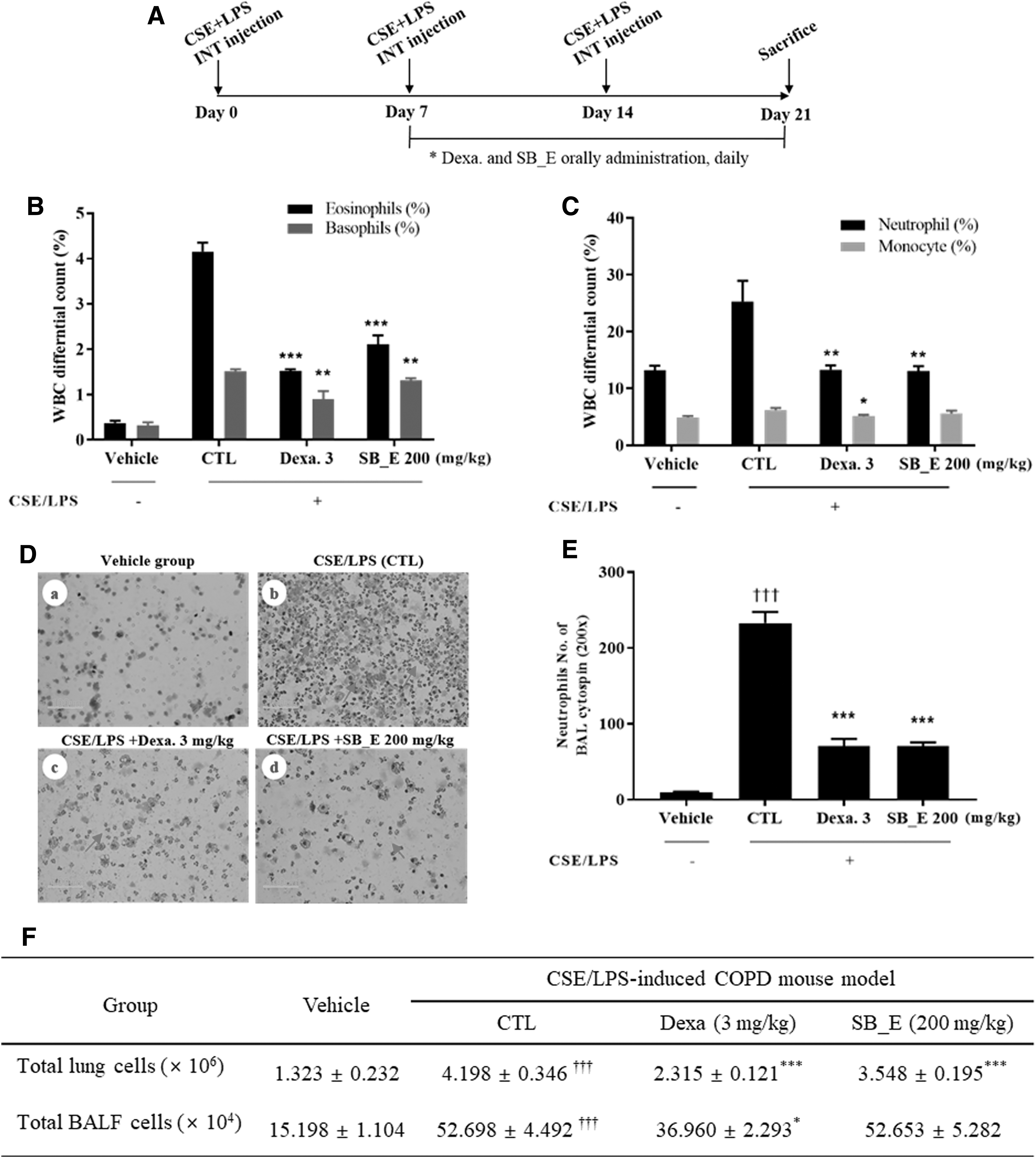
A COPD model was established by exposing mice to the CS extract (CSE) plus LPS (Fig. 2A). In brief, mice were administered LPS (Sigma-Aldrich, St. Louis, MO, USA) or saline (vehicle group) by intranasal injection. COPD was induced by intranasal instillation of LPS and CSE according to a method described by Mizutani et al. 15 The mice were randomly divided into the following four groups: vehicle (no treatment), control (CTL, CSE/LPS), dexamethasone (CSE/LPS + Dexa), and SB_E (CSE/LPS + SB_E) groups. The mice were administered a single-dose intratracheal instillation of SB_E or vehicle, and the control group was administered saline using bronchial tubes. Dexa was administered as a positive control at a dose of 3 mg/kg, and SB_E was administered to mice orally at a dose of 200 mg/kg for 14 days. After intraperitoneally (i.p.) injecting an anesthetic, LPS (100 μg/mL) and CSE (1 mg/mL) were administered by intratracheal injection three times with a 7-day interval. Mice were anesthetized by i.p. injections of ketamine hydrochloride (Yuhan, Korea) and xylazine hydrochloride (Rompun®, Bayer, Korea). The general condition of all the mice was observed daily, including the physical appearance, behavior, hair condition, liveliness, sensitivity, and respiratory murmur.
Collection of BALF
To separate BALF from the trachea and lungs, a syringe was inserted into the trachea and airways of the anesthetized mice, and the fluid was aspirated. The trachea was cannulated, and the left bronchus was tied for a histological experiment. The BALF supernatant was stored at −25°C for the determination of cytokines, and we also examined the accumulation of neutrophils in the BALF.
Flow cytometric analysis
The antibodies against CD4 (RM4–5, rat IgG2a), CD8 (53–6.7, rat IgG2a), CD69 (H1.2F3, rat IgG), and granulocytic marker GR1 (RB6–8C5, rat IgG2b) used for the flow cytometric analysis were purchased from Becton Dickinson Pharmingen (San Diego, CA, USA). Cells from the lungs and BALF were incubated with fluorescein isothiocyanate- and phycoerythrin-labeled monoclonal antibodies for 30 min, washed with phosphate-buffered saline (PBS), fixed with 0.5% paraformaldehyde for 20 min, washed again with PBS, and then stored at 4°C until the analysis using two-color flow cytometry with a Fluorescence-activated cell sorting (FACS) Calibur™ instrument (BD Biosciences, Mountain View, CA, USA).
Enzyme-linked immunosorbent assays of inflammatory mediators
The trachea was cannulated, and BALF samples were obtained by washing the airway lumina, and the BALF supernatant was stored at −20°C for determination of cytokine levels. The levels of MIP2, TNF-α, IL-17A, and CXCL-1 in BALF were determined using enzyme-linked immunosorbent assay (ELISA) kits according to the manufacturer's instructions (R&D Systems, Minneapolis, MN, USA).
Isolation of total RNA from tissue and quantitative real-time polymerase chain reaction
To study the anti-COPD effects of SB_E on cytokine gene expression in lung cells, quantitative real-time polymerase chain reaction (qPCR) was performed. Total cellular RNA was extracted from the lung tissue using a phenol–chloroform-based method (RNAsolB; Tel-Test Co., Inc., Friendswood, TX, USA) according to the manufacturer's instructions. Real-time qPCR was performed using an Applied Biosystems 7500 Fast real-time PCR system in the manual mode (threshold: 0.05, baseline: 6–15 cycles). Gene expression was analyzed using a SYBR Green PCR Master Mix (Applied Biosystems, Grand Island, NY, USA) at a final primer concentration of 200 nM, using β-actin as the endogenous control. The following PCR conditions were used: 2 min at 50°C, 10 min at 94°C, 40 cycles of 1 min at 94°C, and 1 min at 60°C. The primer sequences used for quantitative real-time PCR were as follows: mouse IL-1β: 5′-TGATGGCTTATTACAGTGGCAATG-3′ and 5′-GTAGTGGTGGTCGGAGATTCG-3′; mouse IL-6: 5′-GTCTTGCCTGCTGCCTTC-3′ and 5′-AGTGCCTCTTTGCTGCTTTC-3′; mouse TNF-α: 5′-CGTCAGCCGATTTGCTATCT-3′ and 5′-CGGACTCCGCAAAGTCTAAG-3′; mouse mucin 5AC (MUC5AC): 5′-AGAATATCTTTCAGGACCCCTGCT-3′ and 5′-CACCAGTGCTGAGCATACTTTT-3′; and mouse glyceraldehyde-3-phosphate dehydrogenase (G3PDH): 5′-CATGTTCCAGTATGACTCCACTCACG-3′ (VIC).
Lung histopathological study
Mice were perfused with saline, and the whole lungs were inflated with fixative. The lung tissue was fixed in a 10% formaldehyde solution for 24 h, soaked in water for 8 h, and then dehydrated and embedded in paraffin using standard methods. Tissue sections (5-μm thick) were stained with hematoxylin and eosin (H&E) and Masson's trichrome (Sigma-Aldrich, Korea). Three separate H&E-stained sections were evaluated for each mouse using light microscopy at 100 × magnification. To determine the severity of inflammatory cell infiltration, peribronchial cells, mucus production, and goblet cell hyperplasia were quantified in the airway epithelium in a blinded manner using the five-point (0–4) grading system described by Tanaka et al. 16 The scores were as follows: 0, no cells; 1, a few cells; 2, ring of cells two cell layers deep; 3, ring of cells two–four cell layers deep; and 4, ring of cells four cell layers deep.
Statistical analysis
Data are expressed as the mean ± standard error of the mean (SEM). The data were analyzed using a one-way analysis of variance (ANOVA), and statistical significance of differences between groups was calculated using a nonparametric Mann–Whitney test, followed by Dunnett's multiple comparison test (SPSS version 19.0; IBM, USA). A P < .05 was considered statistically significant.
Results and Discussion
HPLC analysis
Four major constituents were identified in SB_E by comparing retention times and UV absorbance of major peaks, using reverse-phase HPLC chromatography, with those of commercial standards. The SB_E chromatographic profile contained peaks with retention times of 6–11 min. Ion chromatogram analysis of SB_E revealed the presence of baicalin, oroxylin A 7-O-glucuronide, baicalein, and wogonin (Fig. 1). S. baicalensis is one of the constituents of traditional medicine prescriptions. In this experiment, boiling water was used as a traditional extraction method. As a result, hydrophobic phenolic compounds were detected. These components are nonpolar but are not completely similar to oils, and they are not completely soluble in water. However, as shown in Figure 1, very small amounts were soluble in boiling water.
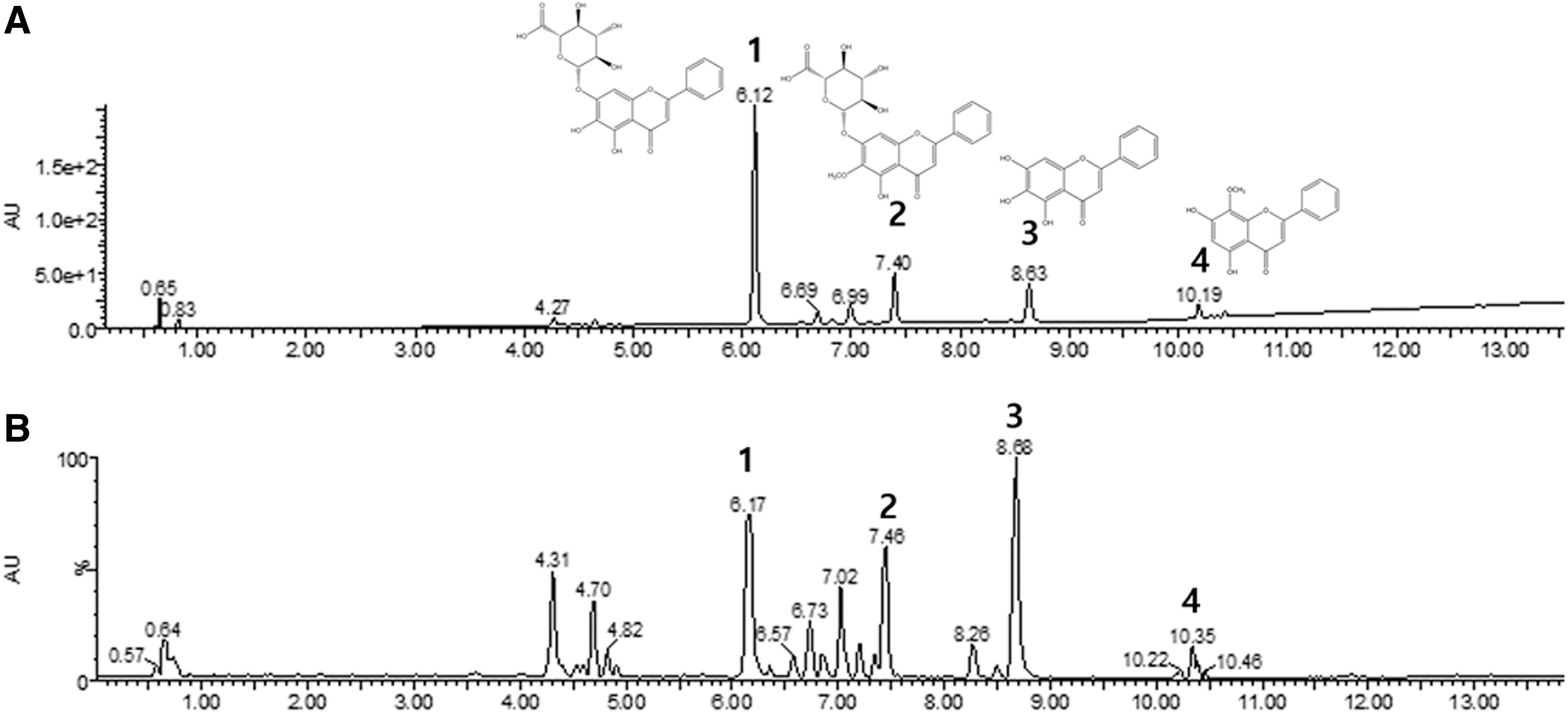
HPLC chromatograms of a mixture of four standards (
Inhibitory effect of SB_E on airway neutrophil accumulation in BALF of COPD-induced mice
Changes in the cell types during airway inflammation in asthmatic mice, particularly neutrophils, are an important course in the development and pathogenesis of COPD. 17 Neutrophil, eosinophil, and basophil numbers were reduced in the blood of the SB_E-treated mice compared to those in the control group (Fig. 2B, C). To evaluate the effects of SB_E and Dexa on the recruitment of cells to the airway, we investigated neutrophils in the BALF. The number of neutrophils was significantly higher in the BALF cytospin of the COPD-induced mice than in that of the control group. However, the absolute number of neutrophils in the BALF decreased in the SB_E- and Dexa-treated groups (Fig. 2D, E). The changes in airway eosinophil accumulation and influx of inflammatory cells into the lung and BALF in COPD mice with or without intratracheal instillation of CSE/LPS for 21 days are shown in Figure 2F. The number of total leukocytes in the BALF from the SB_E-treated group was significantly lower than that in the BALF from the CSE/LPS-induced group (Fig. 2F). In the present study, we investigated the potential of an orally administered herbal medicine, SB_E, for the treatment of CSE/LPS-induced COPD by inhibiting inflammation. According to the definition of the Global Initiative for Chronic Obstructive Lung Disease, COPD is a complex chronic airway disease characterized by persistent airflow limitation, which is usually progressive. 18 This disease is now the third leading cause of death worldwide. 19 The worldwide morbidity and mortality of COPD is expected to increase in the coming decades and, therefore, COPD poses a significant burden on the economy and society. 20 The strongest and most common risk factor for COPD is exposure to CS, 21 which leads to alveolar wall destruction with airspace enlargement, one of the major pathologies of COPD. 18,22 It is thought that CS-induced inflammation, 23 apoptosis, 24 and oxidative stress, 25,26 are the major drivers of emphysema. Neutrophils are key mediators of COPD as they migrate to the airway under control of chemotactic factors and become activated. 27,28 In addition, increased numbers of neutrophils in the airway lumen and BALF in individuals with COPD are correlated with the disease severity. 29,30 The COPD model used in this study involved the inhalation of LPS to help induce emphysematous changes. 31,32
Inhibition of airway inflammation by SB_E in a CSE/LPS-induced COPD mouse model. (
Effects of SB_E on lung inflammation in COPD-induced mice
The lungs of the COPD mice showed histopathological features typical of inflammatory lung tissues, such as distinct infiltration of leukocytes, goblet cell hyperplasia, and erosion in peribronchial and perivascular areas. Infiltrating inflammatory cells such as neutrophils, eosinophils, and basophils were mainly observed in the peribronchial regions of the lung. In contrast, histological sections from the oral Dexa- and oral SB_E-treated mice revealed reduced airway inflammation in lung tissues (Fig. 3). SB_E treatment markedly attenuated eosinophil-rich leukocyte infiltration when compared to that of the CSA/LPS-induced mice (Fig. 3A, panels b and f, panels d and h, Fig. 3B).
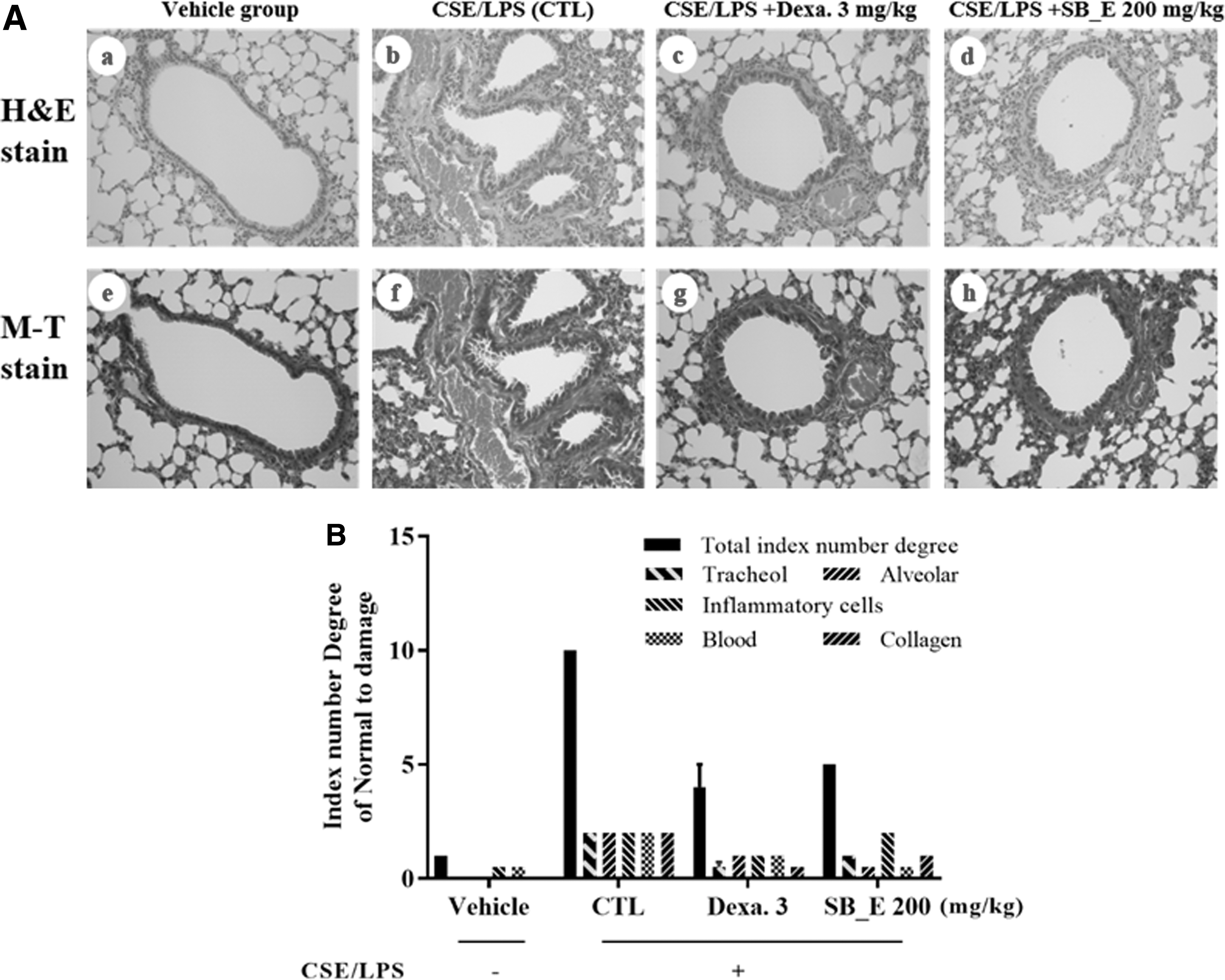
Effects of SB_E on histopathological changes and histology scores in the lung of a CSE/LPS-induced murine model of chronic obstructive pulmonary disease. Untreated mice received the vehicle, whereas model mice were induced by intratracheal injection of CSE/LPS and then treated with the vehicle (CTL), Dexa (3 mg/kg), or SB_E (200 mg/kg) for 21 days. Lungs were fixed, sectioned, and stained with H&E and Masson's trichrome (M-T) stains. (
Inhibitory effects of SB_E on inflammatory cytokines in BALF of COPD-induced mice
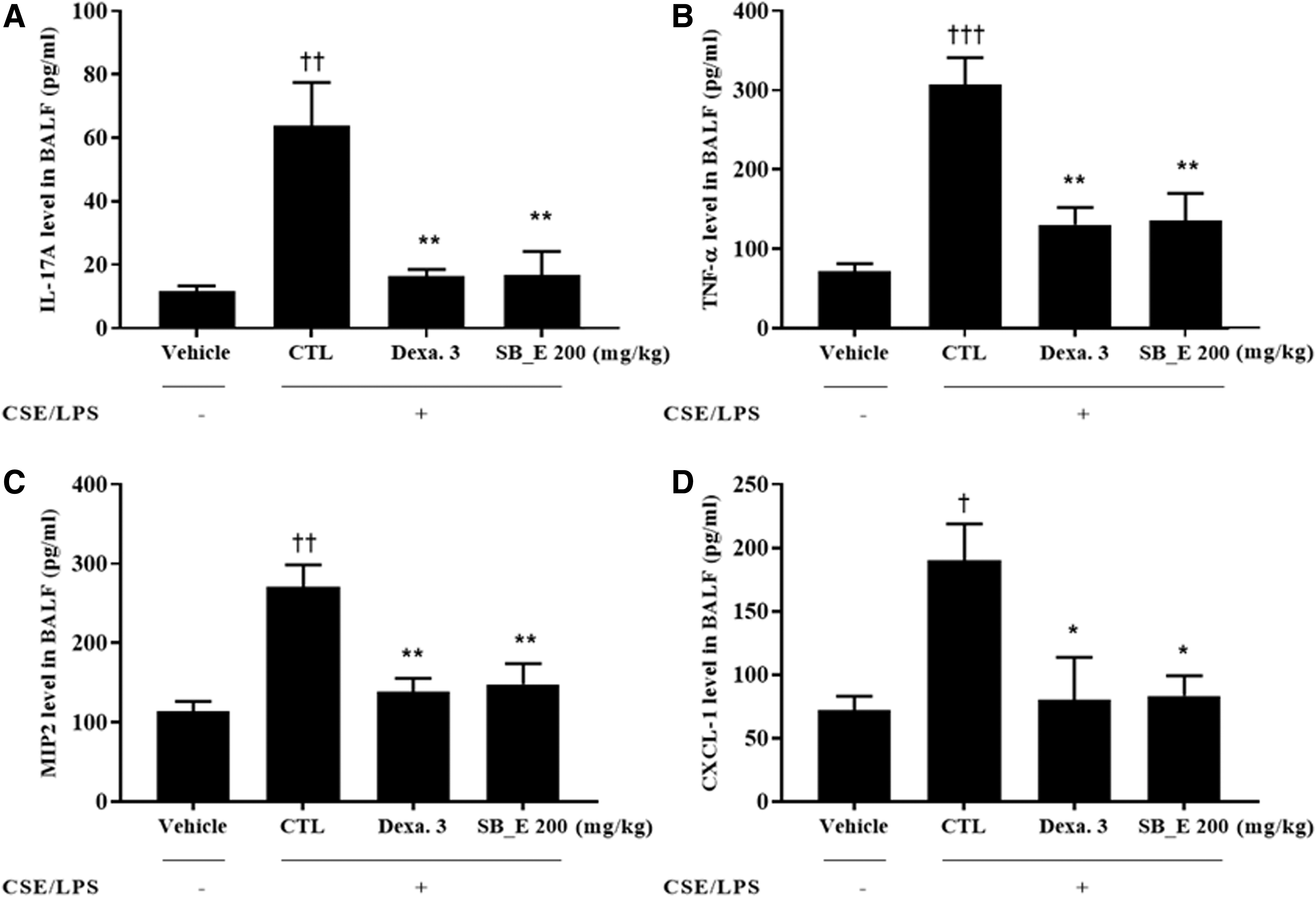
A previous study showed that in a T helper 2 (Th2) environment, continuous production of eosinophils occurs in a mouse model of allergic inflammation. 33 However, the T-cell diversity has been expanded to several subpopulations, including Th17 cells, suggesting that the mechanism is more complicated. 34 Interestingly, cotransfer of Th17 and Th2 cells enhanced the expression of eotaxin-1 in the lung, whereas neutralization of eotaxin-1 before challenge with an inhaled antigen decreased eosinophil recruitment into the airways of mice inoculated with a combination of Th2 and Th17 cells. 35 We measured the levels of COPD-accompanying cytokines in BALF to determine the mechanisms underlying the SB_E-mediated inhibition of airway inflammation. In animals exposed to LPS and CSE, the levels of TNF-α, IL-17A, MIP2, and CXCL-1 in BALF were higher than those in the untreated group. However, administration of SB_E significantly suppressed the increases in TNF-α, IL-17A, MIP2, and CXCL-1 in BALF compared with those of the COPD-induced untreated mice (Fig. 4). Increased expression of CXCL-1, CXCL-2, and their receptor CXCR2 following in vitro exposure to CSE suggests that they play pivotal roles by, at least in part, attracting inflammatory cells. 36 Chemotactic signals for neutrophil recruitment include leukotriene B4, CXCL-1 (previously known as growth-related oncogene-α), and chemokine (C–X–C motif) ligand 8 (CXCL-8), whose expression increases in COPD and is likely derived from alveolar macrophages and epithelial cells. 37 IIL-17A plays a functional role in airway remodeling, mucus hypersecretion, and acute exacerbation in response to COPD-associated inflammatory stimuli. 38 –40 IL-17 enhances the release of IL-1β-mediated CXCL-8 from human airway smooth muscle cells. 41 In a mouse model, SB_E prevented lung and airway inflammation and decreased the levels of Th2 cell-type cytokines in the BALF. Therefore, SB_E effectively inhibited airway inflammation by regulating the expression of inflammatory cytokines through blockade of MIP2 and CXCL-1 secretion.
Effects of SB_E on IL-17A
Inhibitory effects of SB_E on mRNA expression of inflammatory cytokines in lung tissue of COPD-induced mice
The COPD-induced mice showed increased expression of the genes encoding IL-1β, IL-6, TNF-α, and MUC5AC in lung tissues compared with that in the vehicle group. Administration of SB_E significantly suppressed TNF-α expression compared with that in the control group (Fig. 5). IL-1β, IL-6, and MUC5AC showed a tendency to decrease in the SB_E-treated group, but the difference was not significant. Our results showed that SB_E inhibited lung inflammatory responses in the CSE/LPS-induced COPD model. TNF-α and IL-1β have long been known to be classical proinflammatory cytokines contributing to the development of COPD. 42 –44 IL-6 is stimulated by TNF-α and IL-1β and plays a critical role in the pathogenesis of emphysematous changes. 45 Therefore, inhibition of proinflammatory cytokines is one of the most promising approaches for treating COPD. 37

Expression of IL-1β
Inhibitory effects of SB_E on absolute numbers of immune cell subtypes in BALF and lungs of COPD-induced mice
Flow cytometric analysis was used to evaluate the effects of SB_E on immune cell subtypes. The numbers of CD4+, CD8+, CD69+, and CD11b+/GR1+ cells in the BALF and lungs of the COPD-induced mice were higher than those in the untreated group, but were significantly lower in the SB_E-treated mice than in the control mice (Table 1). Thus, SB_E profoundly inhibited airway inflammation in the mouse model of COPD, and these effects were caused by suppression of Th2-type cytokines and neutrophil infiltration. Therefore, we propose that SB_E could be used for the treatment of pathologic inflammatory airway disorders such as COPD. These findings enhance our understanding of the pathogenesis of COPD and, thus, may have important implications in the clinical treatment of COPD. Four reference phytochemicals were identified as major active constituents of SB_E. Baicalin, a major flavonoid compound isolated from the root of S. baicalensis, has been shown to significantly protect pulmonary function and attenuate CSE/LPS-induced inflammatory responses by decreasing the number of inflammatory cells and production of TNF-α, IL-8, and matrix metalloproteinase 9. 46 Moreover, baicalin and baicalein have been found to exert significant anti-inflammatory effects in CS-induced COPD rat models and CSE/LPS-induced cell models by decreasing IL-8, IL-6, and TNF-α expression and activation of nuclear factor-κB. 47,48 Baicalein and wogonin, major flavonoids of S. baicalensis, may have therapeutic potential in allergic asthma by reducing the levels of TNF-α, IL-1β, IL-4, IL-5, and IL-13 as well as histamine release from mast cells. 49,50 Takagi et al. 51 have shown that wogonin attenuates ovalbumin antigen-induced neutrophilic airway inflammation by inhibiting Th17 differentiation. Our results are consistent with those previously reported, indicating that these major active constituents of SB_E have anti-inflammatory and potent inhibitory effects on TNF-α and IL-8 release. Although the standard reference constituents of SB_E are known to exhibit anti-inflammatory effects, the explicit mechanisms need to be further elucidated.
Quantification of Immune Cell Subtypes in the Lungs and Bronchoalveolar Lavage Fluid of Mice by Fluorescence-Activated Cell Sorting Analysis
Results are expressed as total absolute numbers (mean ± SEM; n = 8 mice per group).
P < .001 versus the vehicle group.
P < .001 versus the CTL group.
BALF, bronchoalveolar lavage fluid; COPD, chronic obstructive pulmonary disease; CSE, cigarette smoke extract; CTL, control; Dexa, dexamethasone; FACS, fluorescence-activated cell sorting; LPS, lipopolysaccharide; SB_E, Scutellaria baicalensis root extract; SEM, standard error of the mean.
In conclusion, our data show that the water extract of S. baicalensis suppressed proinflammatory cytokines, which are key characteristics of COPD, in a CSE- and LPS-induced COPD model by suppressing Th2 responses. Currently, a study is ongoing to determine the exact composition of SB_E and the factors involved in these inhibitory effects. The data of this study suggest that inhalation of the herbal formula may be a promising strategy for the treatment of COPD.
Footnotes
Acknowledgment
This research was supported by a grant from the Daejeon University R&D fund.
Author Disclosure Statement
No competing financial interests exist.