




Abstract
Aging is an inevitable and ubiquitous progress that affects all living organisms. A total of 18 strains of lactic acid bacteria (LAB) were evaluated on the activation of adenosine monophosphate-activated protein kinase (AMPK), an intracellular energy sensor mediating lifespan extension. The cell-free supernatant (CFS) of Lactobacillus fermentum DR9 (LF-DR9), Lactobacillus paracasei OFS 0291 (LP-0291), and Lactobacillus helveticus OFS 1515 (LH-1515) showed the highest activation of AMPK and was further evaluated. The phosphorylation of AMPK by these three LAB strains was more evident in U2OS and C2C12 cells, compared to the other cell lines and control (P < .05). Using premature senescent Sprague-Dawley rats induced by D-galactose (D-gal), the administration of LAB (10 log CFU/rat/day) for 12 weeks prevented the shortening of telomere length in D-gal-treated rats compared to the untreated control (P < .05). LF-DR9 lowered gene expression of p53, a known senescent biomarker, in gastrocnemius muscle and tibia compared to the control. The selected LAB strains also enhanced lipid, renal, and liver profile of rats, suggesting added potential of the strains in preventing aging-induced metabolic diseases. Strain LP-0291 and LH-1515 showed ability to adhere to mucin, no antibiotic resistance, tolerated and proliferated under gastric and intestinal simulated conditions, and inhibited the growth of pathogens Escherichia coli, Staphylococcus aureus, and Staphylococcus epidermidis, comparable to commercial probiotic LF-DR9 and Lactobacillus sakei Probio 65. This study provided an insight into the potential of LAB for exhibiting antisenescence effects, with potentials as new medicinal foods for targeted antiaging therapies.
Introduction
Aging is a multifactorial process featuring time-dependent functional decline and characterized by a progressive loss of physiological integrity affecting almost all living organisms. 1 Globally, 617 million (8.5%) of people are aged above 65, and that number is expected to reach 1.6 billion by the year of 2050. 2 Aging is associated with morbidity and mortality, and increased risks of cancer, diabetes, cardiovascular disorders (CVD), osteoporosis, sarcopenia, and neurodegenerative diseases. 1 One of the major factors of aging is the deterioration of metabolism due to constant exposure to oxidative stress, which further leads to mitochondrial dysfunction. 3
Probiotics are defined as “live microorganisms when administered in adequate amounts exert health benefits to the host.” 4 Lactic acid bacteria (LAB) is the largest group of probiotic microorganisms. In the past decades, probiotics' benefits were mainly on the prevention and treatment of irritable bowel syndrome, constipation, and diarrhea. 5 Recently, the probiotic health effects have seen diverted to areas beyond gut, in aspects of mental health, cancer modulation, as well as obesity. 5,6
Emerging evidence has linked dysbiosis to age-related metabolic diseases during aging. 7 In the elderly, a reduced gut microbial diversity is seen as a specific feature in metabolic diseases, which leads to increased inflammation and a compromised immune system. 8 Thus, it is crucial that the establishment of a stable gut microbiota could lead to healthier aging. As probiotics play an important role in gut modulation and improving the barrier function of the gut, we believed that it can potentially alleviate symptoms related to aging. This study aims to evaluate the potential of LAB as an antiaging approach. We are also reporting the possible mechanism of the strains to serve as a targeted antiaging therapy by the modulation of metabolic markers and senescence genes in musculoskeletal system.
Materials and Methods
Bacteria strains and media
All strains of LAB, unless stated otherwise, were obtained from School of Industrial Technology, Universiti Sains Malaysia (Penang, Malaysia), listed in Supplementary Table S1; Supplementary Data available online at
Cell culture and media
Human neuroblastoma cell line SH-SY5Y (ATCC), human colorectal cell line Caco-2 (ATCC), human keratinocytes cell line HaCaT, human prostate cell line DU145 (ATCC), human bone osteosarcoma cell line U2OS (ATCC), and mouse muscle cell line C2C12 (ATCC) were cultured in Dulbecco's modified essential medium (DMEM) (Invitrogen, Waltham, MA, USA), except for human ovary cell line OVCAR3 (ATCC), which was cultured in Roswell Park Memorial Institute (RPMI) 1640 medium (Invitrogen) in the presence of 10 μg/mL insulin (MP-Bio, Santa Ana, CA, USA). All cells were supplemented with 10% fetal bovine serum, 2 mM L-glutamine, 100 U/mL penicillin, and 50 μg/mL streptomycin at 37°C and 5% CO2.
Screening for putative antiaging potential from CFS
Screening was based on the activation of adenosine monophosphate-activated protein kinase (AMPK) by CFS. The AMPK (A1/B1/G1) Kinase Enzyme System coupled with the ADP-GLO assay kit was used, according to the manufacturer's protocol (Promega, San Luis Obispo, CA, USA).
Cell-based enzyme-linked immunosorbent assay for AMPK activation study
All cell lines were seeded to a final concentration of 5 × 10 4 cells/mL per well onto a 96-well plate before the cells were treated with CFS at 25% (v/v) for 24 h. The AMPK phosphorylation was measured using an In-cell ELISA Colorimetric Detection Kit (Thermo Scientific, Waltham, MA, USA) according to the manufacturer's protocol.
Animal models and diets
All experimental procedures were conducted under GLP condition and facility, according to the National Institutes of Health (NIH) Public Health Service Policy and approved by the Animal Ethics Committee USM [USM/Animal Ethic Approval/2016/(103)(806)]. Eight-week-old male Sprague-Dawley rats were obtained from the BRIMS Animal Facility of Monash University Malaysia (Selangor, Malaysia). Standard laboratory chow diet and water were provided ad libitum. Upon acclimatization (1-week), the animals were divided into six groups (n = 6; 3 rats/cage). Premature senescence induction in rats was adapted from a previous study. 9 All treatment groups received 600 mg/kg D-galactose (D-gal) (Sigma-Aldrich, Saint Louis, MO, USA) subcutaneous injection daily, except for naive control that received 0.9% saline. Treatment groups include the following: (1) LF-DR9, (2) Lactobacillus paracasei OFS 0291 (LP-0291), (3) L. helveticus OFS 1515 (LH-1515), and (4) metformin. Each LAB [10 log colony-forming units (CFU)] and metformin (300 mg/kg) were freshly prepared in 1 g of the feed pellet daily. 10 All rats were housed individually during feeding to ensure complete consumption of the pellet. The rats were sacrificed by carbon dioxide asphyxiation after 12 weeks.
Measurement of telomere length
Genomic DNA was extracted from the whole blood collected by heart puncture. Telomere length was measured using quantitative polymerase-chain reaction method as previously described. 11 Briefly, 20 ng of DNA sample was added to the SensiFAST SYBR® mix (Bioline, Cricklewood, London, UK) with telomere and single copy gene (SCG) primers as listed in Table 1.
Primer Sequences Used in the Study
SCG, single copy gene.
Treadmill exhaustion test
The rats were trained on an exercise treadmill (Le 8710 treadmill; Panlab Harvard Apparatus, Catalonia, Barcelona, Spain) for three consecutive days before exhaustion test, according to a previous study. 12 The speed, time, and distance attained before the rat was removed from the treadmill upon exhaustion was recorded. Work and power were measured according the study by Nie et al. 13
Senescence gene p53 expression
Soleus and gastrocnemius muscle, tibia, and femur from the hind leg of rats were stored in RNAlater (Ambion, Austin, TX, USA) at −80°C. Total RNA was extracted from homogenized samples using TRI Reagent® (Sigma-Aldrich) and first-strand cDNA was synthesized using ReverTra Ace-α-® kit (Toyobo, Kita-ku, Osaka, Japan). The mRNA expression levels were determined with Agilent AriaMx Realtime PCR System (Agilent Technologies, Santa Clara, CA, USA). The PCR consisted of SensiFAST SYBR mix (Bioline), 20 ng of cDNA, and 10 μM of p53 primers (Table 1). Gapdh was used as an endogenous control. 14
Blood metabolic parameters
Biochemistry tests were performed using an autoanalyzer AU5822 (Beckman Coulter, Brea, CA, USA). Whole blood samples collected in clot activator tubes were centrifuged at 1500 g for 15 min and the serum was collected. Total cholesterol, triglycerides, high-density lipoprotein (HDL), and low-density lipoprotein (LDL) were quantified based on enzymatic color test. Enzyme aspartame aminotransferase (AST); alanine aminotransferase (ALT); and alkaline phosphatase (ALP) were determined according to the method recommended by International Federation for Clinical Chemistry (IFCC). The quantification of chloride and phosphate was performed using ion-selective electrode, while creatinine was based on the formation of creatinine picrate complex. 15
Safety characteristics of LAB strains
Mucin adhesion assay
Sterile mucin (Sigma-Aldrich) with a final concentration of 10 mg/mL was prepared in phosphate-buffered saline. Coating was performed using 100 μL of the mucin solution onto each well (96-well plate) and incubated at 4°C for 20 h. The adherence of LAB strains onto mucin was assayed according to the protocol by Tham et al. 16
Antibiotic resistance
Minimum inhibitory concentration (MIC) against individual antibiotics was assayed using the microdilution broth method (European Food Safety Authority [EFSA], 2012; ISO, 2010) as previously described. 17 Antibiotics were prepared in a 96-well plate as follows: ampicillin and clindamycin (0.032–16 mg/L), kanamycin (2–1024 mg/L), erythromycin (0.016–8 mg/L), tetracycline and chloramphenicol (0.125–64 mg/L), and streptomycin and gentamicin (0.5–256 mg/L) (MP Biomedicals, Santa Ana, CA, USA). Standardized culture of LAB strains (OD600nm; 0.16–0.2) in LAB susceptibility test medium consisting of 90% Iso-Sensitest medium (OXOID, Basingstoke, Hampshire, UK) and 10% MRS broth were added to each well and incubated at 37°C for 48 h. MIC value was recorded as the lowest concentration of antibiotic that prevents visible bacterial growth.
Antimicrobial activity of LAB
Antimicrobial activity was tested against pathogens E. coli ATCC 25922, S. aureus ATCC 6538P, and S. epidermidis ATCC 12228 in a 96-well plate. Briefly, 100 μL of CFS from LAB strains was added to 100 μL of standardized cultures of pathogens (OD600nm; 0.3–0.35) and incubated at 37°C for 24 h, with growth measured as absorbance (OD600nm) at every 2-h intervals. Pathogens growing in its culture media without any treatment were designed as negative controls. Antibiotics amoxicillin (1.5 μg/mL) and vancomycin (2 μg/mL) were used as positive controls. 18
Survival in simulated gastric and small intestinal juice
Sterile simulated gastric fluid (SGF) was prepared by suspending pepsin (Sigma-Aldrich) in saline solution to a final concentration of 3 mg/mL and adjusted to pH 2.5. Sterile simulated intestinal fluid (SIF) contained 0.3% bile salt (Sigma-Aldrich) and 1 mg/mL pancreatin (Sigma-Aldrich) in saline solution was adjusted to final pH 8.0. 19 The LAB strains were assayed using 1% (v/v) inoculum size in SGF and SIF and incubated at 37°C for 3 h. 20 To test for simultaneous resistance to gastric and intestinal environment, LAB strains were first incubated in SGF (37°C; 3 h) and centrifuged (12,000 g for 5 min) to obtain cell pellets, which were then inoculated into SIF and incubated for another 3 h at 37°C.
Statistical analysis
Data were expressed as means plus or minus standard error of means and analyzed using SPSS version 20.0 (IBM, USA). Significant differences between sample means were studied using one-way analysis of variance (ANOVA) with post hoc analysis by Tukey's test. Independent t-tests were used for in vivo experiments comparing between treatment groups. Two-way repeated measure ANOVA was used for time-based experiments. A P < .05 was deemed statistically different.
Results
CFS with antiaging potential
Strain LF-DR9 exhibited the highest AMPK activation followed by LP-0291 and LH-1515 compared to the positive control and other strains studied (Fig. 1A; P < .05). However, in the presence of AMPK inhibitor (dorsomorphin), only LF-DR9 and Lactobacillus casei OFS 393 showed higher AMPK activation compared to the positive control (Fig. 1B; P < .05). Therefore, strains LF-DR9, LP-0291, and LH-1515 were selected for subsequent analyses.
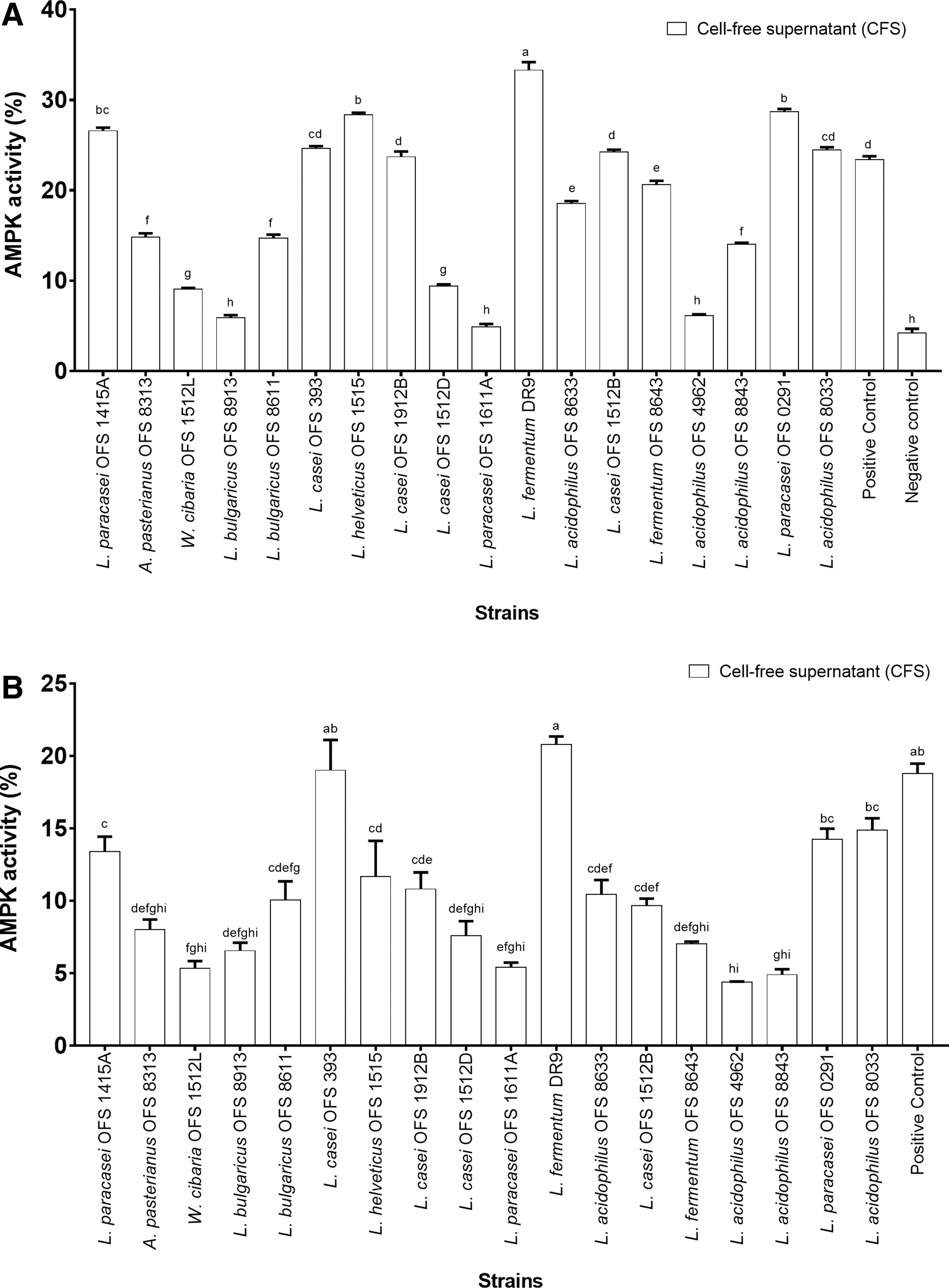
Screening for AMPK activation. Percentage of
AMPK activation in cell lines
The AMPK phosphorylation by the CFS of LF-DR9, LP-0291, and LH-1515 was further tested on seven different cell lines (Fig. 2). The CFS of LF-DR9 was effective in promoting AMPK phosphorylation in U2OS cells, with AMPK activity higher than the untreated and positive control, respectively (P < .05), whereas the CFS of LP-0291 was more effective in C2C12 cells (P < .05). The CFS of all strains showed insignificant AMPK phosphorylation compared to the untreated control (P > .05) when tested on SH-SY5Y, OVCAR3, and DU145 cells.
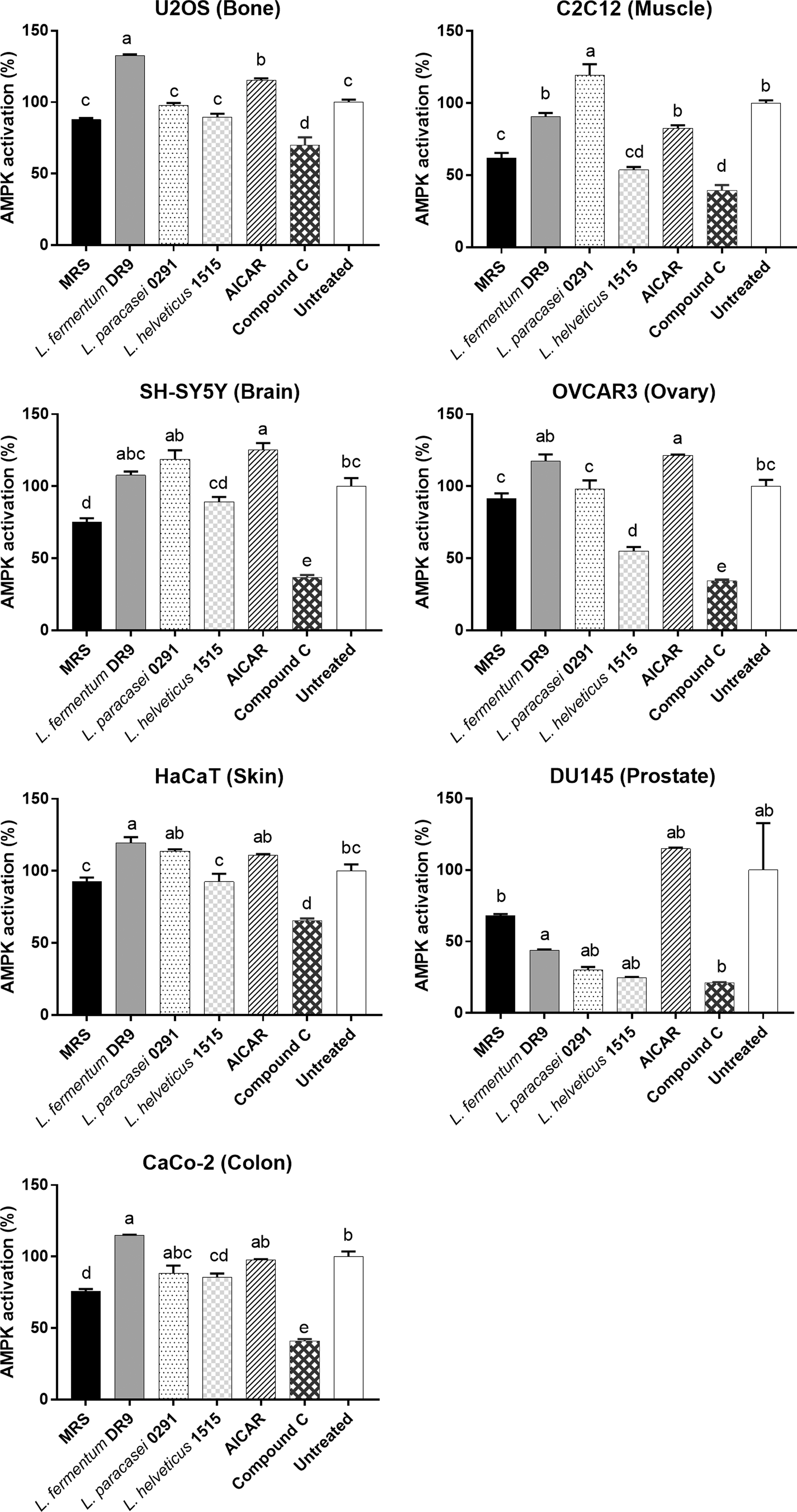
AMPK phosphorylation in cell lines. Human bone osteosarcoma U2OS, mouse muscle C2C12, human neuroblastoma SH-SY5Y, human ovary OVCAR3, human keratinocytes HaCaT, human prostate DU145, and human colorectal CaCo-2 cell lines were treated with CFS from strains LF-DR9, LP-0291, and LH-1515 and unfermented MRS both for 24 h. AICAR and compound C (20 μM) were used as positive control and negative control, respectively. AMPK phosphorylation was determined using cell-based ELISA. Percentage of activation is expressed in relation to untreated control. Error bars represent SEM (n = 3). a–eMeans among different treatments with different superscripted lowercase letters are significantly different (P < .05). Statistical analysis was performed with one-way ANOVA. LF-DR9, Lactobacillus fermentum DR9; LH-1515, Lactobacillus helveticus OFS 1515; LP-0291, Lactobacillus paracasei OFS 0291. ELISA, enzyme-linked immunosorbent assay; MRS, Mann, Rogosa, Sharpe.
The antiaging effect of LAB in premature senescent rat
LAB prevented telomere shortening
Upon D-gal senescence induction, the telomere length of aged rats was significantly shortened (P = .024) compared to the young rats (Fig. 3). LAB treatment of the senescence-induced rat showed an improvement in telomere length, with the highest from groups receiving LF-DR9 (P < .001), followed by LP-0291 (P < .001) and LH-1515 (P = .002), when compared to the aged rats. Metformin also significantly prevented the shortening of telomere length compared to the aged rats.
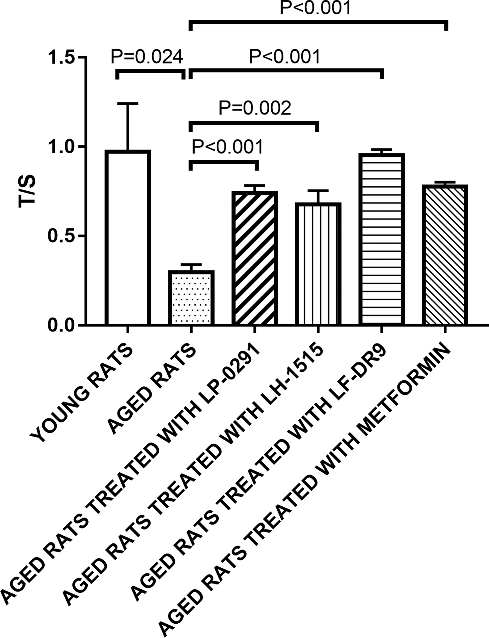
Measurement of telomere length. Telomere length was determined in blood of senescence-induced rats. Young rats: untreated rats receiving 0.9% saline subcutaneous injection daily (n = 6); aged rats: rats subjected to 600 mg/kg D-gal subcutaneous injection daily (n = 6); aged rats treated with LP-0291: rats subjected to 600 mg/kg D-gal subcutaneous injection daily and treated with LP-0291 (1 × 10¹⁰ CFU/day; n = 6); aged rats treated with LF-DR9: rats subjected to 600 mg/kg D-gal subcutaneous injection daily and treated with LF-DR9 (1 × 10¹⁰ CFU/day; n = 6); aged rats treated with LH-1515: rats subjected to 600 mg/kg D-gal subcutaneous injection daily and treated with LH-1515 (1 × 10¹⁰ CFU/day; n = 6); and aged rats treated with metformin: rats subjected to 600 mg/kg D-gal subcutaneous injection daily and treated with metformin (300 mg/kg/day; n = 6). Telomere length standardized to SCG was quantified using qPCR. The data are expressed as the means ± SEMs. Statistical analysis was performed with independent t-tests. CFU, colony-forming unit; D-gal, D-galactose; qPCR, quantitave polymerase-chain reaction; SCG, single copy gene.
LAB enhanced exercise capacity
The effect of LAB on physical performance related to aging was tested with treadmill exhaustion tests (Fig. 4). Running distance was not affected by all treatments, but the running capacity (time, speed, work, and power) was greatly affected by senescence induction. The running time, speed, work, and power were significantly lower in aged rats compared to the young rats. The LAB strains, especially LP-0291 and LF-DR9, were able to ameliorate the adverse effects of aging on exercise capacity, with improved performance in running time, speed, work, and power compared to the aged rats.
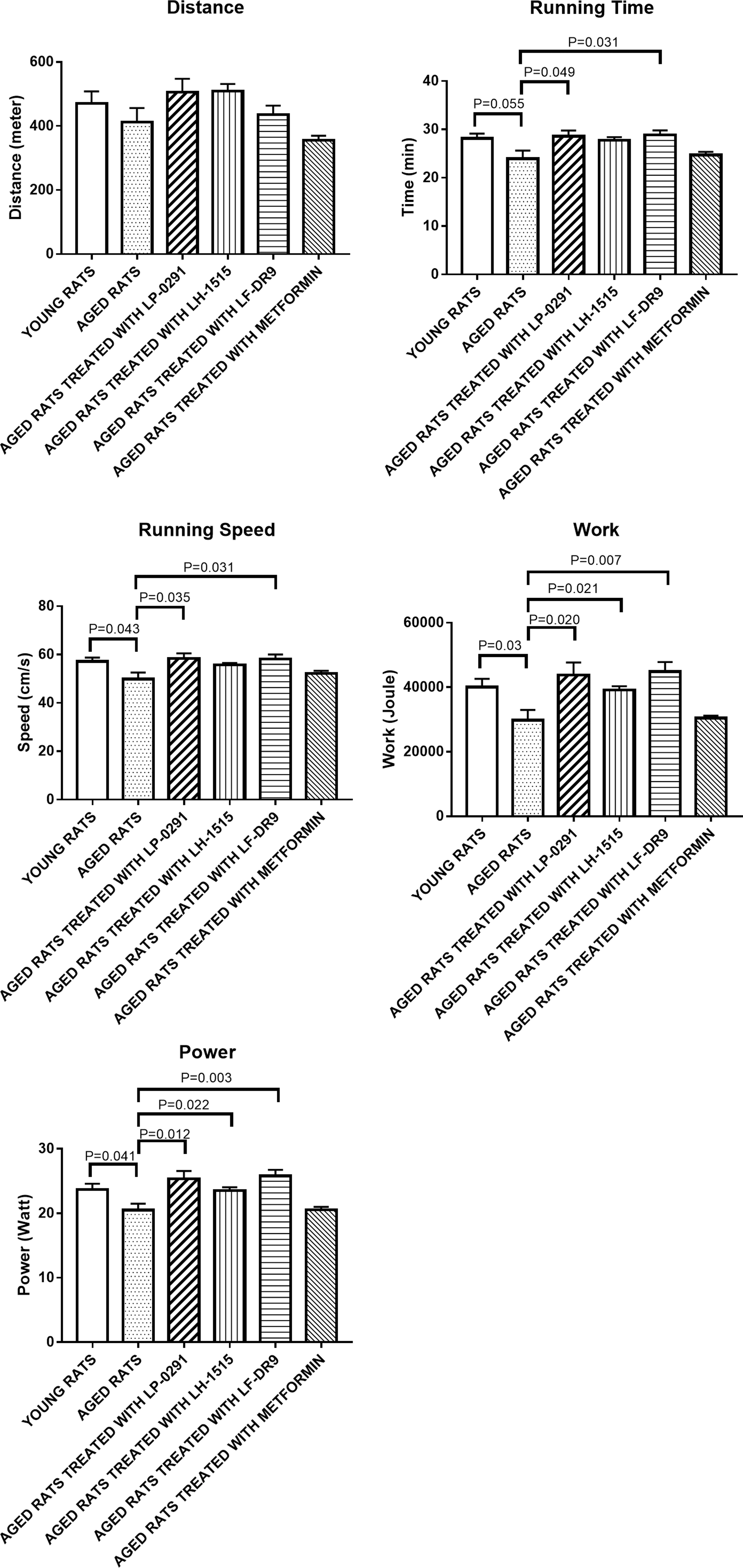
Treadmill exhaustion test. Uphill treadmill performance evaluated on senescence-induced rats. Young rats: untreated rats receiving 0.9% saline subcutaneous injection daily (n = 6); aged rats treated with LP-0291: rats subjected to 600 mg/kg D-gal subcutaneous injection daily and treated with LP-0291 (1 × 10¹⁰ CFU/day; n = 6); aged rats treated with LF-DR9: rats subjected to 600 mg/kg D-gal subcutaneous injection daily and treated with LF-DR9 (1 × 10¹⁰ CFU/day; n = 6); aged rats treated with LH-1515: rats subjected to 600 mg/kg D-gal subcutaneous injection daily and treated with LH-1515 (1 × 10¹⁰ CFU/day; n = 6); and aged rats treated with metformin: rats subjected to 600 mg/kg D-gal subcutaneous injection daily and treated with metformin (300 mg/kg/day; n = 6). Data recorded in distance, time running speed, work, and power are expressed as the means ± SEMs. Statistical analysis was performed with independent t-tests.
LAB ameliorated p53 gene expression in bone and muscle
Figure 5 showed that in gastrocnemius muscles, the p53 gene was increased significantly (P = .003) in aged rats compared to the young rats, while only treatment group receiving LF-DR9 showed a significant reduction (P = .012) when compared to the aged rats. In bone samples, aged rats attained a significant increase (P = .021) in p53 gene expression in tibia compared to the young rats, whereas all the aged rats treated with LF-DR9, LP-0291, and LH-1515 had significantly reduced p53 gene expressions.
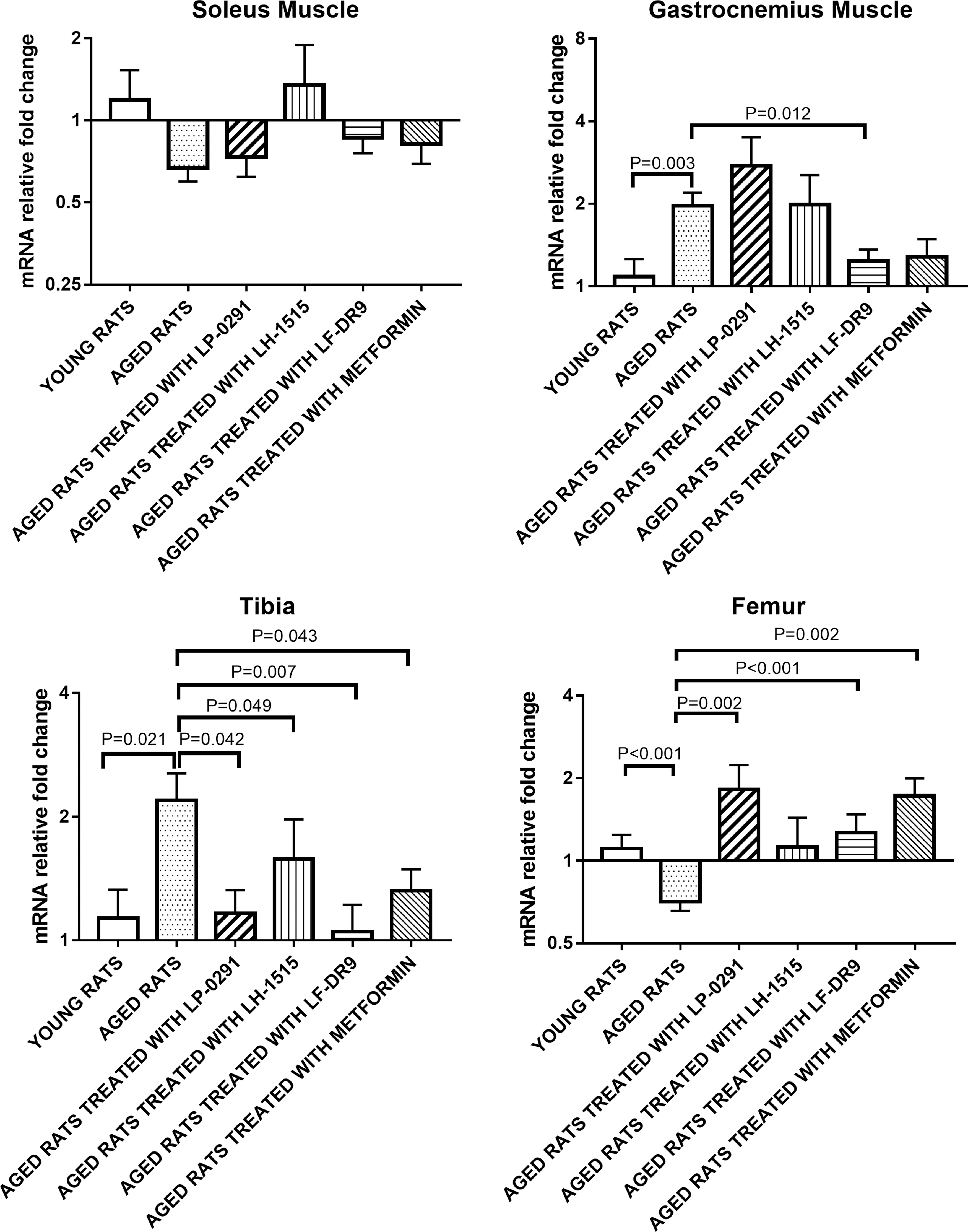
p53 gene expression in muscle and bone. The p53 gene expression was determined in soleus and gastrocnemius muscles and tibia and femur of senescence-induced rats using qPCR. Young rats: untreated rats receiving 0.9% saline subcutaneous injection daily (n = 6); aged rats: rats subjected to 600 mg/kg D-gal subcutaneous injection daily (n = 6); aged rats treated with LP-0291: rats subjected to 60 0 mg/kg D-gal subcutaneous injection daily and treated with LP-0291 (1 × 10¹⁰ CFU/day; n = 6); aged rats treated with LF-DR9: rats subjected to 600 mg/kg D-gal subcutaneous injection daily and treated with LF-DR9 (1 × 10¹⁰ CFU/day; n = 6); aged rats treated with LH-1515: rats subjected to 600 mg/kg D-gal subcutaneous injection daily and treated with LH-1515 (1 × 10¹⁰ CFU/day; n = 6); and aged rats were treated with metformin: rats subjected to 600 mg/kg D-gal subcutaneous injection daily and treated with metformin (300 mg/kg/day; n = 6). The data are expressed as the means ± SEMs. Statistical analysis was performed with independent t-tests.
Effects of LAB on metabolic profiles
There were no significant changes between the aged and young rats in lipid, renal, and liver profile measured in serum samples (Fig. 6). Rats treated with LH-1515 showed lower triglyceride, while LP-0291 and LF-DR9 demonstrated reduced cholesterol, HDL, and LDL compared to the aged rats. For liver profile, AST tended to be lower in rats receiving LP-0291 (P = .052); ALT was increased by LP-0291 (P = .019) and LF-DR9 (P = .002); and ALP was enhanced by LF-DR9 (P = .004). In renal profiles, strain LP-0291 tended to increase both chloride (P = .053) and phosphate (P = .057), while reducing creatinine (P = .007).
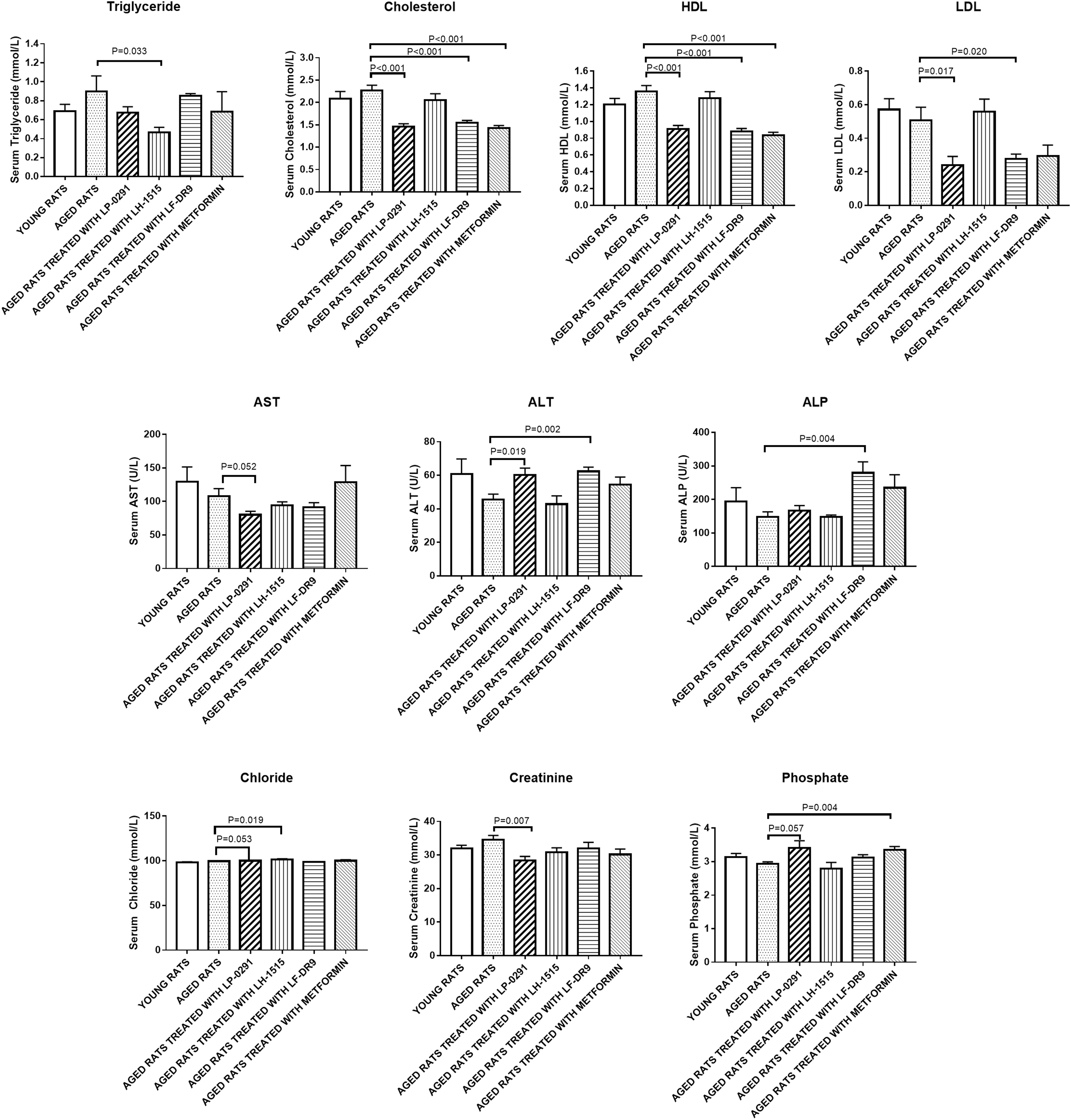
Serum biochemical profile of senescence-induced rats. Lipid profile is tabulated as triglyceride, cholesterol, HDL, and LDL. Liver function profiles were accounted by AST, ALT, and ALP. Kidney profiles are presented as chloride, creatinine, and phosphate. Young rats: untreated rats receiving 0.9% saline subcutaneous injection daily (n = 6); aged rats: rats were subjected to 600 mg/kg D-gal subcutaneous injection daily (n = 6); aged rats treated with LP-0291: rats subjected to 600 mg/kg D-gal subcutaneous injection daily and treated with LP-0291 (1 × 10¹⁰ CFU/day; n = 6); aged rats were treated with LF-DR9: rats subjected to 600 mg/kg D-gal subcutaneous injection daily and treated with LF-DR9 (1 × 10¹⁰ CFU/day; n = 6); aged rats were treated with LH-1515: rats subjected to 600 mg/kg D-gal subcutaneous injection daily and treated with LH-1515 (1 × 10¹⁰ CFU/day; n = 6); and aged rats treated with metformin: rats subjected to 600 mg/kg D-gal subcutaneous injection daily and treated with metformin (300 mg/kg/day; n = 6). The data are expressed as the means ± SEMs. Statistical analysis was performed with independent t-test. ALP, alkaline phosphatase; ALT, alanine aminotransferase; AST, aspartame aminotransferase; HDL, high-density lipoprotein; LDL, low-density lipoprotein.
Fulfillment of LAB strains as probiotics
The health benefits of LAB strains discussed in our study were of economic importance; thus the industrial applicability of both strains LP-0291 and LH-1515 was compared to the local commercial probiotic strain LF-DR9 and foreign commercial probiotic strain LS-Probio65. In Figure 7A, LP-0291 and LH-1515 exerted significantly higher mucin adherence properties compared to LS-Probio65, while LF-DR9 performed as good as LS-Probio65 in mucin adhesion test (P > .05).
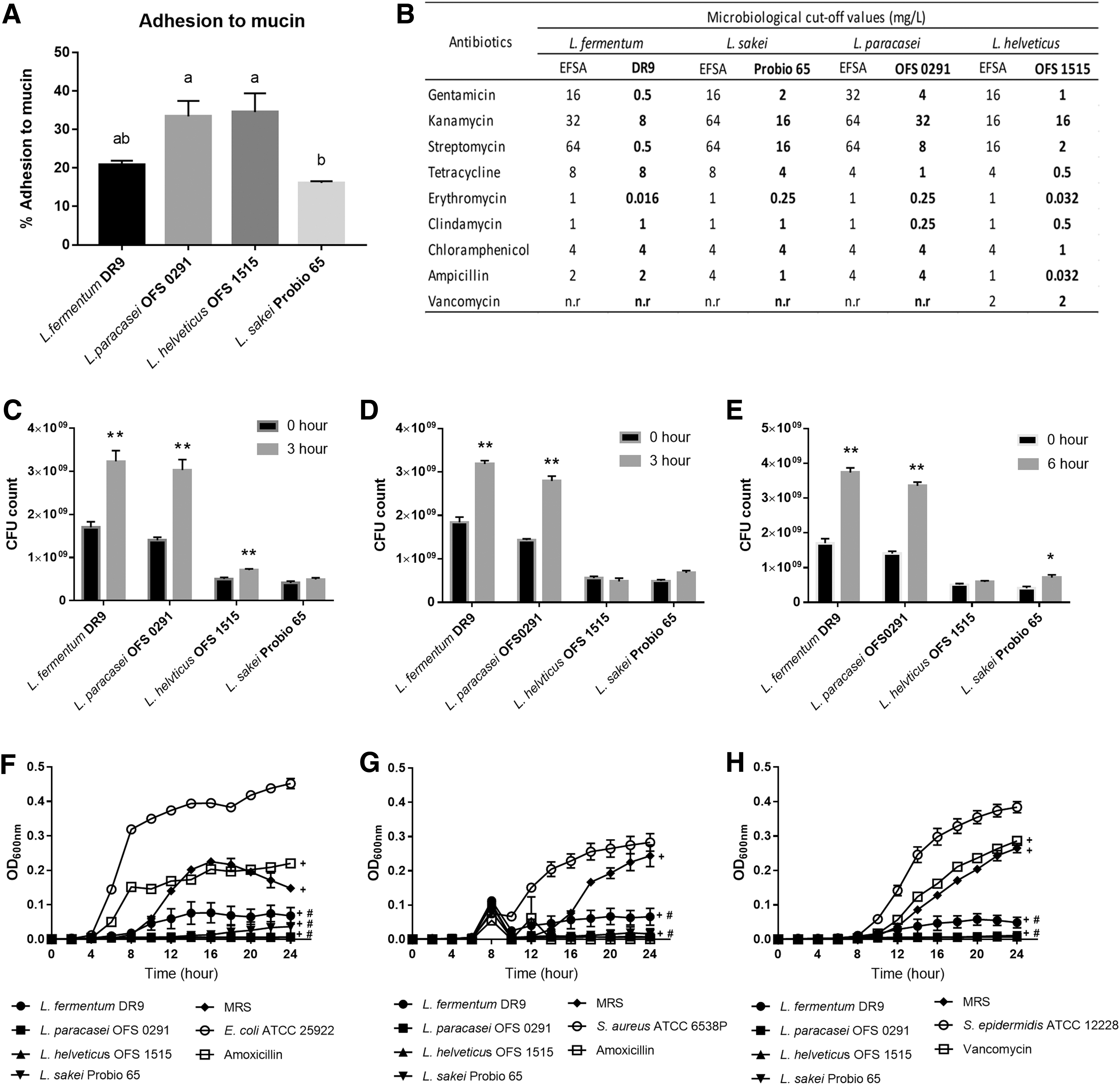
Probiotic properties of selected LAB strains.
Both LP-0291 and LH-1515 were tested for antibiotic resistance against nine antibiotics namely gentamicin, kanamycin, streptomycin, tetracycline, erythromycin, clindamycin, chloramphenicol, ampicillin, and vancomycin according to the EFSA guidelines (Fig. 7B). Microbiological cutoff values were defined differently according to the specific taxonomical unit such as species and genus of the strains studied. Both LP-0291 and LH-1515 exhibited lower and/or equivalent MICs compared to the established EFSA cutoff value, in a similar manner of the two commercial strains, thereby confirming that these strains did not exhibit antibiotic resistance properties.
The survivability of LAB strains in the gastrointestinal tract was experimented using simulated gastric and intestinal environments. LP-0291, LH-1515, and LF-DR9 displayed good survivability in acidic gastric environment, with a statistically higher bacterial count recorded after 3 h of incubation (Fig. 7C). However, only strain LF-0291 showed an enhanced growth rate upon incubation in intestinal environment for 3 h, similar to the commercial strain LF-DR9 (Fig. 7D). In the test for simultaneous gastric and intestinal tolerance, a statistically significant increase in CFU count was recorded for LF-0291, LF-DR9, and LS-Probio65 upon 6 h of treatment (Fig. 7E).
The antimicrobial activity of the LAB strains was evaluated against common pathogens, that is, E. coli ATCC 25922 (Fig. 7F), S. aureus ATCC 6538P (Fig. 7G), and S. epidermidis ATCC 12228 (Fig. 7H). The CFS of LP-0291 and LH-1515 had significantly suppressed the growth of all three pathogens tested, compared to the respective negative control and MRS treatment groups. The inhibitory effect of these strains was comparable to the commercial strains LF-DR9 and LS-Probio65.
Discussion
Calorie restriction (CR) has remained the most researched antiaging intervention that protects against deterioration of biological functions, oxidative stress, and metabolic rate. 21 –23 The hallmarks of CR include a reduction of glucose and insulin levels, which in turn activates the energy-sensing network regulators such as AMPK. 24 Thus, AMPK was chosen as the longevity marker. Among all the tested strains, the CFS of LF-DR9, LP-0291, and LH-1515 were good AMPK activators. AMPK functions by transcriptionally reprogramming cellular metabolic reactions to extrinsic stress and also acts as a switch for cellular energy homeostasis. 23,25 In this study, we showed that LAB strains could exert mimetic antiaging effects of CR without an actual reduction in calorie intake. They could also be regarded as novel AMPK activators, useful in the therapy of metabolic diseases related to the aging process.
D-gal mimetic aging was first documented by the use of a low-dose injection in mice to accelerate the aging process. 26 The aging model demonstrated neurological impairment, reduced antioxidant levels, and weakened immune system as seen in the normal aging process. 27 D-gal is also closely associated with metabolic syndrome as it causes lipid metabolic disorders and insulin resistance. A previous study using a metabonomics approach demonstrated that D-gal induced dyslipidemia and abnormal increases of metabolites in the liver of mice. 28
Premature senescence was induced using chronic D-gal injection to mimic the age-related metabolic imbalances. The aging model was validated by measuring telomere length, a biological indicator that determines the lifespan of an organism. 29 In normal cellular process, each cell division results in loss of a small portion of telomeric DNA and if continued, leads to chromosomal degradation and/or cell death. Telomere length in humans decreases at a rate of 24.8–27.7 base pairs per year, and it is related to increased incidence of age-related diseases and decreased lifespan. 29 In this study, D-gal reduced telomere length, demonstrating the efficacy of D-gal in inducing mimetic aging effects. Metformin was selected as the positive control as it was shown, in previous study, to lengthen the lifespan of mice by increases in the AMPK activity as well as antioxidant protection. 30 However, this drug causes adverse effects such as hypoglycemia and stomach upset. 24 Metformin treatment of D-gal rats also showed an improved telomere length, justifying the antiaging effect of the drug. To our knowledge, we were the first to report that LAB treatment of senescence-induced rats could prevent shortening of telomeres, with antiaging potential comparable to that of metformin. Such data open up an insight on LAB and/or probiotics as a natural source of food supplements for a healthy aging approach.
LAB strains improved lipid profile, and thus could potentially reduce risks of CVD, obesity, and diabetes during aging. The liver and renal profile were also positively altered, implying the protective effects of LAB in metabolic and excretory systems, which may prevent age-related chronic kidney disease and nonalcoholic fatty liver. 31 It is postulated that protective roles of these strains on metabolic perturbation were due to the modulation of AMPK. As a master switch for cellular energy homeostasis, AMPK regulates the dynamics of mitochondrial to overcome oxidative stress by minimizing energy consumption and exhaustion of resources, thereby restoring metabolic functions. 32
Apart from metabolic disorders, aging is also associated with loss of bone strength and decreased muscle mass. Age-related muscle wasting is termed sarcopenia, which involves decline in muscle functional capacity. 33 Sarcopenia increases the risk for falling, which further increases the susceptibility for bone fracture; thus, muscle and bone are very much interrelated during aging. 33 Upon D-gal senescence induction, rats experienced intolerance to exercise endurance. This is consistent with the aging phenomena, where increase in age reduces locomotor ability due to muscle weakness, joint stiffening, and lack of flexibility.
In addition to improving exercise performance in D-gal rats during uphill exercise test, LAB strains also reduced the senescence markers in bone and muscle. The p53 gene, which encodes a tumor suppression protein, acts as a critical marker for telomeric stress-induced senescence. 34 It regulates cell cycle arrest, DNA repair, apoptosis, and cellular senescence. 34 LP-0291 and LF-DR9 reduced p53 gene expression in localized gastrocnemius muscle and tibia, correlating with the promotion of AMPK phosphorylation in both U2OS (bone) and C2C12 (muscle) cells in vitro. AMPK agonist (5-aminoimidazole-4-carboxamide ribonucleotide [AICAR]) reportedly upregulated mitochondrial related genes in the muscle, correlating to improved motor function of aged mice. 35 We illustrate that LAB strains potentially protected bone and muscle from senescence in an AMPK-dependent manner.
It is a prerequisite for LAB strains to fulfill probiotic criteria and is generally regarded as safe for application and commercialization as food ingredients. The selection criteria for probiotics include the following: comprising resistance to gastric acidity and intestinal tolerance, adherence to mucosal surfaces, antibiotic resistance, and antimicrobial properties against potentially harmful pathogens. 4,20 Both strains LP-0291 and LH-1515 fulfilled the requirements with lower/equal values than the cutoff MIC values for all the antibiotics tested, with similar characteristics as local and foreign commercial strains, LP-DR9 and LS-Probio65, and complied with reduced tendency of horizontal gene transfer as per the EFSA safety guidelines. 36 Live probiotic cells must first pass through the upper gastrointestinal tract by surviving the harsh stomach acidic pH, bile salts, and pancreatic fluid before adherence onto intestinal lining. 20 LP-0291 survived and proliferated with comparable effects to the local commercial strain LF-DR9 in both gastric and upper intestinal conditions. Both LP-0291 and LH-1515 also adhered to mucin, which was employed to mimic the effect of the mucus-secreting cells on the human intestines. 16 In addition, both LP-0291 and LH-1515 exerted powerful antimicrobial properties against three potentially harmful pathogens, E. coli, S. aureus, and S. epidermidis, comparable to LP-DR9 and LS-Probio65. LAB have been widely reported to produce various organic acids, particularly lactic acid and acetic acid, bacteriocin, hydrogen peroxide, diacetyl, and antifungal peptides. 16,18,37 Our data justified that LP-0291 and LH-1515 fulfilled the requirements as probiotics.
In conclusion, altogether, the LAB strains successfully alleviated aging symptoms in D-gal-induced rats by protecting against telomere shortening and age-related metabolism disorders. LF-DR9 and L. paracasei OFS 0219 also reduce age-related tissue atrophy, specifically by targeting the musculoskeletal sites. LAB strains evaluated in this study also fulfilled all the criteria as probiotics, and thus could be utilized and safe to commercialize as next emerging medicinal food supplements (probiotics) for antiaging-targeted therapies.
Footnotes
Acknowledgments
The authors would like to acknowledge the financial assistance obtained from Basic Science Research Program of the National Research Foundation of Korea (NRF-2017R1D1A1B03031112), Clinical Nutrition Sdn. Bhd., the URICAS grant (1001/PTEKIND/870030) from Universiti Sains Malaysia, and MyBrain15–MyPhD from Ministry of Higher Education (MOHE).
Author Disclosure Statement
Y.Y.H. and M.T.L. conceived and designed the experiments. Y.Y.H., C.H.O., and M.T.L. performed the study and analyzed the data. B.Y.K., S.B.C., A.S., S.S., C.E.O., K.L.O., and W.S.J. contributed reagents, materials, and analysis tools. Y.Y.H., M.T.L., and W.S.J. wrote the article.
References
Supplementary Material
Please find the following supplemental material available below.
For Open Access articles published under a Creative Commons License, all supplemental material carries the same license as the article it is associated with.
For non-Open Access articles published, all supplemental material carries a non-exclusive license, and permission requests for re-use of supplemental material or any part of supplemental material shall be sent directly to the copyright owner as specified in the copyright notice associated with the article.
