
Abstract
Pleurotus eryngii var. ferulae (PEF) is traditionally used in the prevention and treatment of lifestyle-related diseases. In this study, we investigated the ability of PEF extract to prevent obesity and metabolic diseases and explored the underlying mechanism. Mice were fed a high-fat diet (HFD) containing PEF extract for 12 weeks, and their body weight, adipose tissue and liver weights, and lipid profiles and blood glucose levels, were monitored. Fecal triglyceride (TG) levels were also measured and olive oil-loading tests were performed. Furthermore, the effect of PEF extract on pancreatic lipase (PL) activity was examined in vitro. Treatment with PEF extract for 12 weeks resulted in a significant decrease in the HFD-induced increases in body weight, white adipose tissue weight, liver weights, and lipid profiles, and improved glucose tolerance and insulin sensitivity. To assess the mechanism underlying the effect of PEF extract on obesity and diabetes, we investigated its role in inhibiting lipid absorption. Consumption of an HFD containing PEF extract significantly increased the TG level in feces compared with the controls, suggesting inhibition of TG absorption in the digestive tract. Furthermore, PEF extract suppressed the increase in serum TG levels resulting from oral administration of a lipid emulsion to mice, confirming inhibition of TG absorption. Moreover, PEF extract inhibited PL activity in vitro. Our combined results indicate that the anti-obesity and antidiabetic effect of PEF extract in mice fed an HFD may be caused by inhibition of lipid absorption as a result of reduced PL activity.
Introduction
Obesity involves the accumulation of excessive body fat resulting from an energy imbalance. 1 Moreover, obesity leads to secondary chronic diseases such as dyslipidemia, cardiovascular disease, and type 2 diabetes. 2,3 Therefore, obesity is an increasing issue in health care, and methods of controlling obesity have been actively studied worldwide. 4,5 Several approaches to the prevention and treatment of obesity involve the inhibition of dietary triglyceride (TG) absorption through inhibition of pancreatic lipase (PL). 6 PL plays a key role in the digestion of TG. 7,8 PL is secreted into the duodenum through the duct system of the pancreas and is responsible for hydrolysis of 50–70% of total dietary fats. Therefore, inhibition of PL results in blocking of lipid absorption in the digestive tract.
Pleurotus eryngii var. ferulae (PEF) is an edible mushroom of the family Pleurotaceae and order Agaricales. 9 –11 PEF is a richer source of protein, dietary fiber, and amino acids than other edible mushrooms. 12,13 PEF produces various biologically active components, such as triterpenoids, beta-glucan, saponins, and steroids. 13,14 Furthermore, PEF extract, reportedly, has anti-tumor, antioxidant, anti-inflammatory, and anti-hypercholesterolemic effects. 10,11 However, few investigations have focused on the pharmacological effects of PEF on obesity and metabolic diseases. In this study, we used a model of obesity induced by feeding a high-fat diet (HFD) to mice for 12 weeks to evaluate the preventive effect of PEF extract on features of metabolic disease. Furthermore, we elucidated the mechanism underlying the effect of PEF extract on lipid absorption. We demonstrate that the PEF extract prevents obesity and its related metabolic complications, such as hyperlipidemia, fat accumulation in tissues, and diabetes. Furthermore, the PEF extract significantly increased TG levels in feces and decreased serum TG levels by inhibiting PL. Therefore, PEF extract has potential as an ingredient of functional foods for preventing obesity and diabetes.
Materials and Methods
Reagent
Bovine serum albumin fraction V fatty acid-free was purchased from Calbiochem. Tween 20 was purchased from Affymetrix, Inc. Orlistat, p-nitrophenyl butyrate (p-NPB), PL (Type II from porcine pancreas), taurocholic acid sodium salt hydrate, olive oil, carboxymethylcellulose sodium salt, and a serum TG determination kit quantitation kit were purchased from Sigma-Aldrich Chemical (St. Louis, MO, USA). All reagents were of the highest grade available.
Preparation of water extract
PEF powder was obtained from the Gyeongsangbuk-Do Forest Environment Research Institute (Gyeongju, Korea). Dried PEF powder material was fully extracted in distilled water at 100°C for 1 h. After cooling to room temperature, the extracts were filtered. The filtrates were then evaporated in a rotary vacuum evaporator and freeze-dried to powder. Dried water extracts were stored at −20°C and dissolved in distilled water before use. The yield (w/w) of the water extracts was 30–40%. The concentration of PEF extract was calculated based on the dry weight of water extracts.
Animals and diets
C57BL/6 male mice were maintained on a 12 h light/dark cycle with access to water ad libitum. After acclimatization for 1 week on a normal chow diet (NCD), the mice were fed one of the following two diets for 12 weeks: (1) HFD containing 60% fat (HFD group), (2) HFD supplemented with 10% PEF extract (HFD+PEF), or (3) HFD supplemented with 0.0088% orlistat (HFD+Orlistat). The compositions of the diets are given in Table 1. Body weights were recorded weekly throughout the experimental period. Food intake was measured on a per-case basis every 2 or 3 days throughout the study. Food intake (g/mouse/day) was determined by subtracting the remaining food weight from the initial food weight of the previous feeding day and dividing by the number of mice housed in the cage. All animal experiments were approved by the Pohang University of Science and Technology Institutional Animal Care and Use Committee (POSTECH IACUC; Approval No. POSTECH-2014-0050). All animal experiments were performed in our animal facility according to the IACUC guidelines and regulations.
Composition of the Experimental Diets (g/100 g Diet)
HFD: No. D12452 (Research Diets, Inc., New Brunswick, NJ, USA).
HFD, high-fat diet; PEF, Pleurotus eryngii var. ferulae.
Blood chemistry
At the end of the 12-week feeding period, mice were killed and blood was collected. Plasma was obtained by clotting of blood for 1 h and centrifugation for 1 h at 900 g. Blood chemistry was analyzed using an Auto Chemistry Analyzer (BS-380; Mindray).
Histological analysis
Epididymal fat and livers were fixed in 4% paraformaldehyde, prepared as paraffin blocks, sectioned at 4 μm, and stained with hematoxylin and eosin (H&E). The specimens were examined using a light microscope at 200 × magnification.
Intraperitoneal glucose tolerance test
An intraperitoneal glucose tolerance test (IPGTT) was performed near the end of the experiments, after 11–12 weeks as follows. Mice were subjected to fasting overnight, followed by intraperitoneal administration of 1 g/kg glucose. Blood glucose concentrations at the indicated time points were measured using an Accu-Chek Performa (Roche, Mannheim, Germany).
Intraperitoneal insulin tolerance test and insulin levels
An intraperitoneal insulin tolerance test was performed at the end of the experiments and after 11–12 weeks as follows. Mice were subjected to fasting for 4 h, followed by intraperitoneal administration of 0.5 U/kg insulin, and measurement of blood glucose levels at the indicated time points. Fasting plasma insulin levels were determined using an insulin enzyme-linked immunosorbent assay kit (ALPCO, Salem, NH, USA).
Fecal lipid extraction
C57BL/6 male mice were housed under a 12-h light/dark cycle with access to water ad libitum. After acclimatization for 1 week on an NCD, the mice consumed the HFD containing 5% PEF extract, 10% PEF extract, or HFD containing 0.0088% orlistat for 1 day. Fecal lipids were extracted using the method of Bligh and Dyer with slight modifications. 15 In brief, all feces were collected within 24 h after starting the experiment. The collected feces were dried at 60°C and weighed. The dried fecal samples were added to deionized water and stored at 4°C overnight. The following day, the dried feces mixture was homogenized by vortexing, and lipids were extracted using methanol:chloroform (2:1, v/v). The lower chloroform phase was withdrawn and the lipid in the phase was dried. The TG concentrations were measured using a commercial kit (Sigma-Aldrich Chemical).
Olive oil-loading test
Age- and sex-matched C57BL/6 mice (25 g) were divided into five groups, with each group matched for body weight (n = 3–5). A lipid emulsion containing 30% olive oil, 1% Tween 20, and 0.5% methylcellulose solution (w/w) was prepared. After fasting for 12 h, each mouse was orally administered 0.5 mL of the lipid emulsion or lipid emulsion plus PEF extract (final concentrations, 0.04, 0.8, or 2 g/kg). Blood samples were collected from the orbital sinus at 0, 2, 4, and 6 h after administration of the lipid emulsion or lipid emulsion plus PEF extract. Serum was separated by centrifugation at 4°C and stored at −20°C for the TG assay. Serum TG concentrations were measured using a serum TG determination kit (Sigma-Aldrich Chemical).
PL activity assay
PL activity was determined with p-NPB as a substrate using a modification of a method described previously. 16
Statistical analysis
Data are presented as mean ± standard error of the mean. The data were evaluated by one-way analysis of variance followed by Tukey's post hoc analysis. P values <.05 were considered to indicate statistical significance.
Results
Effect of PEF extract on body weight gain
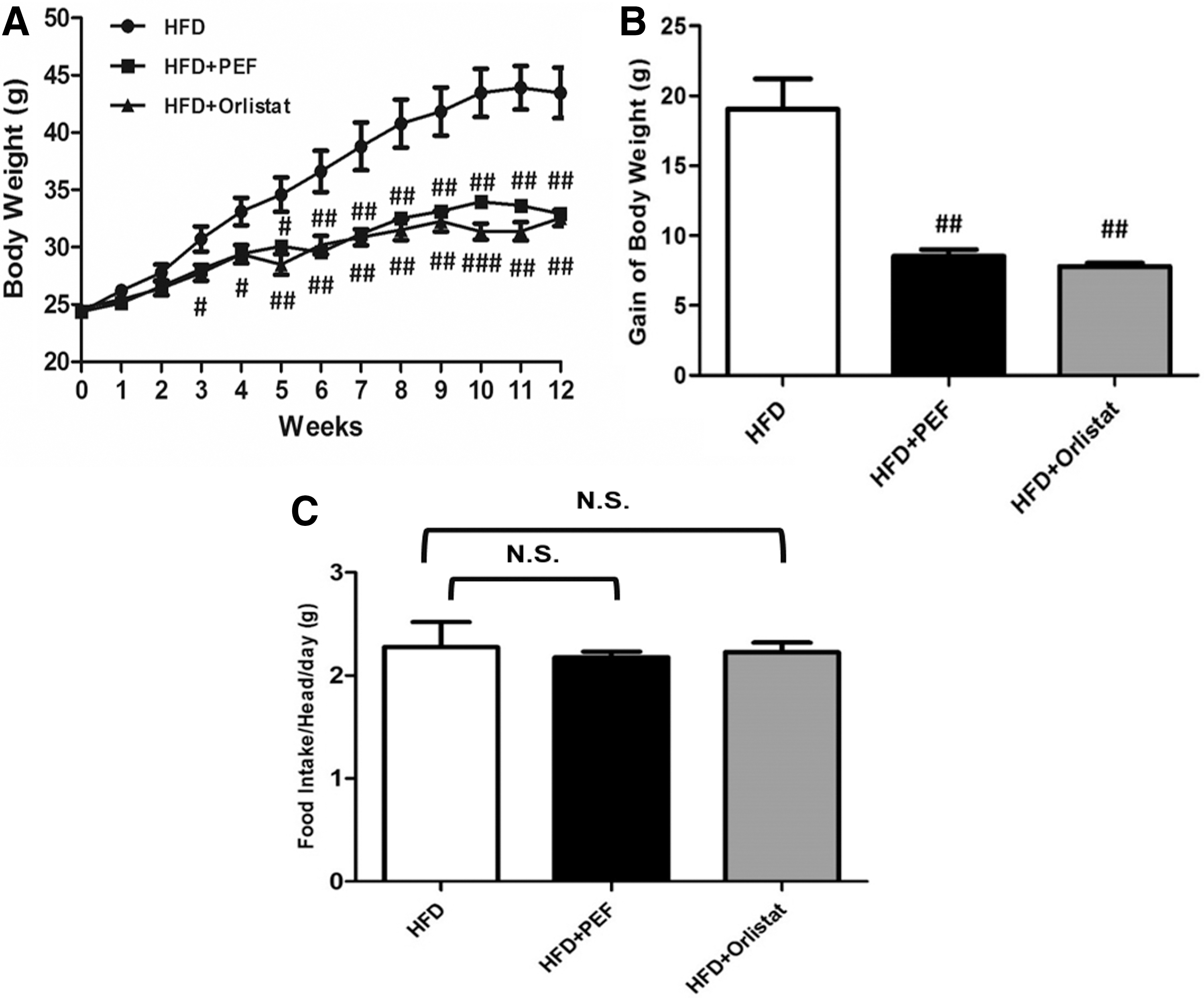
Mice were fed an HFD for 12 weeks to induce obesity. The treated groups were supplied with PEF and the positive control group was supplied with the drug, orlistat. The body weight gain in the HFD+PEF group (32.92 ± 0.7 g) was significantly lower than that in the HFD group (43.46 ± 2.2 g) (Fig. 1A–C). There was no significant difference in food intake among the groups. Therefore, PEF extract reduced the HFD-induced gain in body weight.
Effect of PEF extract on body weight
Effects of the PEF extract on fat accumulation in tissues, liver deterioration, and hyperlipidemia
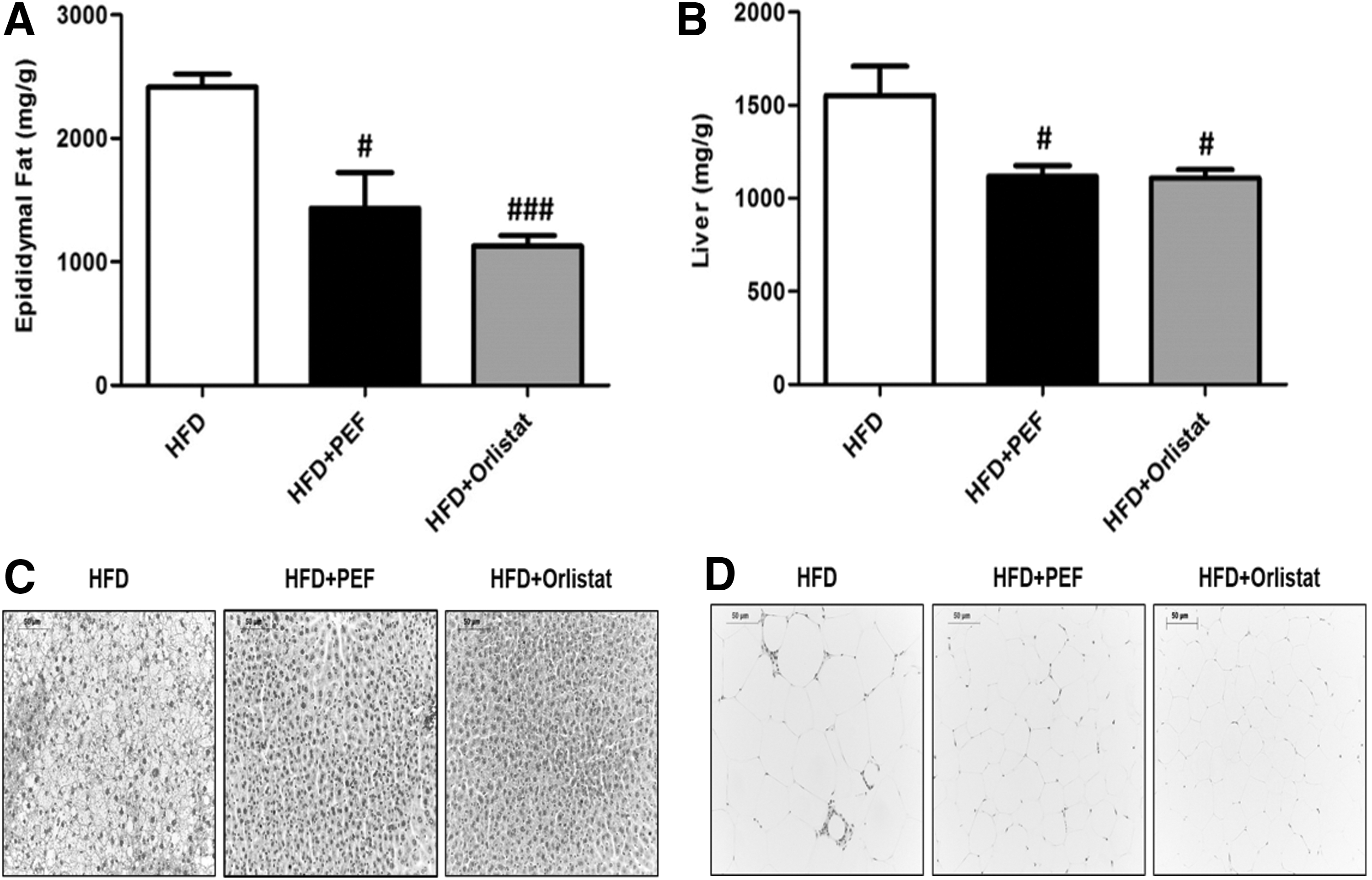
The epididymal fat weight in the PEF group (1434.3 ± 289 mg) was significantly lower than that in the HFD group (2416.8 ± 105.2 mg) (Fig. 2A). Histological analysis showed that adipocytes of mice were smaller in the HFD+PEF group than those in the HFD group (Fig. 2B). The liver weight in the HFD+PEF group (1117.1 ± 58.1 mg) was significantly lower than that in the HFD group (1552.9 ± 155.5 mg) (Fig. 2C). Moreover, mice in the HFD+PEF group exhibited reduced formation of lipid droplets (Fig. 2D). In addition, to determine the damaging effects of the HFD on the liver, aspartate transaminase (AST) and alanine aminotransferase (ALT) activities levels were measured. In liver disorders, AST and ALT activities increase with HFD-induced liver damage. Therefore, AST and ALT are useful indicators for evaluating liver deterioration caused by obesity. As given in Table 2, the AST and ALT levels were significantly lower in the HFD+PEF group than those in the HFD group. These results suggest that PEF extract has a beneficial effect on the liver damage induced by HFD.
Effect of PEF extract on epididymal adipose tissue and liver weights in HFD-induced obese mice
Effects of Pleurotus eryngii var. ferulae Extract on Plasma Triglyceride, Total Cholesterol, Low-Density Lipoprotein Cholesterol, High-Density Lipoprotein Cholesterol, Aspartate Transaminase, and Alanine Aminotransferase Levels in High-Fat Diet-Induced Obese Mice
Data are given as mean ± SEM (n = 5).
Differences were subjected to one-way ANOVA (for three or more groups; * P < .05; ** P < .01; and *** P < .001).
ALT, alanine aminotransferase; ANOVA, analysis of variance; AST, aspartate transaminase; LDL-C, low-density lipoprotein cholesterol; SEM, standard error of the mean; TC, total cholesterol; TG, triglyceride.
We next examined whether the altered adipose tissue and liver weights in the HFD+PEF group were correlated with changes in blood lipid levels. The plasma TG, total cholesterol, and low-density lipoprotein cholesterol levels in the HFD+PEF group were significantly lower than those in the HFD group (Table 2). Taken together, these results show that PEF extract prevented the development of obesity, fat accumulation in tissues, and hyperlipidemia in HFD-fed mice.
Effect of PEF extract on insulin sensitivity
Because PEF extract intake resulted in remarkable improvements of several indicators of metabolic syndrome in HFD-induced obese mice, we also tested its effects on diabetes-related indicators, such as the concentration of glucose and insulin in the blood, and performed IPGTT and intraperitoneal insulin tolerance test. As given in Figure 3, mice in the HFD+PEF group showed significantly improved glucose tolerance after intraperitoneal injection with glucose. Moreover, the HFD+PEF exhibited significantly decreased blood glucose concentrations at 60, 90, and 120 min after insulin injection (Fig. 3B) and significantly lower plasma insulin concentration levels, suggesting enhanced insulin sensitivity (Fig. 3C).
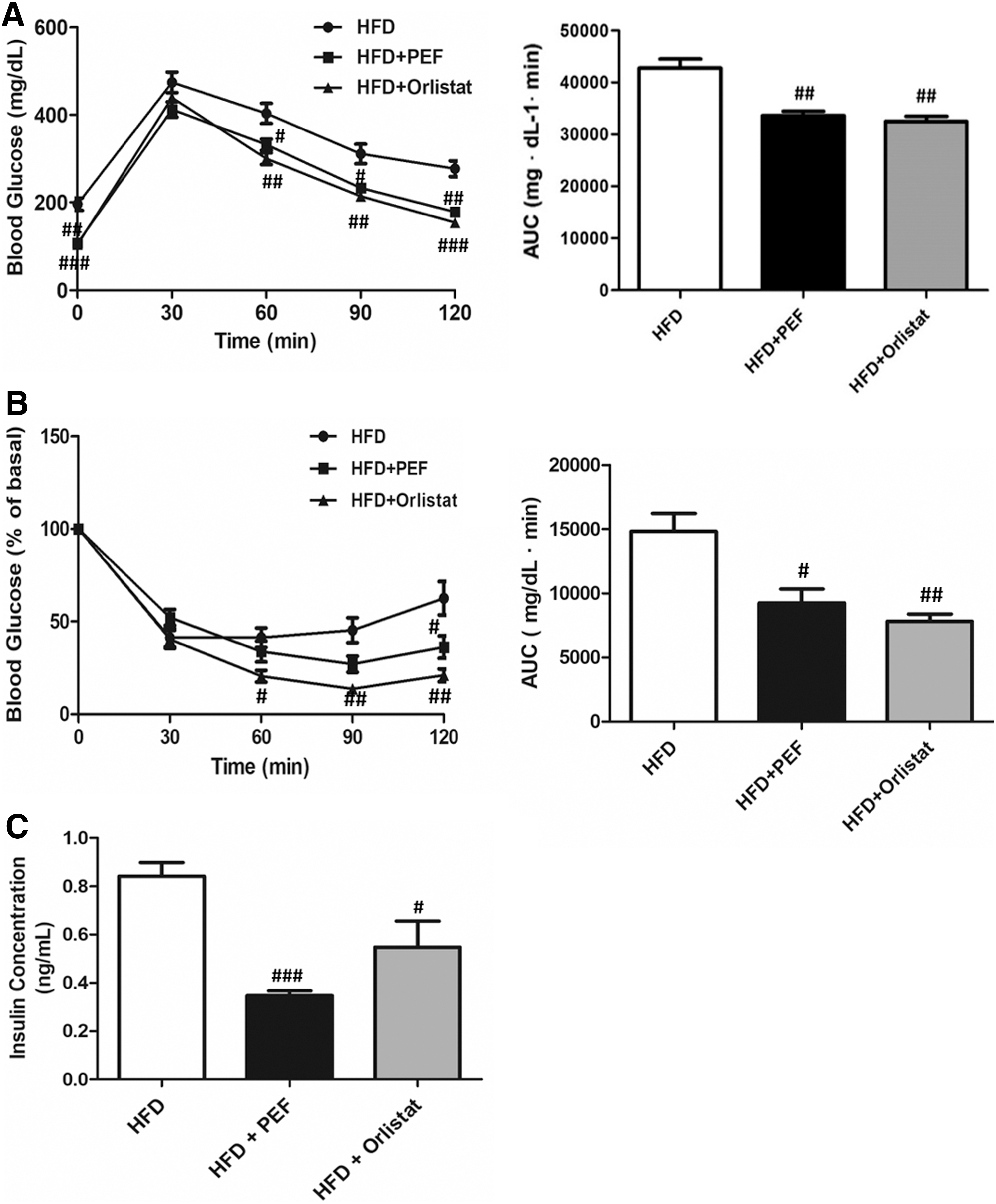
Effect of PEF extract on glucose and insulin tolerance in HFD-induced obese mice. (A) Glucose tolerance (AUC values), (B) insulin tolerance (percentage of the baseline and AUC values), and
Effect of PEF extract on fecal lipid contents
PEF extract prevented obesity and diabetes in HFD-fed mice (Figs. 1 –3). However, the underlying mechanism is unclear. Inhibiting absorption of dietary lipids can lead to weight loss, because excessive lipid intake is associated with obesity. Therefore, to investigate the effects of PEF extract on the absorption of dietary lipids, we measured fecal TG levels. The fecal TG level was significantly higher in the HFD+PEF extract group than that the HFD group (Table 3), indicating that the PEF extract inhibited the uptake of dietary lipids.
Effects of Pleurotus eryngii var. ferulae Extract on the Dried Feces Weight and Fecal Lipid Content
TG concentration in dried feces from mice in the HFD, HFD+PEF, or HFD+Orlistat groups. Data are given as mean ± SEM (n = 3–4).
Differences were subjected to one-way ANOVA (for three or more groups; ** P < .01).
Effect of PEF extract on serum TG levels and PL activity
The effect of PEF extract on intestinal absorption of TG was investigated. Compared with the control group, the lipid emulsion (TG) group exhibited significantly increased serum TG levels after administration of 30% olive oil. However, this increase was inhibited by oral administration of PEF extract in a dose-dependent manner. Oral administration of PEF extract (0.04, 0.8, and 2 g/kg) resulted in reduced serum TG levels at 2, 4, and 6 h (Fig. 4A). The activity of PL is required for absorption of dietary fat. 17 Incubation of 0.5, 1, 5, 10, 50, and 100 mg/mL PEF extract inhibited PL activity by 70.7, 65.7, 55.3, 53.1, 20, and 5.3%, respectively (Fig. 4B). The IC50 value of PEF extract was 10 mg/mL. Taken together, these data suggest that PEF extract reduces lipid uptake by inhibiting PL activity.
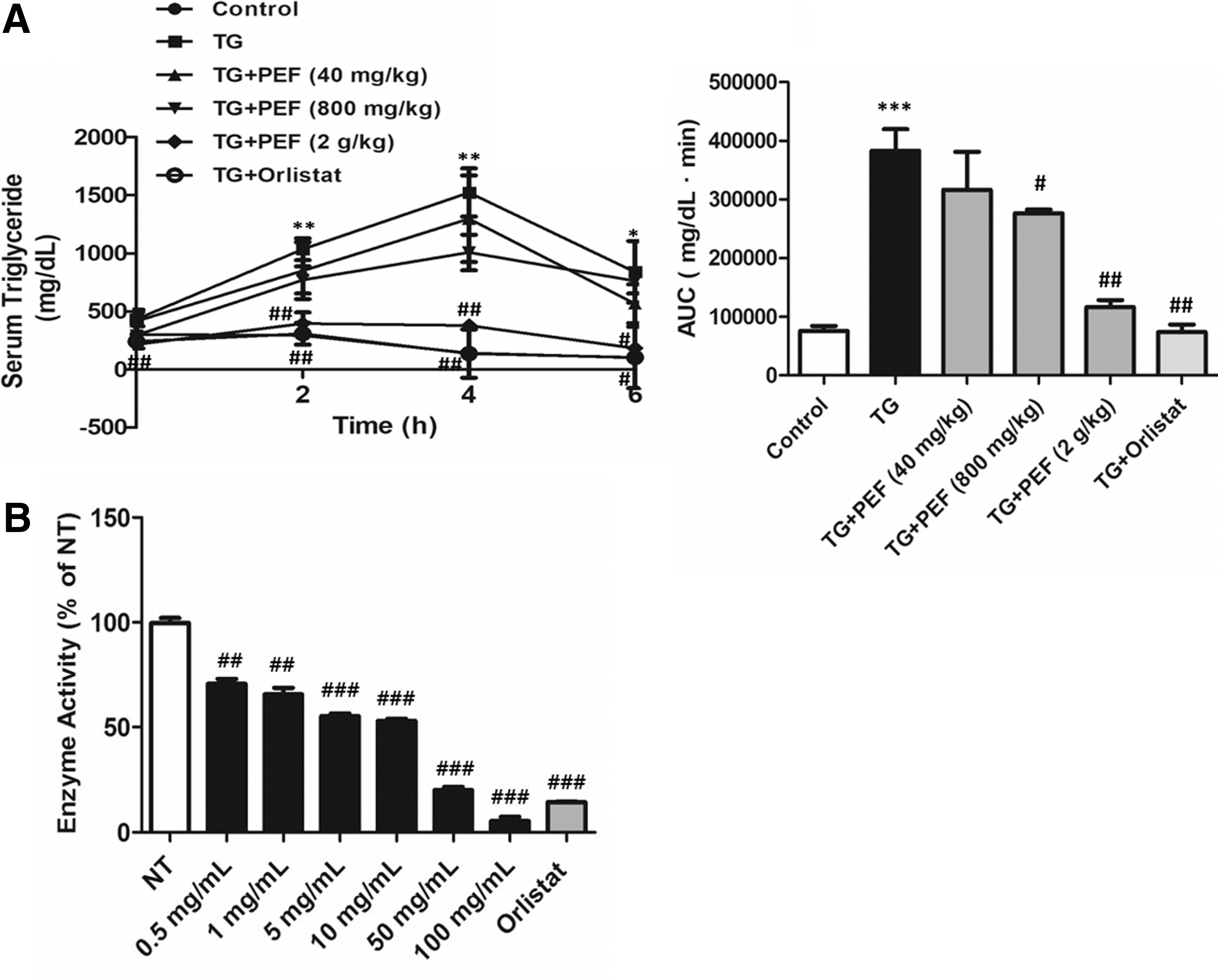
Effect of PEF extract on serum TG levels. C57BL/6 mice were orally administered a lipid emulsion only or a lipid emulsion containing 0.04, 0.8, or 2 g/kg PEF extract, and serum TG levels were analyzed
Discussion
PEF is a widely consumed edible mushroom, and its anti-tumor, antioxidant, anti-inflammatory, and antimicrobial activities have been well investigated. 11 Moreover, feeding a diet containing PEF to hypercholesterolemic rats reduced plasma lipids and body weight. 10 However, the mechanism by which PEF exerts its anti-obesity and antidiabetic effects remains elusive. Specifically, how PEF extract inhibits PL activity has not been elucidated. In this study, we demonstrated for the first time that PEF extract prevented HFD-induced obesity and metabolic diseases including diabetes, reduced serum TG levels after oral administration of a lipid emulsion to mice, and inhibited PL activity. Taken together, based on these results, PEF extract may prevent obesity and its related complications by inhibiting intestinal absorption of dietary fat through inhibition of PL activity. Therefore, PEF can potentially be consumed to prevent obesity.
Inhibition of dietary lipid absorption may be an effective means of regulating obesity. 18,19 Indeed, PL activity is required for intestinal absorption of dietary lipids. 20,21 PL plays a key role in digestion of TG into monoacylglyceride and fatty acids that facilitates absorption of dietary lipid in the digestive tract. 6,7,22 Therefore, we determined the effect of PEF extract on PL activity, based on its suppression of TG absorption in mice (Table 3 and Fig. 4). PEF extract inhibited PL activity, which prevented development of obesity and metabolic disease. This effect is similar to that of orlistat. 23,24 However, the anti-obesity effect of PEF extract cannot be explained solely by inhibition of PL; other mechanisms related to adipogenesis are involved. 13 PEF inhibits adipogenesis in 3T3-L1 preadipocytes by suppressing production of adipogenic factors, including PPAR-α and C/EBP-β. 13 Therefore, PEF may exert anti-obesity effects by inhibiting both lipid absorption and adipogenesis, indicating a high potential as an anti-obesity food.
Obesity is associated with insulin resistance, which is characterized by high glucose intolerance 25 ; therefore, improved insulin resistance is important for preventing the development of diabetes. This study showed that treatment with PEF extract significantly improved insulin sensitivity in obese mice (Fig. 3). The improved insulin sensitivity is probably because of a reduction in the accumulation of white adipose tissue (Fig. 2). It has been reported that the accumulation of white adipose tissue is an important predictor of insulin resistance, hyperglycemia, and other metabolic risk factors. 26 An increase in the white adipose tissue weight has been found to be accompanied by an induction of inflammatory cytokines involved in insulin resistance 27,28 ; therefore, inhibition of fat accumulation by PEF may also prevent obesity-induced diabetes.
PEF is rich in bioactive constituents such as polysaccharides, saponins, beta-glucan, triterpenoids, organic acids, and steroids. 13,14 Among them, polysaccharides can be used to ameliorate obesity. Polysaccharides from Ganoderma lucidum mycelia prevent diet-induced obesity and alleviate inflammation. 13 Han et al. reported that saponins isolated from Panax japonicas rhizomes and Platycodon grandiflorus prevent HFD-induced increases in body and adipose tissue weights by reducing intestinal absorption of dietary lipid through inhibition of PL activity. 22,29 –34 Furthermore, beta-glucan has been reported to decrease the fat mass and improve the plasma lipid profile. 35 Although the active constituents in PEF extract that are responsible for its anti-obesity and antidiabetic effects have not been identified, the active constituents such as polysaccharides, oligosaccharides, beta-glucans, and saponins may be water soluble. 13,14,22,29 –39 Therefore, we plan to investigate the antilipase effect of other bioactive constituents of PEF extract, which will lead to the development of novel natural therapeutic agents.
In conclusion, the results demonstrate that PEF extract may be used to prevent the development of obesity and metabolic diseases, including diabetes. These effects are mediated by reducing dietary lipid absorption through inhibition of PL activity. Our findings suggest that PEF has potential as an ingredient in functional foods for the prevention of obesity and metabolic diseases.
Footnotes
Acknowledgments
This research was supported by the Gyeongsangbuk-Do Forest Environment Research Institute.
Author Disclosure Statement
No competing financial interests exist.