
Abstract
GS-E3D is an enzymatically modified ginseng extract by pectin lyase. In this study, we evaluated the preventive effects of GS-E3D on blood-retinal barrier (BRB) leakage in a rat model of diabetes. To produce diabetes, rats were injected with streptozotocin. GS-E3D was orally gavaged at 25, 50, and 100 mg/kg body weight for 6 weeks. We then compared the effect of GS-E3D with that of an unmodified ginseng extract (UGE) on retinal vascular leakage. The administration of GS-E3D significantly blocked diabetes-induced BRB breakdown. Immunofluorescence staining showed that GS-E3D reduced the loss of occludin in diabetic rats. In TUNEL staining, the number of apoptotic retinal microvascular cells was dose dependently decreased by GS-E3D treatment. GS-E3D decreased the accumulations of advanced glycation end products in the retinal vessels. In addition, the inhibition potential of GS-E3D on BRB breakage was stronger compared with UGE. These results indicate that GS-E3D could be a beneficial treatment option for preventing diabetes-induced retinal vascular injury.
Introduction
Diabetes-induced retinal dysfunction is the most well-established ocular complication of diabetes mellitus. 1 Hyperglycemia induces blood-retinal barrier (BRB) leakage and microvascular injury in the retinas. The pathologic features in the retinas under diabetic conditions include the following: vascular hyperpermeability, pericyte death, microaneurysm, acellular capillary, and abnormal angiogenesis. 2 Macular edema can develop due to the BRB breakdown. 3 The strict glycemic management successfully delays the onset of diabetic retinopathy. Although many classes of antidiabetic agents have been introduced, the global incidence of diabetic retinopathy is still increasing. 4
Advanced glycation end products (AGEs) are responsible for the development of diabetic retinopathy. 5 –7 The generation of AGEs is enhanced under hyperglycemic conditions. 8 High concentrations of AGEs accumulate in the diabetic retinal microvessels 9 and trigger retinal vascular leakage. 10 Administration of exogenous AGEs led to the breakdown of BRB in normoglycemic animals. 11
Previous reports have demonstrated a preventive role of AGE inhibitors against diabetic retinopathy in several experimental diabetic animals. 12 –14 Aminoguanidine, a well-known AGE inhibitor, inhibited the development of microaneurysm and pericyte apoptosis in the diabetic retinas. 15 In our previous study, aminoguanidine reduced retinal AGE accumulations and ganglion cell loss in Zucker diabetic fatty rats. Several botanical and synthetic agents have been introduced as AGE inhibitors. 16
Ginseng is a popular herbal supplement to improve vitality and has numerous pharmacological activities. 17 –19 Ginseng also has been show to possess potent antidiabetic properties in animal experiments 20,21 and in patients with diabetes. 22 Some transformation methods using enzymatic conversion 23 and fermentation 24 have been introduced to enhance the pharmaceutical activities of herbal extracts. Recently, we newly developed an enzymatic modified ginseng extract (GS-E3D) using pectin lyase. This product has an increased concentration of ginsenoside Rd compared to an unmodified ginseng extract (UGE). 25 Our recent report showed that GS-E3D exhibits the stronger inhibition potential on glycation compared with UGE. 26 In this study, the preventive properties of GS-E3D on diabetes-induced retinal vascular dysfunction were evaluated in a rat model of diabetes. We also compared the effect of GS-E3D with that of UGE on retinal vascular leakage. We identified the possible mechanism of GS-E3D on AGE accumulation associated with retinal microvascular hyperpermeability in the experimental animals.
Materials and Methods
Preparation of GS-E3D
GS-E3D and UGE were produced based on previously reported methods. 25 The ginsenoside contents of GS-E3D and UGE are summarized in Table 1.
Ginsenoside Contents in GS-E3D
UGE, unmodified ginseng extract.
Animal experiments
To induce diabetes, 7-week-old male SD rats (Koatech, Pyeongtaek, Korea) were intraperitoneally injected with streptozotocin (STZ, 60 mg/kg) in citrate buffer. Rats in the control group were given an equivalent amount of buffer only. One week after these injections, rats with normoglycemia or hyperglycemia (> 300 mg/dL blood glucose) were randomized into the following 6 groups of 10 rats: control group (NOR), diabetic group (DM), UGE-treated group (100 mg/kg body weight), and three doses of GS-E3D-treated groups (25, 50, and 100 mg/kg body weight). UGE and GS-E3D were given orally once a day for 6 weeks. All animal experiments were performed in accordance with the IACUC approved protocol (Protocol No. 15–100)
Fluorescein-dextran microscopy
At necropsy, rats were anesthetized by isoflurane inhalation. Fluorescein-dextran (50 mg/kg; Sigma, St. Louis, MO, USA) was given by an intravenous injection. The fluorescence dye was perfused for 15 min, and the fluorescein-dextran-perfused retinas were then incised and flatly mounted on a glass slide. The slides were examined by fluorescence microscopy. Plasma was collected from rat blood before sacrifice followed by centrifugation. The plasma levels of fluorescein-dextran were determined with a fluorometer (Tecan, Männedorf, Switzerland). The signal intensity of fluorescein-dextran-perfused retinas was measured using Image J software (Image J; NIH, Bethesda, MD, USA). The obtained intensity values were then normalized to the plasma levels of fluorescein-dextran.
TUNEL staining
The retinas were fixed in 4% paraformaldehyde for 4 h. Apoptotic cells were detected and visualized using a TUNEL staining kit (Roche, Mannheim, Germany) according to the directions of the manufacturer. The images were quantified by counting the number of TUNEL-positive cells.
Immunofluorescence staining
The retinas were incubated with a blocking buffer. Slides were reacted with the anti-occludin antibody (Invitrogen, Carlsbad, CA, USA) or anti-AGE antibody (Transgenic, Inc., Kobe, Japan) for 1 h and then incubated with a rhodamine-conjugated anti-mouse IgG antibody (Santa Cruz Biotechnology, Santa Cruz, CA, USA). The positive signals were detected by fluorescence microscopy. The signal intensity was quantified using Image J software (Image J; NIH).
Western blot analysis
The retinas were homogenized in RIPA lysis buffer with 1% protease inhibitors cocktail. Western blot was conducted according to the published protocol. 27 Antibodies used in immunoblotting were the anti-occludin antibody (Invitrogen) and anti-β-actin antibody (Sigma). The densities of the bands of interests were analyzed using the Luminograph II system (Atto Corp., Tokyo, Japan).
Statistical analysis
Statistical analysis was performed using one-way analysis of variance (ANOVA) followed by Tukey's multiple comparison test between groups using Prism 7.0 software (GraphPad, San Diego, CA, USA).
Results
High-performance liquid chromatography analysis of GS-E3D and UGE
To compare the contents of ginsenosides in the GS-E3D and UGE, we performed high-performance liquid chromatography (HPLC) analysis. Figure 1 showed the changes in ginsenoside contents after the enzymatic modification by pectin lyase. Ginsenoside Rb1, Rb2, Rb3, and Rc were enzymatically converted to ginsenoside Rd in the GS-E3D.
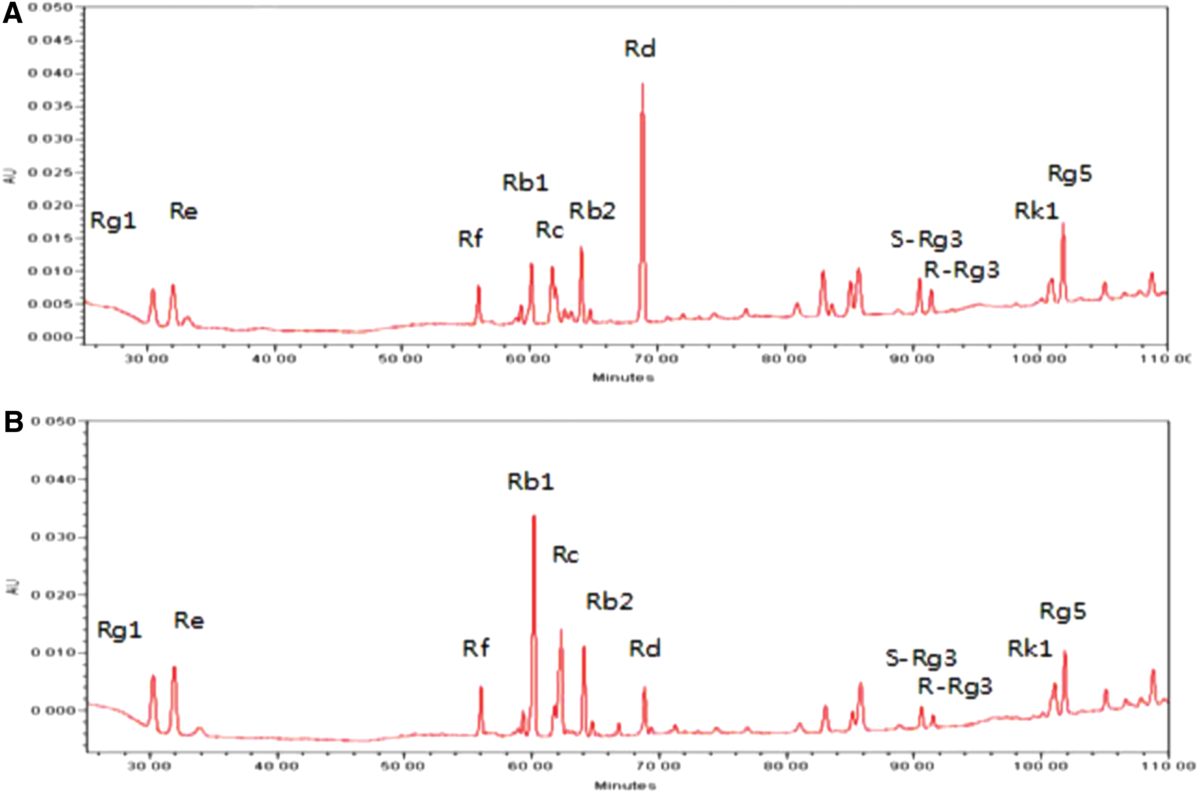
HPLC chromatograms of GS-E3D
Effect of GS-E3D on hyperglycemia
Hyperglycemia was induced in the STZ-injected rats (335.3 ± 93.0 mg/dL). The blood glucose levels of GS-E3D-treated rats were 327 ± 146.1, 330.8 ± 75.8, and 349.8 ± 43.4 mg/dL at doses of 25, 50, and 100 mg/kg/day, respectively. UGE also failed to reduce the blood glucose level (341.7 ± 89.4 mg/dL). No difference was noted between any of the diabetic groups.
Effect of GS-E3D on diabetes-induced retinal vascular hyperpermeability
In the diabetic rats, retinal vessels with an increased fluorescence intensity and decreased delineation of the vessels were highly detected, indicating that the BRB was degraded. However, GS-E3D and UGE prevented these vascular changes (Fig. 2A). The quantitative results showed that GS-E3D dose dependently reduced BRB leakage (Fig. 2B). The inhibitory effect of GS-E3D on vascular hyperpermeability was stronger compared with UGE (P < .05). Therefore, GS-E3D could protect BRB integrity in diabetic rats.
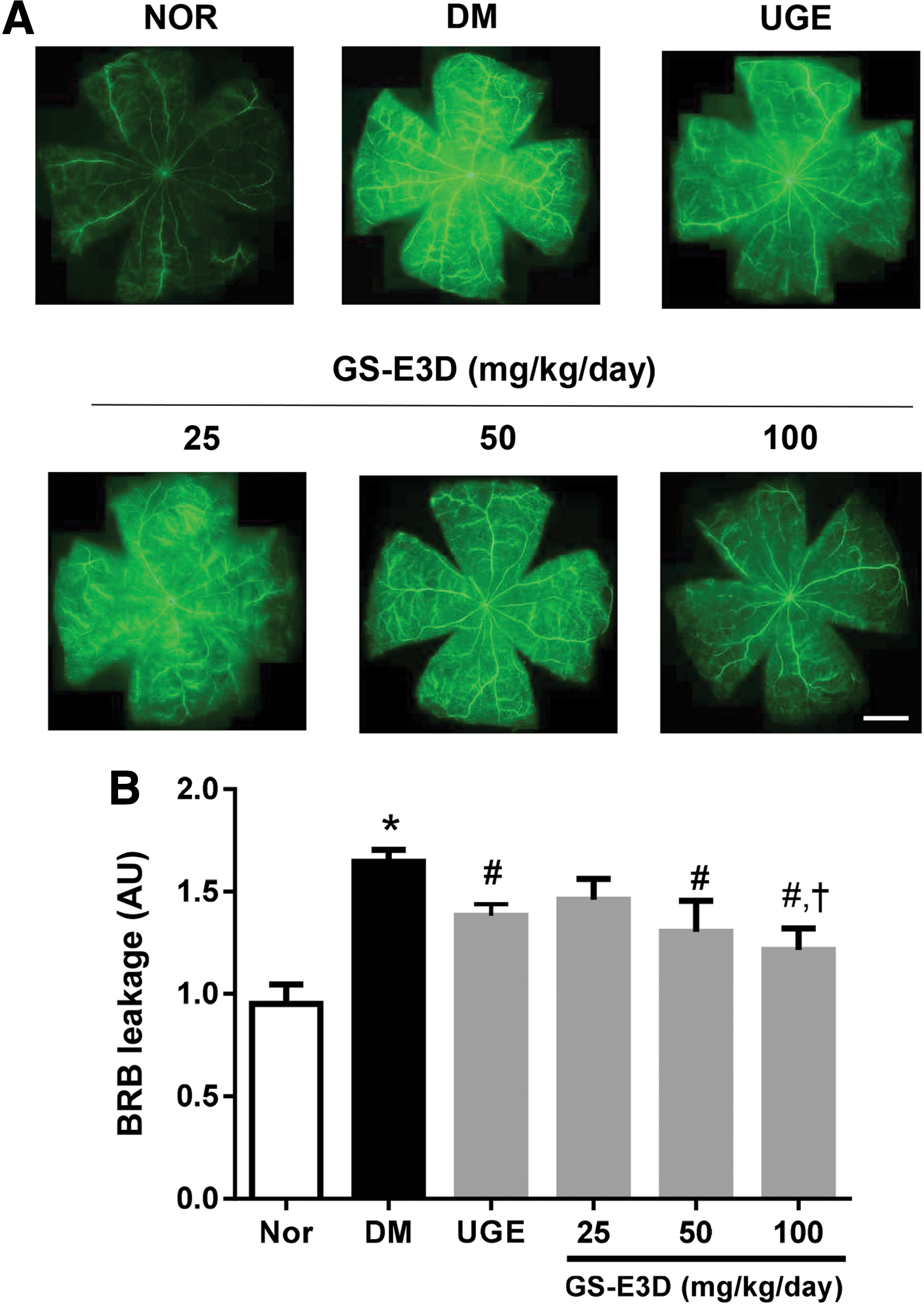
Blood-retinal barrier breakdown.
Effect of GS-E3D on tight junction protein loss
In diabetic retinopathy, increased retinal vascular permeability is closely associated with loosening of the tight junctions and loss of the proteins, occludin and claudins, in the junction. 28 Immunofluorescence staining for occludin showed that GS-E3D and UGE inhibited the loss of occludin in diabetic rats (Fig. 3A). We also examined a Western blot analysis for occludin. The reduction of occludin was ameliorated by GS-E3D treatment in a dose-dependent manner (Fig. 3B). The effect of GS-E3D on tight junction protein loss was stronger compared with UGE (P < .05).

Tight junction protein loss.
Effect of GS-E3D on diabetes-induced retinal vascular apoptosis
In diabetic retinopathy, dysfunction in vascular endothelial cells and pericytes leads to abnormal function of retinal vasculature. 29,30 As shown in Figure 4A, the retinas in the diabetic group had many apoptotic cells in the retinal vessel, whereas GS-E3D- or UGE-treated diabetic rats had few apoptotic cells. GS-E3D dose dependently decreased the number of TUNEL-positive cells, and its antiapoptotic activity was stronger compared with UGE (Fig. 4B, P < .05). These results indicated that several retinal vascular cells were undergoing apoptosis, but GS-E3D could prevent the diabetes-induced apoptotic injury of vascular cells in diabetic rats.
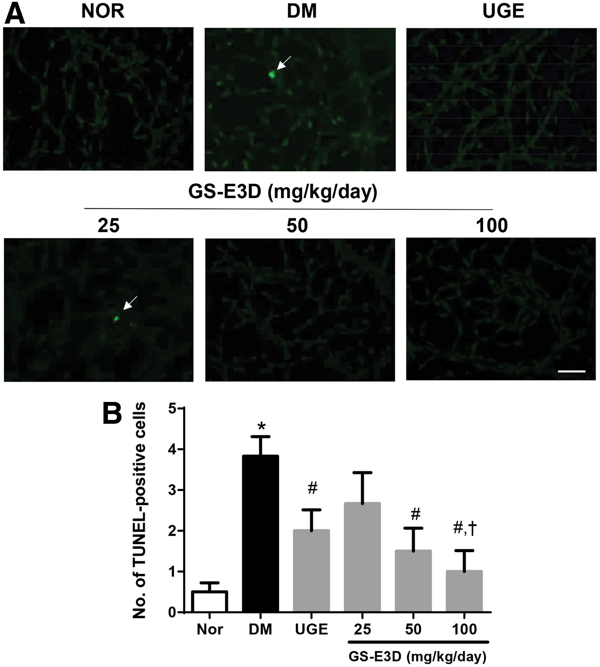
Retinal vascular cell apoptosis.
Effect of GS-E3D on AGE accumulations
We performed immunofluorescence staining for AGEs to identify the retinal AGE accumulations. As shown in Figure 5A, immunofluorescence staining clearly showed AGE accumulation in the retinal vessels of diabetic rats. However, retinal accumulation of AGEs was inhibited by the treatment with GS-E3D or UGE (Fig. 5B). Similarly, UGE treatment also resulted in a decrease in retinal AGE accumulations, but this effect was less for UGE than for that of GS-E3D (P < .05).
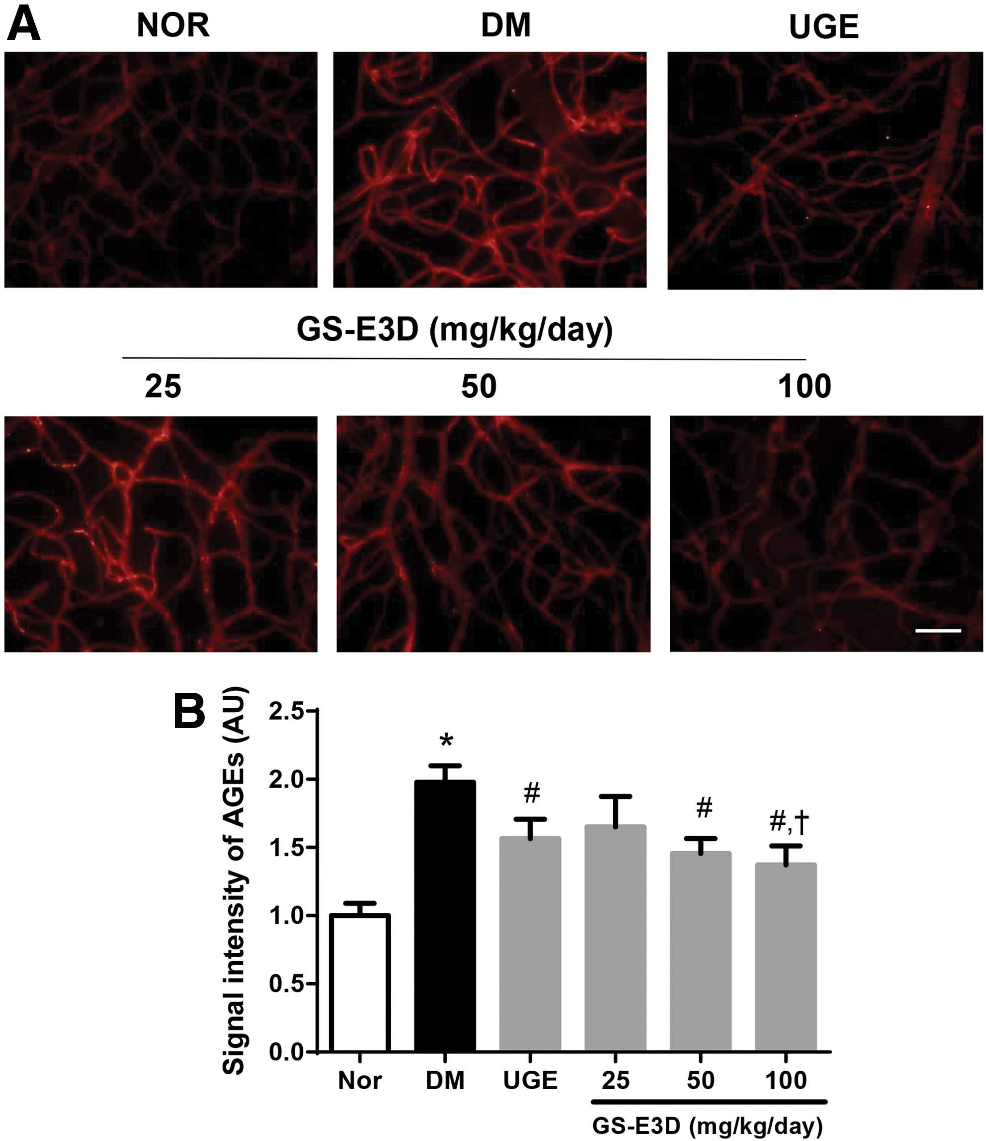
Retinal AGE accumulations.
Discussion
The BRB plays a crucial role in maintaining the retinal microvascular integrity. The BRB breakdown is the earliest characteristic sign of diabetic retinopathy. 31 Occludin participates in the maintenance of vascular permeability and BRB integrity. 32 Diabetic animals induced by an injection of STZ also have increased retinal vascular permeability. 33,34 In this study, we showed that GS-E3D significantly inhibited BRB leakage and occludin loss. These results suggest that GS-E3D can protect the diabetes-induced retinal microvascular leakage.
The enhanced formation of AGEs and its accumulation are considered a causative factor of diabetic retinopathy. 6,7 When exogenous AGEs were injected i.v. repeatedly into normoglycemic animals, vascular basement membrane thickening, 35 the adhesion of leukocytes, 36 and BRB breakdown 11 were developed in the retinas. Several previous studies showed that AGEs activate proapoptotic signaling pathways in the retinal pericytes. 37 –39 The usage of anti-AGEs agents has been proposed as a promising pharmacological strategy for preventing diabetic retinopathy. 40 Several anti-AGE inhibitors can improve diabetic retinopathy. 40 –42 In a previous study, aminoguanidine, a well-known antiglycation agent, inhibited the retinal vascular injury in diabetic animals. 13 Another type of AGE inhibitor, called an AGE breaker, was able to destroy the existing AGE crosslinks. Several potent AGE breakers, such as alagebrium, 43 LR20, LR23, LR90, 44,45 and C36 46 have been described. These AGE breakers reduced the AGE overload in blood vessels 47 and heart. 48 Retinal microvascular cell apoptosis mediated by AGEs plays a crucial role in the major pathological process of diabetes-induced microvascular cell loss. 10,39 Therefore, a prevention of AGE overload can ameliorate diabetes-induced retinal microvascular damage. Collectively, we suggest that antiapoptotic and anti-AGEs activities of GS-E3D might have a retinoprotective effect in diabetic animals.
Recently, we have shown that several herbal extracts derived from Aster koraiensis, 49 or Litsea japonica 25,50 prevented diabetic retinopathy by inhibition of AGE accumulation. Ginseng extract decreased the generation of AGEs in the diabetic rat kidney 51 and reversed the altered expression of genes involved in apoptosis and inflammation in the diabetic rat retina. 52 Ginsenoside Rd, a bioactive compound of ginseng, inhibited methylglyoxal-induced apoptosis in astrocytes. 53 Recently, our study reported that GS-E3D showed a stronger anti-AGE activity (IC50 = 19.65 ± 4.35 μg/mL) compared with UGE (IC50 = 139.46 ± 68.18μg/mL). 26 In addition, GS-E3D also exerted stronger inhibitory potential on BRB breakage compared with UGE. In the HPLC analysis, the contents of ginsenosides in GS-E3D were changed after the enzymatic modification by pectin lyase. Ginsenosides Rb1, Rb2, Rb3, and Rc were enzymatically converted to ginsenoside Rd in the GS-E3D. The oral bioavailability of some ginsenosides such as ginsenosides Rb1 and Rb2 from the intestines is extremely low. 54 Liu et al. reported that ginsenoside Rd was detectable in plasma up to 48 h after oral administration of Panax notoginseng extract, whereas ginsenoside Rc was only detected in nanomolar plasma levels at 6–10 h after oral dosing. 55 Ginsenoside Rb1 upregulated the expressions of glucose transporters and promoted the glucose consumption in adiopcytes. 56 Ginsenoside Rb2 enhanced AMP-activated protein kinase (AMPK) signaling pathways and significantly improved glucose tolerance. 57 Ginsenoside Rb3 increased antioxidant contents and improved insulin action and glucose intolerance in the experimental diabetic animals. 58 Ginsenosides Rd also improved insulin signaling and prevented apoptosis of astrocytes. 53 In our previous report, GS-E3D showed a stronger anti-AGE activity compared with UGE. 26 GS-E3D has potent AGE inhibitory activities in vitro and in vivo, its mechanism of action still remains unclear. Collectively, we showed that GS-E3D had more potent activities than UGE. Its enhanced pharmacological activities may be due to an increased level of ginsenoside Rd.
GS-E3D could protect the diabetes-induced retinal injury without a glucose reduction. In a previous study, the oral administration of UGE (1025 mg/kg/day) ameliorated hyperglycemia in a diabetic mouse model. 59 Although GS-E3D could not improve glucose intolerance, the oral dosage of GS-E3D was about 10-fold less. Moreover, GS-E3D significantly prevented retinal injury through inhibition of retinal AGE overload without a glucose reduction. Similarly, aminoguanidine, a well-known AGE inhibitor, had no effect on fasting blood glucose in STZ-induced diabetic rats, but inhibited AGE deposition in the retinal capillaries and delayed the onset of diabetic retinopathy. 60 AGE production is mainly related to increased serum glucose levels. However, various AGE inhibitors, such as aminoguanidine and LR-90, could prevent the development of diabetic retinopathy without lowering blood glucose. In our previous report, GS-E3D had a potent anti-AGE activity and its inhibitory activity was stronger compared with aminoguanidine and UGE. These results suggest that GS-E3D could prevent retinal vascular injury by inhibiting AGE burden in experimental diabetic animals.
In conclusion, this is the first study to provide several direct evidences that GS-E3D has a retinoprotective effect in vivo. GS-E3D could protect diabetic animals from AGE-induced retinal vascular injury, and this might be due to its anti-AGE activity.
Footnotes
Acknowledments
This research was supported by the Korea Institute of Planning and Evaluation for Technology in Food, Agriculture, Forestry and Fisheries (IPET) funded by the Ministry of Agriculture, Food and Rural Affairs (315049-05-2-CG000). Part of this study was carried out with the support of R&D Program for Forest Science Technology (Project No. 2017040B00-1819-BA01) provided by Korea Forest Service (Korea Forestry Promotion Institute).
Author's Contributions
E.J. performed the experiments and wrote the article; C.S.K., W.J., S.B.P., and M.K.P. performed the experiments and analyzed the data; and J.K. designed and supervised the study.
Author Disclosure Statement
No conflicts of interest, financial or otherwise, are declared by the authors.