
Abstract
Chlorogenic acid (CGA) is a major component of green coffee beans. Surfactin, a cyclic lipopeptide, is produced and secreted by Bacillus subtilis strains. In this study, bioactivities of fermented green coffee bean extract (FGCBE) and the individual compounds, CGA and surfactin. were compared in HepG2 cells. The concentration of surfactin and CGA in the FGCBE and non-fermented green coffee bean extract (NFGCBE) were determined to be 9.2 and 7.33 and 0.72 and 0.53 mg·mL−1, respectively. The FGCBE contained about 20% and 26% more CGA and surfactin than the NFGCBE. Although CGA and surfactin exhibited cytotoxicity at concentrations more than 100 and 20 μg respectively, the FGCBE 50 containing CGA (460 μg·mL−1) and surfactin (720 μg·mL−1) effectively prevented cell death by oxidative stress and also strongly activated the proliferation of cells incubated with under 50 μM H2O2. The CGA and surfactin in FGCBE were 9.2 and 72 times higher than the CGA and surfactin compounds (50 and 10 μg·mL−1). The relative proliferation of the FGCBE-treated cells also was 3.3 and 8.8 times higher than the CGA and surfactin compounds treated the oxidative stressed cells with 50 μM H2O2. These results suggest that the single compounds such as CGA and surfactin generally have cytotoxicity at low concentration of them but FGCBE contained them acted as strong antioxidants, activators of cell proliferation, inhibitors of cell apoptosis. Various bioactive compounds in fermented coffee bean also seem to help cell proliferation and decreasing of cytotoxicity by CGA and surfactin in coffee bean.
Introduction
Chlorogenic acid (CGA) is one of the most abundant polyphenol compounds in the human diet. It is a major phenolic compound that can be isolated from the leaves and fruits of plants. 1 Green coffee beans contain the largest amounts of CGA found in plants, ranging from 6% to 12%. 2 CGA, a known antioxidant, has been reported to have anti-inflammatory properties, anticarcinogenic activity, and protective effects on neuronal cells. 3 –6 CGA is used in various additives for beverages, cosmetics, tea products, and foods, as well as medical substances. 7 Although there are many biological benefits for CGA, CGA has side effects including increasing of blood pressure, plasma homocysteine and cytotoxicity. 8 –10
It consists of the ester of caffeic acid and quinic acid. The main subgroups of CGAs are 3-O-caffeoylquinic acid (n-chlorogenic acid), 5-O-caffeoylquinic acid (neo-chlorogenic acid), and 4-O-caffeoylquinic acid (crypto-chlorogenic acid). n-Chlorogenic acid is the most abundant compound in green coffee bean. 1
Surfactin is a prominent surfactant and is used as an antibiotic. The lipopeptide, surfactin, is synthesized by a bacterial surfactin synthase and aids in the activation of plasminogen in blood. The subtypes of surfactin are characterized by the same peptide length, but different residues. 11,12 In various studies, surfactin has been found to exhibit beneficial functions, including antibacterial, antiviral, antifungal, and anti-mycoplasmal activities. 13,14 Unfortunately, surfactin is not able to be used as a medicine because it has cytotoxicity such as hemolysis in normal cells. 15
Reactive oxygen species (ROS) are produced by the normal metabolism of oxygen and play important roles in cell signaling and homeostasis. 16 ROS are also responsible for the induction of apoptosis, that is, programmed cell death. 17 ROS are chemically reactive molecules containing oxygen. There are various ROS compounds, including the superoxide anion (O2 −), hydroxyl radical (·OH), and hydrogen peroxide (H2O2), which cause oxidative deterioration of deoxyribonucleic acid (DNA), proteins, and lipids, and accelerate aging and mitochondrial death in cells. 18
In this study, green coffee bean was fermented by Bacillus subtilis, and concentrations of CGAs and surfactin of the fermented green coffee bean extract (FGCBE) were compared with non-fermented green coffee bean (NFGCBE). These major compounds and the extract were applied to liver hepatocellular carcinoma (HepG2) cells to evaluate their cytotoxicity, viability and proliferation on oxidative stress. This study also documents the effectiveness of FGCBE for treating oxidatively stressed HepG2 cells through the comparison of medicinal efficacy between the compounds and FGCBE.
Materials and Methods
Materials
CGA (>95%; HPLC grade; p-no. C3878-1G, catalog no. 327-97-9), neo-chlorogenic acid (>98%; HPLC grade; p-no. 94419-10 MG), and surfactin (>98%; HPLC grade; p-no. s3523-10 mg) were purchased from Sigma-Aldrich (St. Louis, MO, USA). Acetonitrile (HPLC grade) was obtained from Merck KGaA (Darmstadt, Germany) and analytical grade phosphoric acid (49–51%; HPLC grade) was procured from Sigma-Aldrich. Trifluoroacetic acid (99%; p-no. T6508-500ML, catalog no. 76-05-1) was obtained from Sigma-Aldrich and methanol was from obtained from Merck KGaA (Darmstadt, Germany). The solvents were filtered through a 0.45 μM membrane filter (Agilent Technologies; Santana Clara, USA). The fermented (Coffea arabica) and non-fermented GCBE were manufactured by Leader Green (Kyeong-gi, Korea). The ground green coffee beans (120 mesh) were diluted with distilled water and fermented by Bacillius megaterium (ATCC 14945) for 30 days at 37°C.
Apparatus and operation conditions
Liquid chromatography
Chromatographic analysis was performed on a Thermo Scientific Dionex Ultimate 3000 system (Thermo Fisher Scientific, Inc., Germering, German) consisting of a binary pump solvent management system and an auto-sampler. Hydrosphere C18 (150 × 4.6 mm, I.D. S-5 μm, 12 nm (p/n HS12S05-1546WT)) and YMC-triart C18 (250 × 4.6 mm I.D. S-5 μm, 12 nm (p/n TA12S05-2546WT)) columns were employed and the column temperature was maintained at 40°C. The UV detector was set at 324 nm for CGA and 205 nm or 210 nm for surfactin. The mobile phase was prepared daily, filtered under vacuum through a 0.45 μm membrane filter, and degassed before use. The flow rate was set at 1.0 mL·min−1.
Standard solution and sample preparation
The respective CGA standards (n-chlorogenic acid and neo-chlorogenic acid; 10 mg·mL−1) were dissolved in deionized distilled water and working standards were prepared at concentrations of 0.1565, 0.3125, 0.625, 1.25, 2.5, 5, 10, and 20 mg·mL−1. The identification and quantitation of cypto-chlorogenic acid were performed in reference to another study. 19 The surfactin standard (10 mg·mL−1) was dissolved in methanol and working standards of surfactin were prepared at concentrations of 0.3125, 0.625, 1.25, and 2.5 mg·mL−1. Each standard was injected (10 μL) by an auto-sampler. FGCBE and NFGCBE were dissolved in deionized distilled water. The respective solutions were filtered through a 0.45 μm membrane filter paper and the clear filtrate (diluted in ratios of 1:100 and 1:500) was used for high performance liquid chromatography (HPLC) analysis.
Cell culture
A human liver carcinoma cell line (HepG2 cells) was cultured in RPMI1640 medium supplemented with penicillin (100 U·mL−1), streptomycin (100 μg·mL−1), and 10% FBS (Invitrogen, USA). The HepG2 Cells were maintained in a humidified atmosphere of 5% CO2 at 37°C. The medium was then replaced and the cells were cultured for an additional 2 days in RPMI. The cells were seeded at a density of 2 × 10 6 cells/well in a 100pi disk or 1 × 10 4 cells/well in a 96-well plate for 24 h before each treatment.
Cell viability assay
The cell viability was determined using the MTT assay (Promega cell proliferation assay; p-no. G3580, Madison, USA). HepG2 cells were treated with CGA, surfactin, FGCBE, NFGCBE, and H2O2 and seeded at a density of 1 × 10 4 cells in a 96-well culture plate and incubated at 37°C with 5% (v/v) CO2 for 24 h. A 20 μL aliquot of MTT was added to each well and incubated for 3 h. After 3 h of incubation, the absorbance was measured at a wavelength of 490 nm using a microplate spectrometer (Chromate, Awareness Technology, Inc., the Netherlands). All experiments were performed in triplicate.
Statistical analysis
All data were analyzed by ANOVA using SPSS software. When the P-values were found to be significant (P < .05), the means were compared using Tukey's method.
Results
Contents of CGAs and surfactin in fermented green coffee bean
The CGA and surfactin in the FGCBE were analyzed by UHPLC (Fig. 1). The retention times of neochlorogenic acid and CGA were 5.93 and 9.19 min, respectively. The seven peaks of the surfactin isomers were detected by UHPLC, and the corresponding peak numbers and retention times were 1–10.36, 2–12.47, 3–16.18, 4–17.27, 5–18.67, 6–20.81, and 7–22.35 min (Fig. 1b). The standard plots of CGA and surfactin are displayed based on analysis of the standards by UHPLC (Fig. 2a, b). The concentrations of surfactin and CGA in the FGCBE and NFGCBE were measured to be 9.2 and 7.33 and 0.72 and 0.53 mg·mL−1, respectively. The FGCBE contained about 20% and 26% more of the respective compounds than NFGCBE (Fig. 2c).

Analysis of chlorogenic acid and surfactin in the extract by UV-UHPLC.
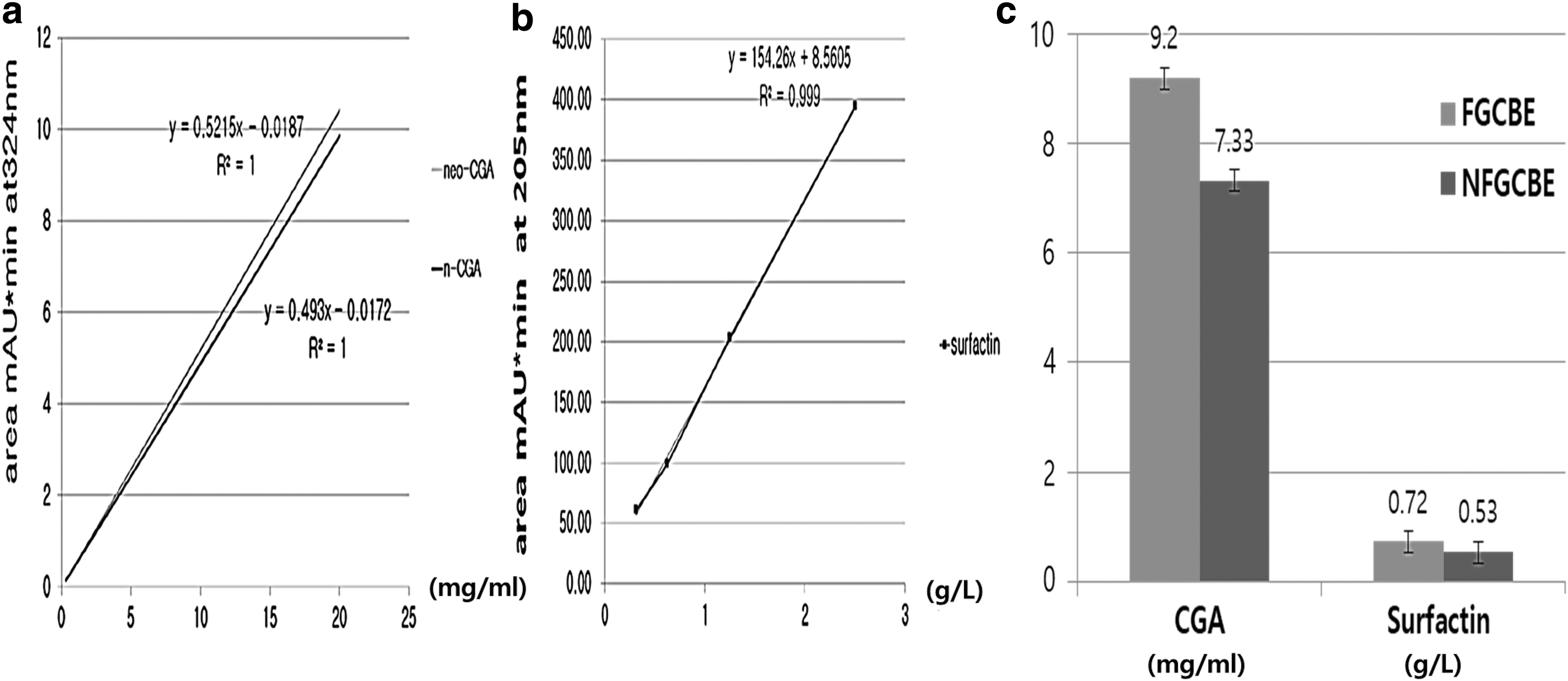
Standard curves and measurement of chlorogenic acid and surfactin in fermented GCBE and non-fermented GCBE.
Viability, proliferation, protection activity and cytotoxicity of CGA and FGCBE for oxidatively stressed HepG2 cells
Based on the cell viability assay (Figs. 3 and 4), the treatment dosages were selected as H2O2 (10–100 μM), surfactin (10 μg·mL−1), CGA (50 μg·mL−1), and FGCBE (50 μg·mL−1). The proliferation and inhibition of the HepG2 cells were measured by MTT assay after 24 h incubation of the cells treated with CGA, surfactin, or FGCBE.

Cell viability determined by MTT assay. The cells were treated with CGA, surfactin, NFGCBE, and FGCBE for 24 h.
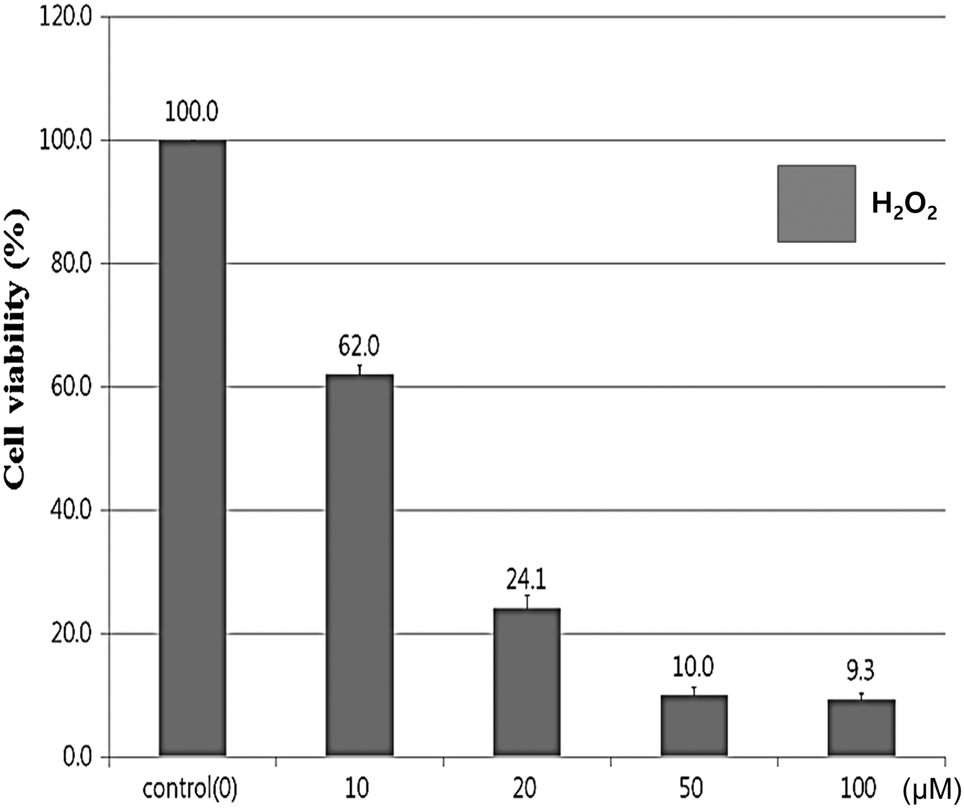
Cytotoxicity of H2O2 toward HepG2 cells. The cells were incubated with different dosages (0–100 μM) of H2O2 for 24 h. The cell viability was evaluated by the MTT assay method. The values are expressed as the average ± SD of triplicate experiments (P < .05).
CGA activated proliferation of the cells and prevented cell death, both in non-stressed and oxidatively stressed environments under 20 μM H2O2 (Fig. 5a). Although the viability of the oxidatively stressed HepG2 cells without CGA declined dramatically, the proliferation of the stressed cells treated with CGA increased dramatically (Fig. 5a, b). In 10–50 μM H2O2, the relative value of the proliferation of the stressed cells was measured at 3.62, 6.47, 3.94 and 2.1, and the peak value was obtained with 20 μM H2O2 (Fig. 5b). Notably, although the cells had cytotoxic damage at over 100 μg·mL−1 CGA (Fig. 3b) the oxidatively stressed HepG2 cells exposed to the low concentrations of CGA increased their viability and proliferation (Figs. 3 and 5).
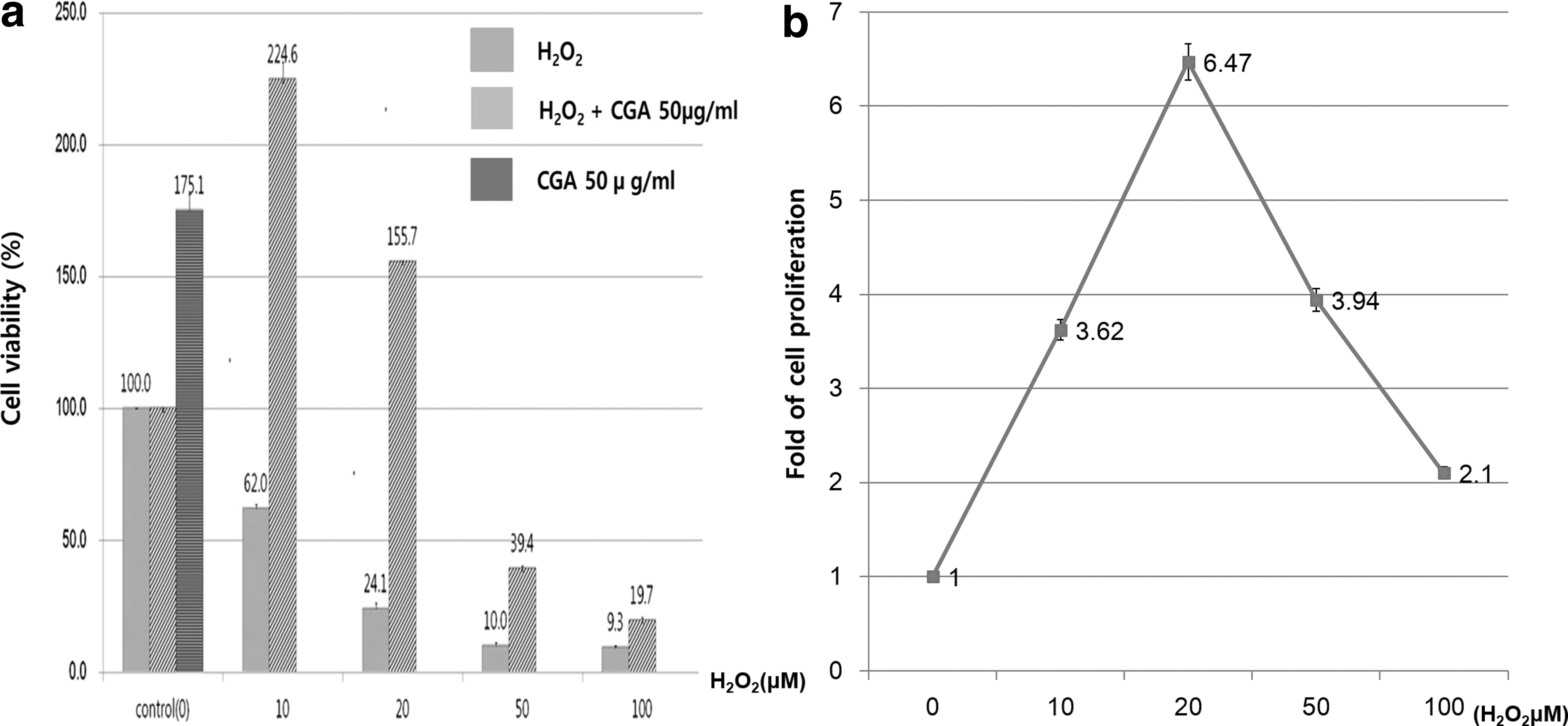
Effect of CGA on oxidatively stressed HepG2 cells. The cells were incubated with H2O2 and CGA for 24 h.
The best efficacy for activation of cellular proliferation as well as prevention of cell death was achieved with 50 μg·mL−1 FGCBE (Fig. 6). CGA in FGCBE 50 were calculated at 460 μg·mL−1 and the HepG2 cells exposed to the FGCBE50 exhibited high cell viability under 50 μM H2O2 (Fig. 6). In 10–100 μM H2O2, the relative value of the proliferation of the stressed cells was measured at 2.9, 7.4, 13.1 and 1.8, and the peak value was obtained with 50 μM H2O2 (Fig. 6b). FGCBE effectively prevented cell death due to oxidative stress and also strongly activated cell proliferation with the use of 50 μM H2O2 (Fig. 6b). Especially, the CGA concentration in FGCBE 50 was 72 times higher in CGA 50 and the value of relative cell viability in FGCBE 50 were highest at 50 μM H2O2 (Fig. 7).
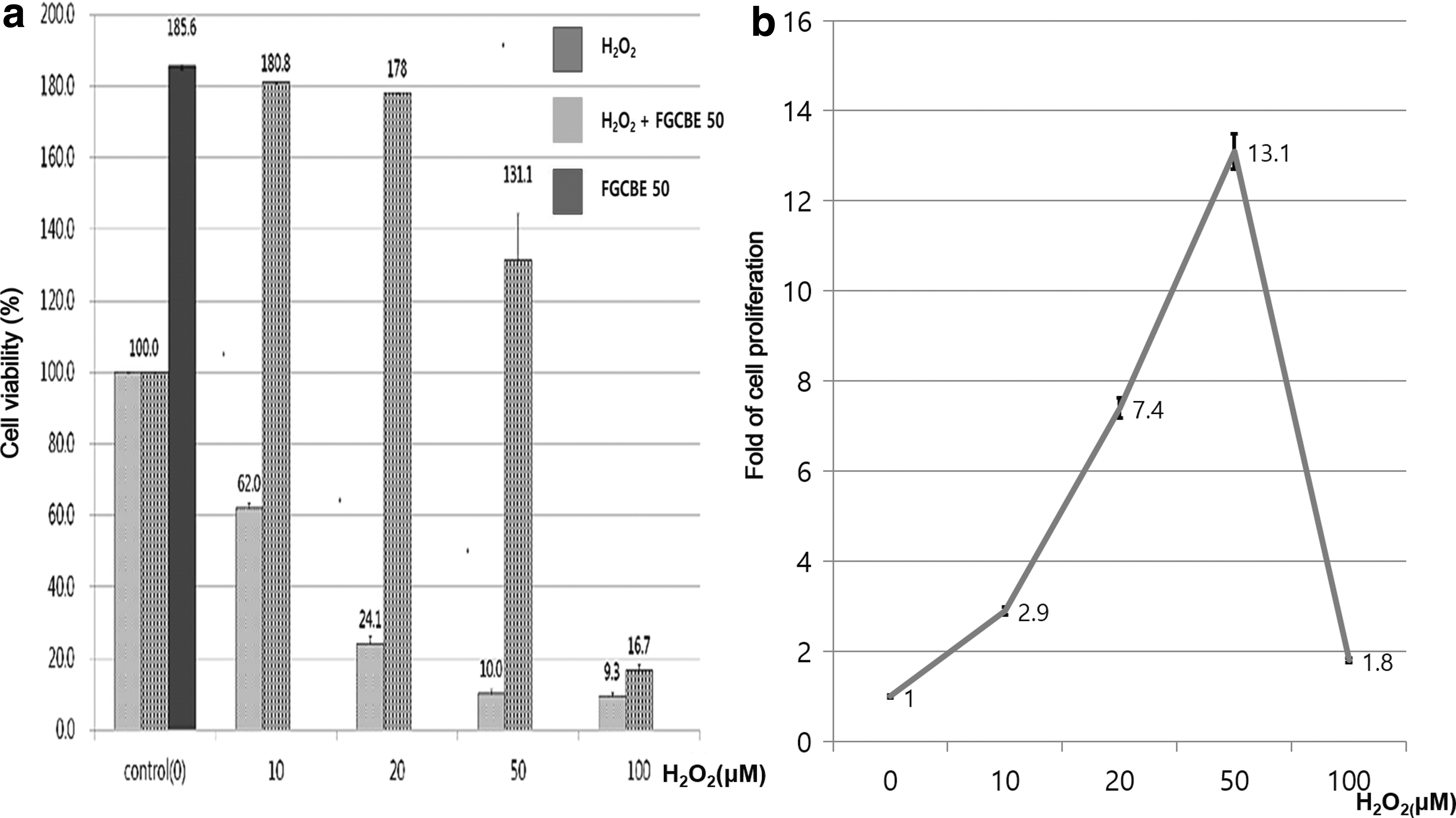
Effect of FGCBE on oxidatively stressed HepG2 cells.

Comparison of cell viability by treating of CGA and FGCBE on oxidatively stressed HepG2 cells.
Cytotoxicity and viability between surfactin and FGCBE for oxidatively stressed HepG2 cells
Surfactin (10 μg·mL−1) synergistically activated cytotoxicity against oxidatively stressed HepG2 cells (Fig. 8a). Notably, 10 μM H2O2 induced strong cytotoxicity, which was 2.7 times higher than that observed with the control when the HepG2 cells exposed to H2O2 were treated with surfactin (Fig. 8b). The concentration of surfactin in FGCBE 50 were measured at 720 μg·mL−1. This value was 72 times higher than the single compound, 10 μg·mL−1 surfactin (Fig. 9a). The more surprising observation was that FGCBE treated cells showed higher viability than the surfactin 10 treated cells under oxidative stress. The relative values for viability were 7.9, 9.8, 8.9 and 1.2 at 10–50 μM H2O2 (Fig. 9b, c).

Cytotoxicity of surfactin for oxidatively stressed HepG2 cells.

Comparison of cell viability by treating of surfactin and FGCBE on oxidatively stressed HepG2 cells.
Discussion
CGA is usually extracted from green coffee bean using hot water, methanol, or ethanol. 20 In contrast with other studies, this study used an extract from green coffee bean fermented by B. subtilis. The efficiency of this fermentation method was determined by quantitation of the CGA content in the green coffee bean extract. Quantitation of CGA was performed with HPLC, ultra high performance liquid chromatography (UHPLC), and liquid chromatography coupled with tandem mass spectrometry (LC/MS/MS). 21,22 UHPLC with UV detection is a sensitive, precise, accurate, and linear method for the routine analysis of GCA. Herein, we analyzed the content of CGA (9.2 mg·mL−1) and surfactin (0.72 mg·mL−1) in the FGCBE. Previous studies that quantified CGA documented nine peaks of CGA isomers in the HPLC spectrum. 23 Three of these isomers, that is, n-chlorogenicacid (3-CQA), neo-chlorogenic acid (5-CQA), and crypto-chlorogenic acid (4-CGA), accounted for over 90%. 23 In this study, two CGA isomers standards were used for calibration, and the total concentration of these isomers in FGCBE and NFGCBE was about 9.2 and 7.33 mg·mL−1, respectively (P < .05). The concentration of CGA in FGCBE was 20% higher than that in NFGCBE. This suggests that bacterial fermentation increases the concentration of CGA in FGCBE via bacterial metabolism. According to other studies, various independent variables, including the extraction temperature and fermentation and extraction time, significantly affected the determined content of medicinal compounds in an extract. 24 –26 Therefore, the processing conditions must be carefully optimized to obtain an excellent extract. H2O2 is known to induce apoptosis through the ROS pathway. 27 Previous reports have demonstrated the cytotoxic effects of CGA on several human cell types, including oral squamous cell carcinoma (HSC-2), salivary gland tumor (HSG), and chronic myeloid leukemia cell lines. 24,25 CGA also induced cell death at concentrations exceeding 200 μM. 26 In this study, HepG2 cell exposed to CGA over 100 mg·mL−1 were increased dramatically their apoptosis. In contrast, HepG2 cell exposed to FGCBE 50 containing high concentration of CGA (460 mg·mL−1) prevented cell apoptosis (Fig. 7). Also, the relative viability of the FGCBE 50-treated cells was higher than that of the CGA treated cells in an oxidative-stress environment (Fig. 7b, c). These results suggest that CGA possesses cytotoxic activity at higher concentrations, but metabolites in FGCBE increase cell viability and decreases the cytotoxicity of high CGA in the cells.
Surfactin is composed of a heptapeptide cycle (L-Glu–L-Leu–D-Leu–L-Val–L-Asp–D-Leu–L-Leu) closed by a C13–C15 hydroxy fatty acid to form a lactone ring system. 28 The C15-surfactin is a major isoform in Bacillus subtilisnatto. 29 In this study, we determined the quantity of C15-surfactin in FGCBE and NFGCBE. Analysis of the surfactin standard by UHPLC showed seven peaks, and the concentration of surfactin in FGCBE and NFGCBE was measured to be 0.73 and 0.53 mg·mL−1, respectively (Fig. 2). The concentration of surfactin in FGCBE was 26% higher than that in NFGCBE. Surfactin has bioactive characteristics, including antibacterial, antifungal, antiviral, anti-mycoplasma, and hemolytic activity. 30 Nevertheless, surfactin has critical drawbacks, such as non-specific cytotoxicity, and causes hemolysis at concentrations of 40–60 μM. Fortunately, the toxic effects of surfactin are not significant at concentrations below 25 μM. 26 In various reports, 31,32 surfactin has been shown to induce apoptosis, but little is known about how the combination of surfactin and other metabolites produced by the bacterial fermentation affect cell viability and cytotoxicity.
This study compares the cytotoxicity and cell viability between surfactin and FGCBE containing surfactin at high concentration in HepG2 cells. Although the surfactin (10 μg·mL−1)-treated cells did display an increasing of cytotoxicity in HepG2 cells (Fig. 8), the cells treated with FGCBE 50 displayed a dramatic reduction in the cytotoxicity (Fig. 9). These results indicate that the single compounds, surfactin has strong cytotoxicity for cancer cells but the strong cytotoxicity also has a side effect of cytoxicity to normal cells. In contrast, metabolites in FGCBE containing high surfactin reduces the cytotoxicity in normal cells as well as increasing of anti-oxidant stress.
Generally, fermented extracts contain more medicinal compounds than the non-fermented congeners. 33,34 In this study, FGCBE was also found to contain more CGA and surfactin than NFCBE (Fig. 2c). The relative value of the proliferation for the FGCBE-treated cells was higher than that achieved with the control. Moreover, the FGCBE-treated cells displayed a dramatic increase in the cell proliferation with the use of 20 and 50 μM H2O2. In a recent study, 35 the NFGCBE (100 μg·mL−1) induced cytotoxicity was examined in various cell types including human lung, colorectal, oesophageal and urinary bladder carcinomal cells. However, treatment with FGCBE (100 μg·mL−1) activated cell viability and proliferation. This result means that FGCBE has potentially positive effects as antioxidant for various organs in human body. Consequently, these results indicate that FGCBE contains effective antioxidants, such as CGA, that are generated by fermentation, and the extract was also effective for preventing cell senescence, death, and apoptosis.
In conclusion, this study demonstrates the improved function, including anti-oxidative, anti-cancer, and activation of cell proliferation, of FGCBE. CGA and surfactin in FGCBE functioned as effective antioxidants and FGCBE exhibited improved anti-cancer efficacy, At adequate concentrations, CGA also activated the proliferation of HepG2 cells.
Footnotes
Acknowledgment
This research was supported by Korea University (K1808591) and the Agriculture, Food, and Rural Affairs Research Center Support Program (714001-07), Ministry of Agriculture, Food, and Rural Affairs.
Author Disclosure Statement
No competing financial interests exist.