
Abstract
To examine the anti-metastatic activities of polysaccharides in broccoli, purified polysaccharides (BCE-I, -II, and -III) were isolated by fractionation of broccoli enzyme extracts and subsequent ethanol precipitation. BCE-I mainly consisted of galactose and arabinose, whereas BCE-II mainly consisted of galacturonic acid and rhamnose, and BCE-III mainly consisted of rhamnose and galactose. Of the three fractions, stimulation of murine peritoneal macrophages by BCE-I showed the greatest enhancement of tumor necrosis factor-α, interleukin (IL)-12, and IL-6 secretion. In addition, intravenous (i.v.) administration of BCE-I enhanced the lethal activity of natural killer (NK) cells on YAC-1 tumor cells significantly and dose-dependently in an ex vivo experiment of NK cell activity. In an experimental model using lung metastasis of Colon26-M3.1 carcinoma cells, prophylactic i.v. and oral administration of BCE-I significantly and dose-dependently inhibited lung metastatic activity. Furthermore, the inhibitory activity of BCE-1 on lung metastasis partially disappeared when NK cell function was removed through treatment of rabbit anti-asialo GM1. These results indicated that BCE-I has potent antitumor metastatic activity, and that its anti-metastatic activity has relevance to the stimulation of NK and other immune cells.
Introduction
Broccoli (Brassica oleracea) is a vegetable belonging properly to the Brassicaceae family, and it is cultivated in multiple locations of the world such as the eastern Mediterranean, Asia, the United Kingdom, and the United States. 1 Broccoli consists of many functional materials and chemical substances such as glucosinolates, sulforaphane, flavonoid, and selenium that are considered to have various health-promoting activities. 2 For example, glucosinolates, mainly found in broccoli, have been shown to decrease the risks of breast cancer, 3 lung cancer, and prostate cancer. 4 Sulforaphane from broccoli has been shown to have positive health effects on hypoglycemia, Alzheimer's disease, Parkinson's disease, and inflammation. 5 In addition, it reduces insulin resistance. 1
In the past, polysaccharides have been recognized only as energy sources; however, recently, plant-base polysaccharides have gained attention because of their multifunctional biological activities such as decreasing inflammation, 6 preserving skin moisture, 7 participating in the complement system, 8 increasing macrophage and lymphocyte proliferation, 9 and exhibiting anti-metastasis activity. 10 Moreover, plant-derived polysaccharides show relatively low toxicity, so they have the potential to be used as therapeutic agents. 11
Many polysaccharides from plants have been known to stimulate the host defense system by activating macrophages 11 and modulating the immune cells such as natural killer (NK) cells, dendritic cells, and T cells. 12 In this study, the activation of macrophages and NK cells has been examined, because of the fact that they are the major constituents of innate immune system. NK cells, especially, have shown antitumor metastatic effects in vivo and in vitro. 13 Therefore, we intensively studied the antitumor metastatic activities of the polysaccharides isolated from broccoli.
In this research, we first isolated crude polysaccharides from pectinase-treated broccoli. Then, we separated crude polysaccharides into three fractions. We identified sugar components of the three fractions and investigated cytokine secretion in murine peritoneal macrophages in response to each fraction. Among the three fractions, BCE-I, exclusively, increased cytokines; therefore, we used BCE-I in further studies. To measure of the antitumor metastatic activity of BCE-I, we used mouse lymphoma YAC-1 cells and mouse Colon26-M3.1 carcinomas. To study the relationship between anti-metastatic activity and NK cells, we used anti-asialo GM1 to block NK cell activity.
Materials and Methods
Materials and reagents
Broccoli (Brassica oleracea var. italica) was cultivated in Jeju, Korea, and harvested in July 2015. Plantase MAX originated from Aspergillus niger was purchased from Bision Co. (Sungnam, Korea). Sephadex G-75 was obtained from GE Healthcare Life Sciences (Uppsala, Sweden). Dulbecco's modified Eagle's medium (DMEM) and hybridoma-serum-free media (SFM) were purchased from Gibco BRL Co. (Grand Island, NY, USA), and fetal bovine serum (FBS) was purchased from Welgene, Inc. (Gyeongsan, Korea). Lactate dehydrogenase (LDH) kit was purchased from Daeil Lab Service Co. Ltd. (Seoul, Korea).
Animals
Specific pathogen-free (SPF) 6-week-old female BALB/c mice were purchased from Orient Bio Co. (Seongnam, Korea). Mice were kept a clean rack in an SPF room at Kyonggi University at a constant humidity (55%) and temperature (24°C ± 1°C), and maintained by light and dark in a 12-h cycle. Water and feed pellets were supplied ad libitum. All animal experiments were conducted according to the guidelines of the Institutional Animal Care and Use Committee at Kyonggi University (2016-004).
Isolation and purification of polysaccharides from broccoli
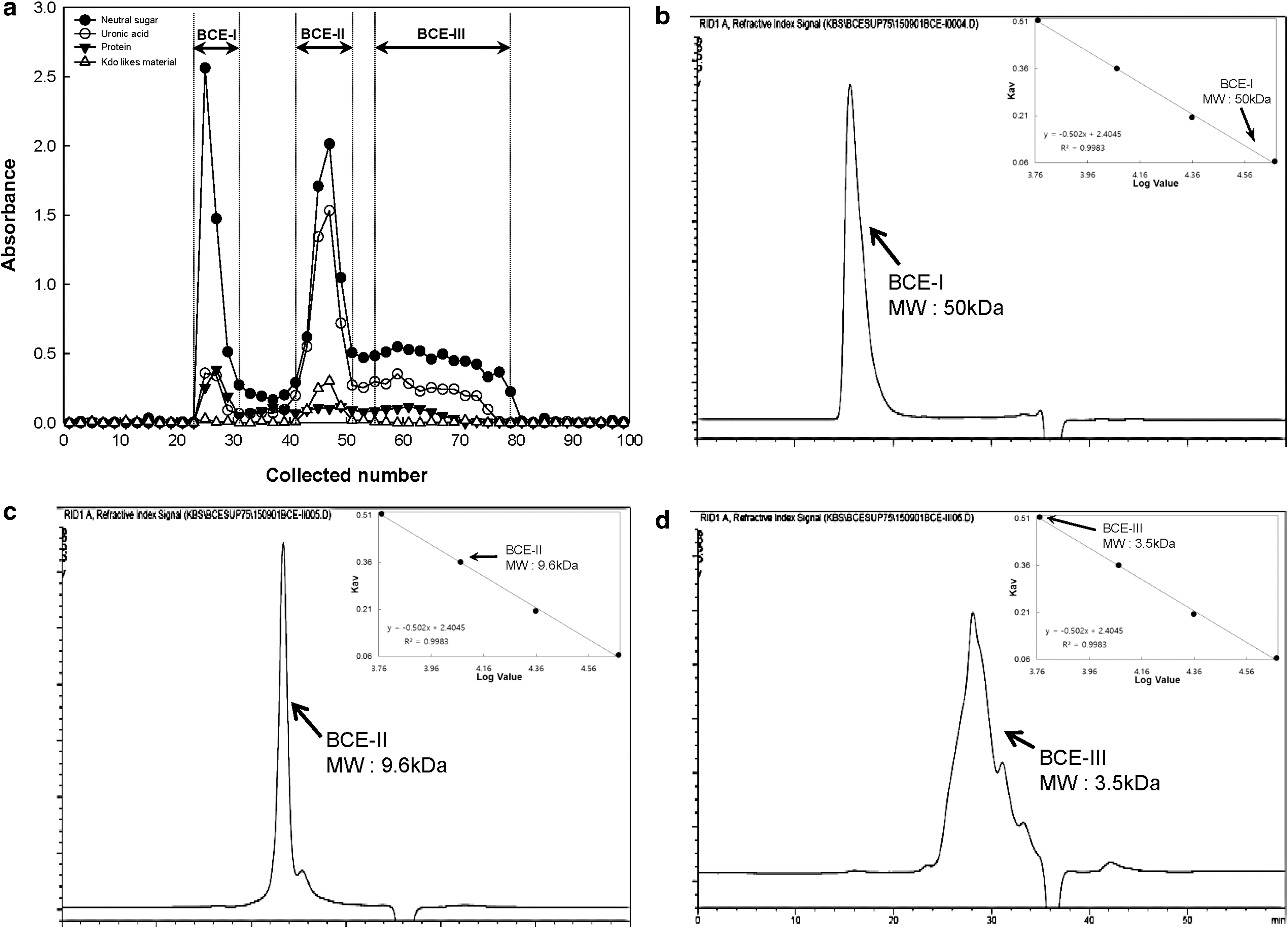
Broccoli (5 kg) was coarsely ground in water (5 L) using a grinder. After adding absolute ethanol (40 L) and homogenization for 2 h, precipitates were dried in an oven to remove ethanol (50°C for 4 days). Dry powder that resulted was suspended in 10 volumes of water and hydrolyzed with 3 mL of Plantase MAX for 2 days at 50°C. The remaining activity was inactivated at 100°C for 15 min. After the supernatant was separated by centrifugation (3000 × g, 30 min), it was precipitated with four volumes of ethanol and dialyzed for 2 days using dialysis cellophane tubing (molecular weight [MW] cutoff: 12–14 kDa; Sigma, St. Louis, MO, USA) and then the solution was lyophilized. Crude polysaccharides were applied to an open column (3 × 90 cm) of Sephadex G-75 (GE Healthcare Life Sciences), equilibrated with 50 mM ammonium formate buffer (pH 5.5), and eluted with the same buffer. Three major purified polysaccharides (BCE-I, BCE-II, and BCE-III) were obtained and desalted using dialysis followed by lyophilization (Fig. 1).
Scheme for isolating and purifying immune-stimulating polysaccharides from broccoli
General analytical methods
Total carbohydrates were identified by a phenol-sulfuric acid method with galactose used as the standard
14
and the m-hydroxydiphenyl method using a galacturonic acid standard.
15
Protein was analyzed by the Bradford method,
16
with bovine serum albumin as the standard (Hercules, CA, USA). The 2-keto-3-deoxy-
Assay of cytokines on murine peritoneal macrophages
Murine peritoneal macrophages were obtained from BALB/c mice treated with 5% (w/w) thioglycollate medium (TG; Sigma) as described previously. 20 Peritoneal macrophages (2.0 × 10 5 cells per well in DMEM-10% FBS) were dispensed into each well of a 96-well culture cluster. After incubation for 2 h, BCE-0 and its subfractions were treated and co-incubated with macrophages in a 5% CO2 incubator at 37°C for 24 h. The culture supernatants were analyzed for the production of various cytokines (interleukin [IL]-6, IL-12, and tumor necrosis factor alpha [TNF-α]) using enzyme-linked immunosorbent assay kits from BD Bioscience (IL-6, IL-12; San Diego, CA, USA) and eBioscience (TNF-α; San Diego) according to the manufacturers' protocols.
Assay of NK cell-mediated tumor cytolytic activity and identification of granzyme B
Three BALB/c mice per group were administered twice intravenously (i.v.) with BCE-I at each concentration (100, 500, or 1000 μg/mouse). Splenocytes were harvested, and the suspensions of these cells [effector cells (E)] were mixed with YAC-1 cells [target cells (T), 1 × 10 4 cells per well] to determine E-to-T cell ratios (E:T) of 25:1, 50:1, and 100:1 in U-bottom 96-well plates.
To examine NK cell-mediated tumor cytolytic activity, we incubated plates in a 5% CO2 incubator for 6 h. After centrifugation for 5 min at 400 × g, 20 μL of supernatant from each well was reacted with LDH solution (Daeil Lab Service Co. Ltd.), and absorbance values were detected at 450 nm by using a microplate reader (Molecular Device, Sunnyvale, CA, USA). The percentage NK cell cytolytic activity was calculated according to the following formula:
Lysis (%) = [(optical density [OD] of the experimental group − OD of E cell spontaneous release − OD of T cell spontaneous release)/(OD of T cell maximum release − OD of T cell spontaneous release)] × 100.
To identify granzyme B, we incubated plates for 24 h in a 5% CO2 incubator. After culture supernatant from each well was assayed using a granzyme B kit (eBioscience) according to manufacturer's protocol.
Inhibitory effect on metastatic tumor cells in vivo
Inhibition effect on lung metastasis by BCE-I was measured after mice were administered BCE-I both orally and i.v. The mice were administered either orally with BCE-I (daily for 20 days) or i.v. with BCE-I (twice, 1 and 3 days before tumor injection). Then, the mice were injected with Colon26-M3.1 cells (4 × 10 4 cells per mouse) i.v. and killed after 14 days. 21 Lungs were excised and fixed with Bouin's solution (Sigma), and tumor colonies were counted under a dissecting microscope.
Depletion of NK cells in mice
Depletion of NK cells was carried out using anti-asialo GM1 sera (Wako Pure Chemicals, Ltd., Japan). The mice were i.v. injected with BCE-I at 1 and 3 days before Colon26-M3.1 carcinoma inoculation and with 50-fold diluted solution of rabbit anti-asialo GM-1 sera (500 μL/mouse, i.v.) 2 days before tumor inoculation. The mice were killed 14 days after tumor cell inoculation, and their lungs were excised and fixed in Bouin's solution. Lung tumor colonies were counted under a microscope.
Statistical analysis
All statistical analyses were carried out using SPSS version 12.0 (SPSS, Chicago, IL, USA). All data were represented as mean ± standard deviation of three independent experiments performed in triplicate. P < .05 was considered significant (Student's t-test). Statistical differences among groups were evaluated by analysis of variance, followed by Duncan's multiple range tests.
Results and Discussion
Preparation and purification of polysaccharides from broccoli
As one of the major polysaccharides present in tracheophytes, pectic polysaccharides are the most abundant component of cell walls in dicotyledons, including broccoli. The pectic polysaccharides consist of three different polysaccharide groups such as homogalacturonan (HG), rhamnogalacturonan (RG)-I, and RG-II, which are covalently bonded to each other. Pharmacological activities have been mainly reported in RG-I and RG-II, rather than in HG. 22 We used pectinase to isolate the active moiety in pectic polysaccharides. Pectinases, usually known as homogalacturonanases, are a group of enzymes that accelerate the hydrolysis of pectic polysaccharides through depolymerization or dimethyl-esterification reactions. Recently, pectinases have been widely utilized in industrial and scientific areas to prepare RG-I and RG-II from various plant sources. 23,24
In this study, we aimed to use the commercial pectinase, Plantase, to increase the immunostimulatory activity of broccoli polysaccharide. After a pectinase hydrolysate was prepared from the broccoli suspension, crude polysaccharides (3.13%) from dried broccoli residues were precipitated in 80% ethanol and separated by centrifugation.
In addition, to characterize the immunostimulatory moiety of broccoli polysaccharides, crude polysaccharides were subjected to a Sephadex G-75 column (Fig. 1a). These fractions were successively separated into BCE-I (50 kDa), BCE-II (9.6 kDa), and BCE-III (3.5 kDa) (Fig. 1b–d). Each fraction was lyophilized after dialysis to obtain dry materials (yield of 0.5% from BCE-I, 1.5% from BCE-II, and 0.5% from BCE-III, relative to the dry base).
Chemical characteristics of BCE subfractions
Pectin is recognized as one of the most complicated polysaccharides, consisting of more than 15 different monosaccharide units. 25 Based on a monosaccharide compositional analysis of the three fractions (Table 1), BCE-I consisted mainly of neutral sugars such as galactose and arabinose. Therefore, we assumed that BCE-I might be a fraction enriched with galactan or arabinogalactan as side chains branched to an RG-I main chain of pectin. In contrast, BCE-II was composed of neutral sugars that consisted mainly of rhamnose and arabinose, as well as uronic acids, mostly of galacturonic acid. Of interest, BCE-II also contained relatively high amounts of a Kdo-like material, in addition to seldom-observed sugars such as apiose, aceric acid, 2-methylfucose, and 2-methylxylose. Together with the large amount of galacturonic acid, rhamnose, and arabinose in BCE-II, the fact that Kdo-like materials and unusual sugars are known indicators of the presence of RG-II polysaccharides 26 indicated that BCE-II is most likely a fraction enriched with RG-II.
Chemical Properties of BCE-I, BCE-II, and BCE-III from Broccoli
Kdo means 2-keto-3-deoxy-
Mole % calculated from the detected total carbohydrate.
Finally, the sugar composition analysis showed that the smallest MW fraction, BCE-III, was mainly made up of rhamnose, galactose, and galacturonic acid. Based on the finding that BCE-III is enriched with galacturonic acid and rhamnose, we speculated that BCE-III is likely a fraction with an RG-I backbone and alternating disaccharide rhamnose and galacturonic acids.
Despite the differences in the proportions of major monosaccharide units of the polysaccharides purified from broccoli, our results were similar to those of a previously published study, 27 which suggested that an anticancer polysaccharide purified from broccoli was mainly made up of arabinose, galactose, and rhamnose at a molar ratio of 5.3:0.8:1.0.
Increased cytokine production by BCE-I
To investigate macrophage activation by the BCE subfractions (I–III), we measured the contents of three cytokines, and TNF-α, IL-6, and IL-12, produced by activated murine peritoneal macrophages. Plant polysaccharides are known to enhance the immune system by modulating macrophage activity. 11 Activated macrophages release proinflammatory cytokines such as TNF-α, IL-6, and IL-12 and initiate an innate immune response. 28 Hence, the amounts of secreted cytokines indicate the relative extent of macrophage activation. Of the BCE subfractions, BCE-I showed the highest cytokine production (Fig. 2). Treatment with BCE-I at concentrations of 8, 40, 200, or 1000 μg/mL enhanced the production of IL-6 by 1.6-, 3.2-, 9.1-, and 16.2-fold, respectively, whereas the level of IL-12 increased by 1.3-, 2.7-, 8.8-, and 16.4-fold, respectively, compared with that of the untreated group (Fig. 2a, b). TNF-α showed the greatest increase, ∼1.7-, 7.7-, 59.4-, and 90.6-fold at 8, 40, 200, and 1000 μg/mL, respectively (Fig. 2c). Therefore, we selected BCE-I for use in subsequent assays.

Effects of BCE fractions from broccoli on production of IL-6
Increased NK cell activity by BCE-I
NK cells are known as innate immune lymphocytes that play a major role in the early host defense against infectious pathogens and tumors. 29 Among the many roles of NK cells, we focused on their antitumor activity, because mice depleted of NK cells are more susceptible to chemically induced tumors. 30 NK cells can destroy tumors directly without previous sensitization by specific antigen receptors through various mechanisms such as secreting cellular granules containing perforin and granzyme B. 31 Granzyme B is cytotoxic protease in CD8+ T and NK cells that is released to kill the target cells. 32
To examine the activation of NK cells by BCE-I, YAC-1 cells were used. YAC-1 is a lymphoma cell derived from Moloney murine leukemia virus that is deficient in major histocompatibility complex class I (MHC-I) expression. YAC-1 cells are highly sensitive cells to cell lysis mediated with NK cell. 33 NK cell-related cytolytic activity was assayed in YAC-1 cell system cocultured with NK cells prepared from BCE-I-treated mice by an LDH release assay. NK cells from mice i.v. administered with 1000 μg/mL of BCE-I showed lysis that was increased ∼50-fold at 100:1 E:T ratio for YAC-1 cells compared with those isolated from nontreated mice (Fig. 3a).
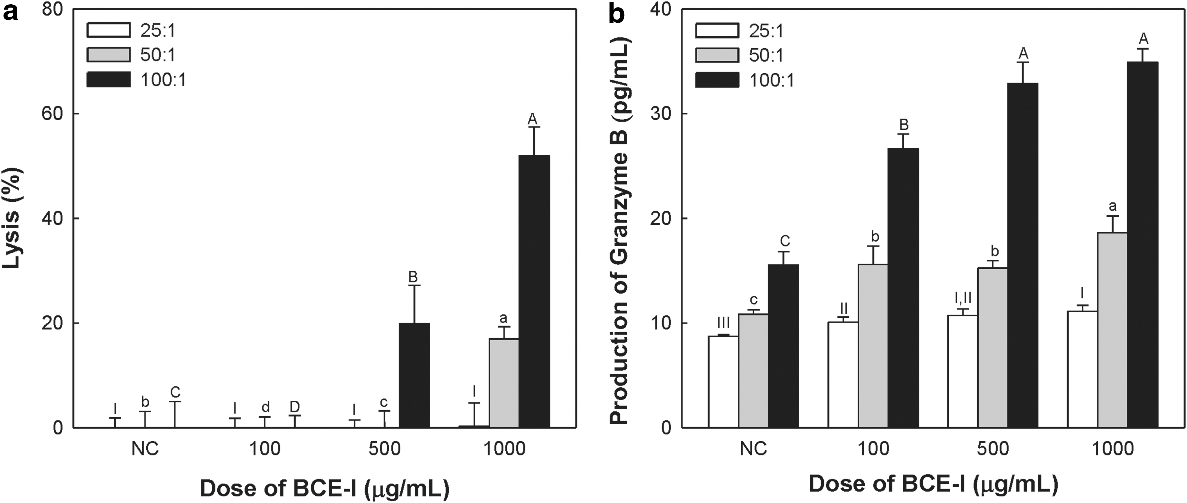
Effects of BCE-I on cytolytic activity
BCE-I also increased granzyme B secretion by NK cells at all E:T ratios. Granzyme B secretion dose-dependently increased after exposure to 100, 500, or 1000 μg/mL of BCE-I. Treatment with BCE-I at concentrations of 100, 500, or 1000 μg/mL showed increased production of granzyme B at 100:1 E:T ratio (Fig. 3b).
Anti-metastatic activity of BCE-I
Metastasis is the spread of a cancer from one organ or part of the body to another. Metastasis of cancer cells has been regarded as an important challenge, because most of the cancer-related deaths (90%) are the result of cancer metastasis in patients with various kinds of cancers. 34 –36 Therefore, blocking cancer metastasis is the best goal for cancer treatment. Based on our assay of NK cells, we determined that BCE-I increases NK cell activity. NK cells have many functions such as regulating tumor cells and virus-infected cells; however, we focused on their anti-metastatic activity in mouse models that allow investigations of the correlation between cancer metastasis and NK cell. 37 It is well known that NK cells play a decisive role in host protection against experimental prostate cancer and melanomas or spontaneous breast cancer-induced lung metastasis. 38 –40
To measure the anticancer metastatic activity by BCE-I, Colon26-M3.1 carcinoma cells were used as a representative lung cancer-metastatic cell line. BCE-I-treated mice, both orally and i.v. showed a dose-dependent decrease in tumor metastasis. BCE-I administered i.v. suppressed metastasis by ∼70% at 1000 μg per mouse of BCE-I, compared with that observed in control mice (Fig. 4a). BCE-I supplemented orally suppressed metastasis by ∼20% at 1000 μg per mouse of BCE-I, compared with that observed in control mice (Fig. 5).

Inhibitory effect on lung metastasis

Inhibitory effect on lung metastasis by BCE-I supplemented orally. BALB/c mice were administered (orally) the indicated doses of BCE-I daily for 20 days before i.v. inoculation of 3 × 10 4 Colon26-M3.1 cells. Mice were killed 14 days after tumor inoculation. Values are expressed as mean ± SD of three independent experiments performed in triplicate. abBars not sharing the same superscript are significantly different from each other. Color images are available online.
To clarify the relationship between tumor metastasis and NK cells, the function of NK cells was blocking by using anti-asialo GM1 sera. Asialo GM1 is a surface antigen of NK cells; therefore, injection of anti-asialo GM1 sera into mice can inhibit NK cell activity and function. 41 As a result, when the function of NK-cell was depleted, tumor metastasis was increased at a high rate, and the antitumor activity of BCE-I disappeared. It points out that the antitumor activity of BCE-I is strongly related to the activation of NK cells (Fig. 4b). Collectively, the above results indicate that NK cells activated by BCE-I may inhibit the growth of local original tumor and tumor metastasis into other tissues and organs.
Footnotes
Acknowledgment
This study was supported by the High Value-Added Technology Development Program (114029-03-2-CG000), Ministry for Food, Agriculture, Forestry, and Fisheries, Republic of Korea (iPET). This research was also supported by Main Research Program (E0170601-02) of the Korea Food Research Institute (KFRI) funded by the Ministry of Science and ICT.
Author Disclosure Statement
No competing financial interests exist.