
Abstract
Hypoxia-induced oxidative stress and disturbed microvascular circulation are both associated with pathogenesis of glaucoma. Ginkgo biloba extract (GBE) has been reported to have positive pharmacological effects on oxidative stress and impaired vascular circulation. This study aimed to investigate the neuroprotective effect of GBE against hypoxic injury to retinal ganglion cells (RGCs) both in vitro and in vivo. The rat RGC line was used, and oxidative stress was induced by hydrogen peroxide (H2O2) in vitro. EGb 761, a standardized GBE, or vehicle was applied to RGCs. Hypoxic optic nerve injury in vivo was induced by clamping the optic nerve of rats with a “microserrefine clip” with an applicator, which was applied without crushing the optic nerve. This method is different from “optic nerve crush model” and does not involve elevation of intraocular pressure, and may serve as a possible normal tension glaucoma animal model. EGb 761 at various concentrations or vehicle was administered intraperitoneally. RGC density was measured to estimate the survival both in vitro and in vivo. The survival of RGCs was significantly (P < .001) higher upon treatment with 1 or 5 μg/mL of EGb 761 compared with vehicle after oxidative stress in vitro. RGC density upon treatment with EGb 761 of 100 mg/kg (1465.6 ± 175 cells/mm2) or 250 mg/kg (1307.6 ± 213 cells/mm2) was significantly higher (P < .01, P < .05, respectively) than that obtained with vehicle (876.3 ± 136 cells/mm2) in vivo. Our results suggest that GBE has neuroprotective effect on RGCs against hypoxic injury both in vitro and in vivo.
Introduction
Ginkgo biloba extract (GBE) mainly consists of flavonoids and terpenoids, including polyphenolic flavonoids, which have antioxidative characteristics. 1,2 The main GBE is EGb 761. 3 EGb 761 has been used widely in the prevention and treatment of dementia, Alzheimer's disease, neurosensory impairment, and peripheral vascular diseases. 4
Research in the area of neuroprotection is expanding rapidly. This is partly because it may represent a new therapy for a challenging disease such as glaucoma. 5 The loss of retinal ganglion cells (RGCs) and their axons is derived from glaucomatous damage. 6 Increased intraocular pressure (IOP) is well known as one of the most important risk factors of glaucomatous injury. It has been recognized that IOP reduction can generally improve future prognosis of all types of glaucoma. 6 However, other risk factors are also associated with the pathogenesis of glaucoma, thus even an optimal IOP might not prevent progression in all glaucoma patients. 6 Other than elevated IOP, vascular injury and hypoxia are frequently associated with the pathogenesis of glaucoma. A mild but repeated reperfusion injury (e.g., IOP fluctuation or disturbed autoregulation) could lead to oxidative stress and result in glaucomatous damage in the long term. 7 Hypoxic conditions could deteriorate or induce RGCs to undergo apoptosis. 8
Ginkgo biloba has a positive influence on oxidative stress and disturbed vascular circulation. 2,3 Oxidative stress and impaired microvascular circulation are both associated with glaucoma. 6 Moreover, antioxidant defense is involved in the aqueous outflow system. 9 –12
The neuroprotective effect of EGb 761 against oxidative damage has been studied in isolated brain mitochondria or dissociated brain cells after treatment with EGb 761 in vitro or in vivo. 13 However, no study has yet reported the in vitro neuroprotective effect of EGb 761 against oxidative stress in RGCs. Several animal models have been suggested for high tension glaucoma, but there has not yet been established animal model for normal tension glaucoma (NTG). 14 –18 In this study, we investigated the neuroprotective effect of EGb 761 on RGCs against oxidative stress in vitro and in vivo using the “microserrefine clip” in rats without elevating IOP, a possible NTG animal model.
Materials and Methods
In vitro
To determine if GBE could have a direct effect on RGC survival, GBE was applied to adult rat RGCs in culture medium. GBE used in this study was EGb 761 (Tanamin iv; YuYu Pharma, Inc., Seoul, Korea), a standardized Ginkgo biloba leaf extract. This EGb 761 contains 24% flavonoids and 6% ginkgolides. To investigate whether EGb 761 could protect RGCs against oxidative damage, hydrogen peroxide (H2O2, 800 μM) was used to induce oxidative stress. Thereafter, either DMSO as vehicle or EGb 761 at a concentration of 1 or 5 μg/mL was used to treat RGCs.
Cell culture
The rat RGC line, RGC-5, was used in this study. They were maintained with Dulbecco's modified Eagle's medium (DMEM; Life Technologies, Rockville, MD, USA) containing 10% fetal bovine serum in addition to streptomycin sulfate (100 μg/mL) combined with penicillin (100 U/mL), with supply of 5% CO2 and 95% air atmosphere in a humidified chamber at 37°C.
Cell proliferation assay
RGC-5 cells were cultured at a density of 1 × 10⁴ cells per well and placed in a 96-well plate. These cells were grown overnight until adherence. The next day, cells were washed and replenished with 100 μL of serum-free media. Hypoxia was induced by oxidative stress-inducible H2O2 (800 μM), and cells were applied with different concentrations of EGb 761 or vehicle as control. After 48 h of application, 10 μL of reagent of Cell Counting Kit-8 (CCK-8; Dojindo, Rockville, MD, USA) per well were added to these cells. After these cells were incubated, cell viability was detected by measuring absorbance value at a wavelength of 490 nm using an xMark microplate spectrophotometer (Bio-Rad, Hercules, CA, USA).
In vivo
Animals and EGb 761 treatment
Male Sprague-Dawley (SD) rats aged 6–8 weeks (150–200 g of body weight) obtained from Orient Bio (Seongnam, Korea) were secured under sterile pathogen-free environment. All experiments were carried out in compliance with the Association for Research in Vision and Ophthalmology Statement for the Use of Animals in Ophthalmic and Vision Research. These animals were maintained with freely accessible food and water, and under 12 h of dark and light cycle. They were divided into four groups randomly. Sixteen SD rats were used in each experiment. A total of five experiments were performed, including three preliminary studies and two main experiments. Animals were intraperitoneally administered with various concentrations of EGb 761 (Tanamin iv; YuYu Pharma, Inc.) in EGb 761-treated group or dimethyl sulfoxide (DMSO) in vehicle-treated control group at 1 h before the induction of hypoxic optic nerve injury. 14,19 The entire experimental procedure was authorized by the IACUC of the Sungkyunkwan University Laboratory Animal Research Center (Permit No. 20160321001).
Induction of hypoxic optic nerve injury
Anesthesia was given to animals through injection of 10 mg/kg xylazine combined with 100 mg/kg ketamine intraperitoneally. Using a surgical microscope, lateral cathotomy followed by temporal conjunctival fornix incision was made, and the optic nerve was exposed. The optic nerve was then clamped for 30 sec using a microserrefine equipped with an applicator (straight, 13 mm; Fine Science Tools, Inc., Canada) at the location of 2 mm behind the optic nerve head. The microserrefine was then taken out. Complete discontinuation and reperfusion of blood flow in the retinal vessels were observed by ophthalmoscopy. In our study, we used microserrefine clip with applicator designed for animal research to clamp the optic nerve. This method allows minimal damage to the optic nerve without crushing the optic nerve, and the degree of clamping can be standardized for a given time. Body temperatures of rats were maintained at 37°C during these procedures. 20 –22
Estimation of RGC density
To visualize live RGCs, retrograde labeling was performed for rats under anesthesia. In brief, after the exposure of the optic nerve, incision was made at the optic nerve sheath with a needle knife at the location of ∼3 mm behind the optic nerve head. The optic nerve was then transected with the needle knife. Then, 10 μL of dextran tetra-methyl-rhodamine (DTMR; Molecular Probes, Eugene, OR, USA) dissolved in phosphate-buffered saline (10 mg/mL) was applied to the transection made at the optic nerve. 23,24 DTMR can move retrogradely to the cell body of RGC by fast axonal diffusion. It stains surviving RGCs only. After 24 h, these eyes were enucleated, and retinas were flattened and fixed with 4% paraformaldehyde for 30 min. 25 These retinas were inspected using a fluorescent microscope (Axiovert 200; Zeiss, Jena, Germany), then photographed using 100 × objective.
RGC count was performed with photomicrographs of 12 standard areas (0.34 × 0.22 mm2) per retina corresponding to three areas in four quadrants of retina at 1, 2, and 3 mm from the optic disk, respectively. The average density was calculated by counting the number of surviving cells from these 12 photographs/mm2.
Statistical analysis
Data of RGC-5 cell count after in vitro experiments were statistically analyzed with one-way analysis of variance (ANOVA) and sequential Tukey's multiple-comparison test. Data of RGC-5 cell count after in vivo experiments were statistically analyzed by nonparametric ANOVA, followed by Kruskal–Wallis test. Statistical significance was considered when P-value was <.05. All statistical analyses were undertaken using SPSS software version 20.0 (SPSS, Inc., Chicago, IL, USA).
Results
In vitro
There was a decrease in RGC numbers accompanied with deformed cellular morphology after oxidative stress was induced with H2O2. However, when RGCs were treated with EGb 761 at a concentration of 1 and 5 μg/mL, cell survival increased significantly, and cellular morphology was normalized (Fig. 1). Colorimetric quantitation of RGCs showed that RGC survival decreased significantly after H2O2 application. Absorbance value of RGCs at wavelength of 450 nm decreased significantly (P < .01) from 1.935 ± 0.34 to 0.721 ± 0.23 after H2O2 application. When 1 or 5 μg/mL of EGb 761 was administered with H2O2, RGC survival increased significantly (P < .001) compared with control (Fig. 1). Absorbance value of RGCs after treatment with 1 and 5 μg/mL of EGb 761 increased significantly (P < .001) to 1.582 ± 0.38 and 1.538 ± 0.41, respectively, compared with control (0.721 ± 0.28).
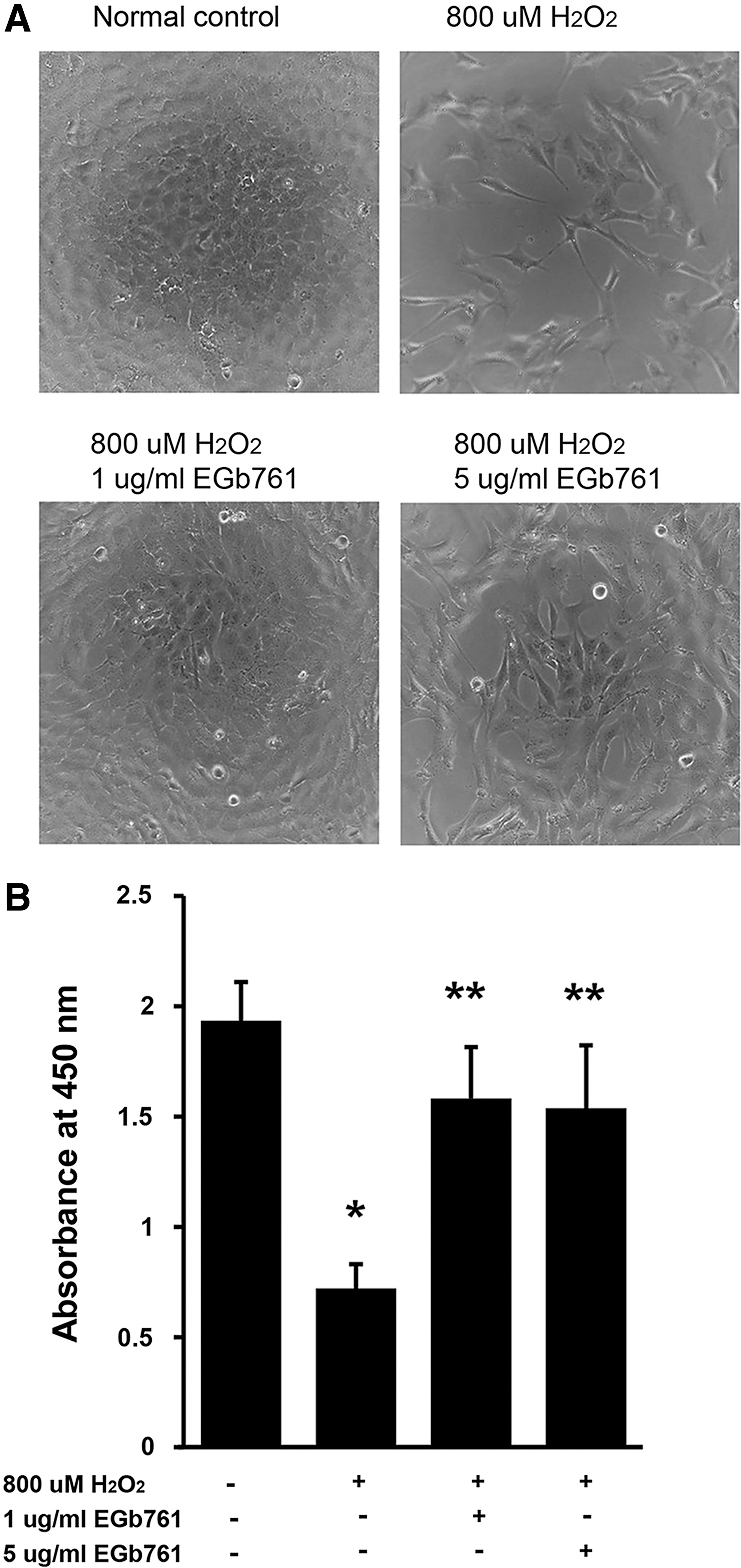
Effect of EGb761 on the survival of RGCs after H2O2 application.
In vivo
Preliminary experiments
First, to determine the time required for retrograde transport of staining dye from transected axon to cell bodies of RGCs, RGCs stained by DTMR solution were observed. It took ∼12 h until almost all RGCs were fully stained with DTMR. RGC density upon staining with DTMR significantly increased from baseline 275.1 ± 183 cells/mm2 to 1837.6 ± 197 cells/mm2 after 12 h of staining (P < .05). RGC density also significantly increased from baseline to 1768.2 ± 226 cells/mm2 after 24 h of staining (P < .05) (Fig. 2). After 6 h of staining with DTMR, RGC density was 617.3 ± 162 cells/mm2, which was not significantly different from the baseline (P > .05).
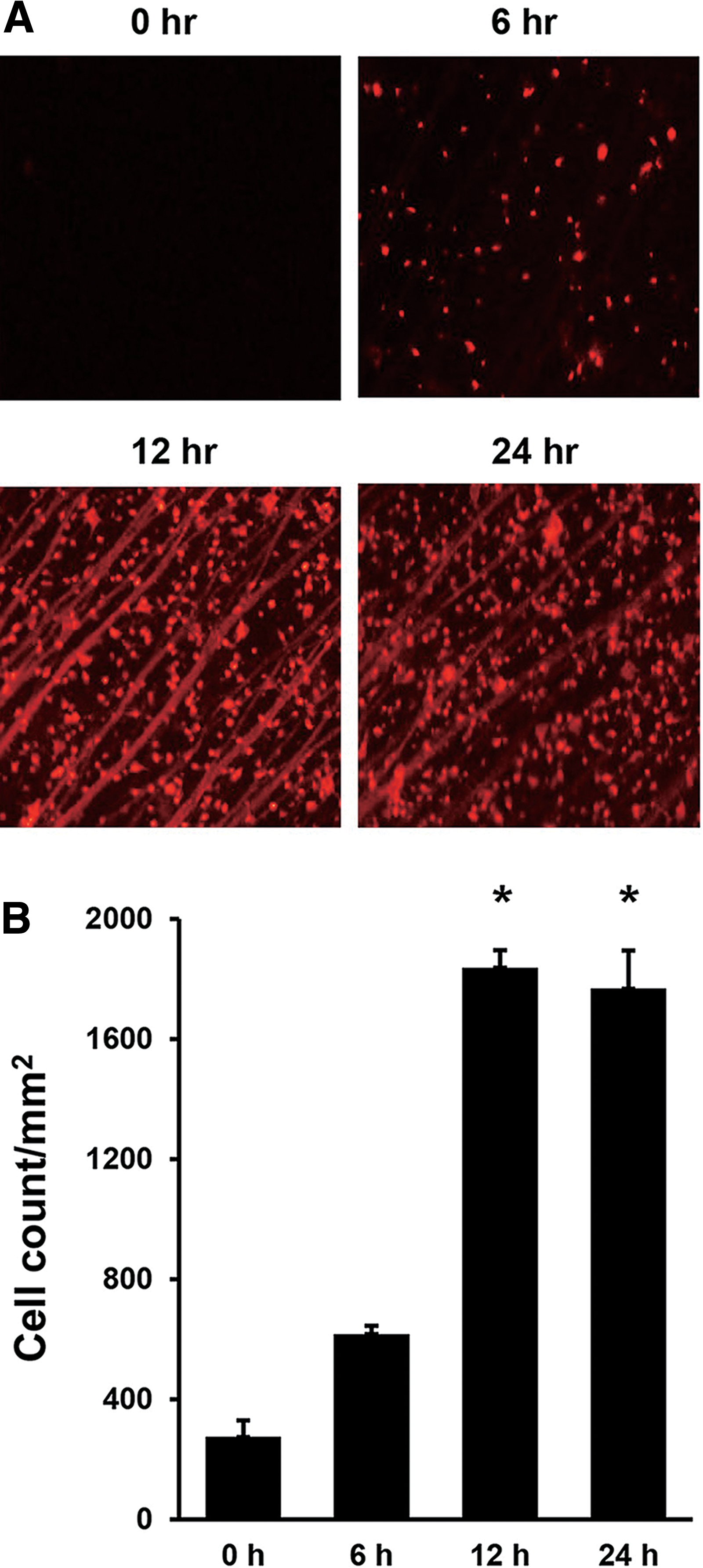
Time course of RGCs stained with DTMR in rats.
Second, to determine the time required for RGCs to die after the clamping of the optic nerve, RGC density was observed after various time periods of clamping. After clamping for 6, 12, 24, and 48 h, RGC densities observed were 1508 ± 154, 1354.6 ± 176, 1114.6 ± 185, and 388.6 ± 191 cells/mm2, respectively. RGC density decreased significantly after 24 and 48 h of clamping (P < .05) compared with the baseline (Fig. 3).

Time course of RGC survival after clamping with microserrefine of optic nerve in rats.
Third, to determine the duration of clamping with microserrefine clip, RGC density was observed after various time periods of clamping. RGC densities after clamping for the duration of 10 sec, 30 sec, 2 min, and 10 min were 1864.2 ± 175, 1028.6 ± 186, 814.6 ± 297, and 384.3 ± 181 cells/mm2, respectively. RGC density decreased significantly (P < .05) after clamping for 30 sec, 2 min, or 10 min compared with the baseline (Fig. 4).
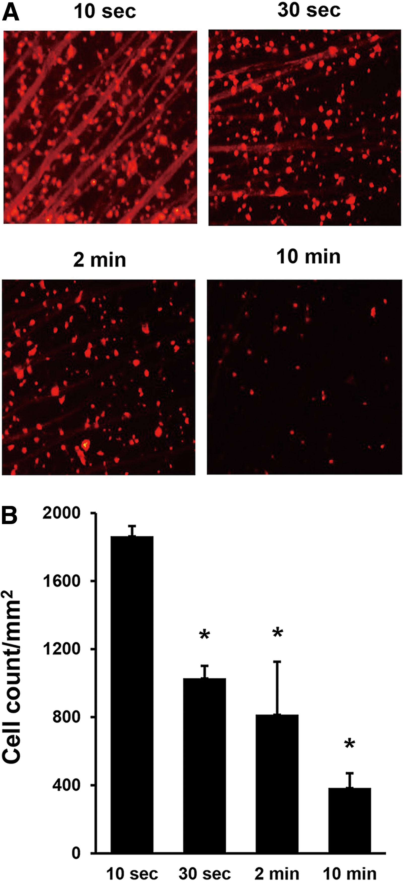
Effect of duration of clamping the optic nerve on RGC survival in rats.
Main experiments
EGb 761 or vehicle was intraperitoneally administered at 1 h before clamping of the optic nerve in experiment I. RGC density was 876.3 ± 136 cells/mm2 in the vehicle-treated group. RGC densities in groups treated with 50, 100, and 250 mg/kg of EGb 761 were 869.6 ± 136, 1465.6 ± 175, and 1307.6 ± 213 cells/mm2, respectively. RGC survival after clamping was significantly higher in the groups treated with 100 or 250 mg/kg of EGb 761 compared with that in vehicle-treated group (P < .01 or P < .05, respectively). However, the neuroprotective effect of EGb 761 at the concentration of 250 mg/kg was not greater than that of 100 mg/kg (Fig. 5).
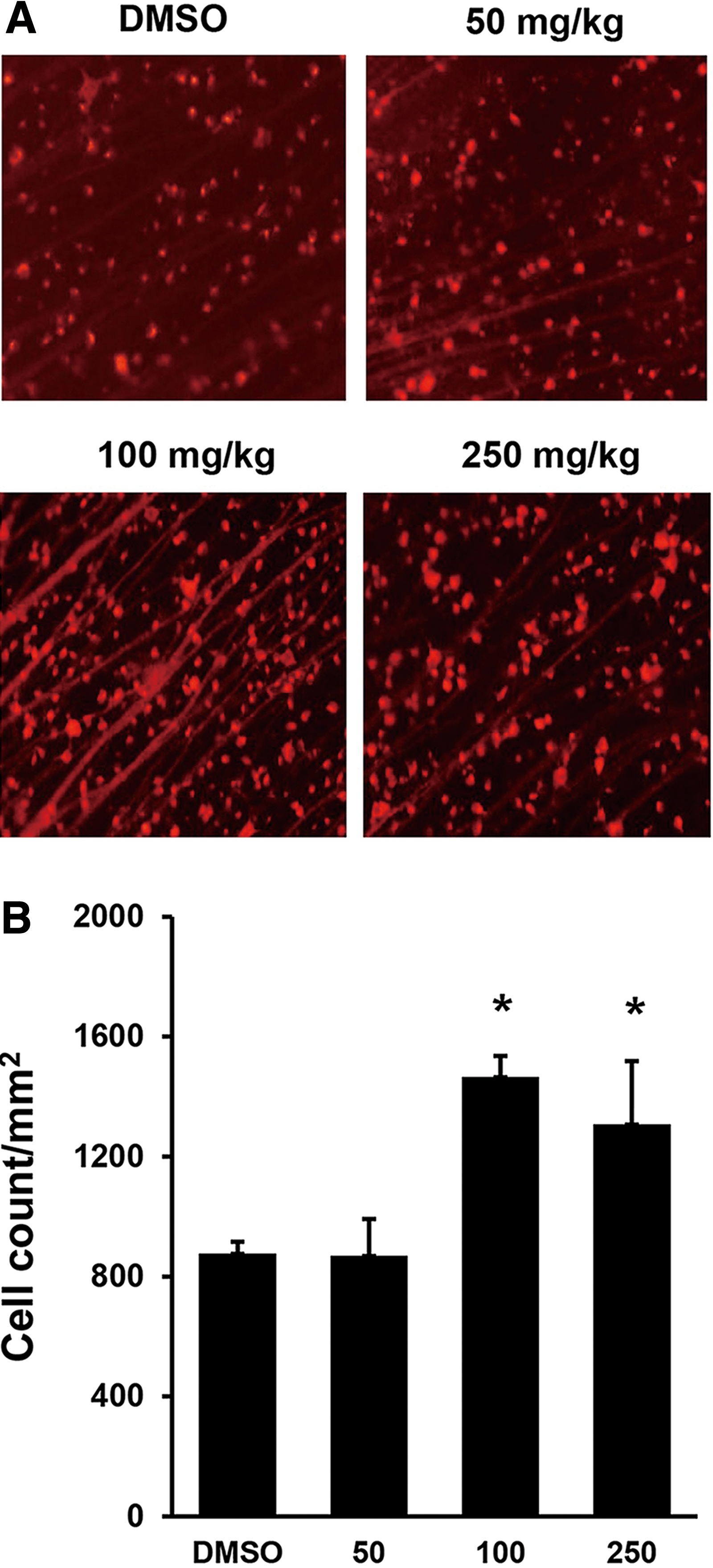
Effect of EGb761 administration before clamping the optic nerve on the survival of RGCs in rats (experiment I).
EGb 761 or vehicle was intraperitoneally administered twice, at 1 h before and immediately after clamping of the optic nerve in experiment II. RGC densities in groups treated with vehicle, 50, 100, and 250 mg/kg of EGb 761 were 953.2 ± 168, 983.4 ± 176, 1582.3 ± 171, and 1382.3 ± 178 cells/mm2, respectively. RGC survival in the group treated with 100 mg/kg of EGb 761 was significantly (P < .01) higher than that in the group treated with vehicle. Neuroprotective effect of EGb761 was not significantly greater at higher dose (250 mg/kg) compared with that at lower doses (50 and 100 mg/kg). Nor the neuroprotective effect was greater when EGb 761 was administered twice compared with one time of administration.
Discussion
In this study, EGb 761 was found to have neuroprotective effects against oxidative stress in RGCs both in vitro and in vivo after optic nerve clamping with a microserrefine clip in rats without elevating IOP. The neuroprotective effect of EGb 761 against oxidative damage in vitro has been revealed in isolated brain mitochondria and dissociated brain cells. 12 However, to our knowledge, no previous study has reported the in vitro neuroprotective effect of EGb 761 against oxidative stress in RGCs yet. We found that 1 and 5 μg/mL of EGb 761 significantly improved the survival of RGCs against oxidative stress induced by 800 μM of H2O2 compared with that treated with vehicle as control in vitro. It is important to confirm the direct neuroprotective effect of EGb 761 on RGCs in vitro before conducting in vivo experiments in rats.
In optic nerve crush models in rats, previous studies have used aneurysm clips and crushed the optic nerve for >60 sec. 17,18,26 However, in this study, we used microserrefine clips with an applicator to clamp the optic nerve without crushing to induce minimal injury to the optic nerve. Optic nerve injury was standardized using this device designed for animal research. Minimal constant amount of force can be engaged compared with tying with nylon or just holding the clip for a duration of several 10 sec. We conducted pilot experiments to find the shortest time needed to properly damage the optic nerve to see RGC death instead of using 60 sec as described in previous studies. 17,18,26
We also hoped to reflect ischemia/reperfusion injury in this animal model. Ischemia/reperfusion injury also contributes to the pathogenesis of glaucoma, especially in NTG. 7,8 We initially tried to dissect and identify the central retinal artery from the optic nerve sheath and clamp the central retinal artery alone. However, the artery was too small and fragile to actually carry out the initial plan. Further studies with better instruments and strategies are needed to make a perfect ischemia/reperfusion injury animal model.
Other than elevated IOP, oxidative stress and disturbed microvascular circulation are both associated with glaucoma. 6 It has been summarized that oxidative stress was certainly involved in the pathogenesis of glaucoma. 27 There are multiple lines of evidence supporting the involvement of oxidative stress as a component of glaucomatous optic degeneration in various subcellular compartments of RGCs. 28 –30 Besides the direct cytotoxic effect resulting in RGC death, it may be possible that reactive oxygen species (ROS) are associated with signaling RGC death. ROS act as a second messenger and/or regulate protein function in this signaling pathway. 31 Modification of oxidative protein in glaucomatous optic degeneration increases neuronal susceptibility to injury and also causes glial dysfunction. 32
Impaired microcirculation plays an important role in the pathogenesis of glaucomatous injury. 6 Unstable ocular perfusion, either due to IOP fluctuation or due to a disturbed autoregulation, leads to a mild reperfusion injury. These conditions generate oxidative stress and as a result, the concentration of superoxide (O2 −) within the optic nerve head axons increases. Nitric oxide (NO) is produced by the activation of neighboring astrocytes by either mechanical or ischemic stress. The combination of O2 − anion with NO produces peroxynitrite (ONOO−), which diffuses within the axons toward retina and causes apoptosis. 33 Primary vascular dysregulation syndrome is best observed in NTG, 34 which is more prevalent in the Asian population than in other races. 35 Moreover, this is one of the reasons we tried to develop an NTG animal model, which accounts for the majority of primary glaucoma in Asians.
GBE was significantly effective in improving RGC survival after optic nerve clamping in rats when administered at 1 h before optic nerve injury at a concentration of 100 mg/kg compared with vehicle as control in our study. According to our results, GBE at concentration of 250 mg/kg was not more effective than that at 100 mg/kg. This might be because the effect of GBE might reach a plateau after a certain concentration, possibly at 100 mg/kg. Moreover, the neuroprotective effect of EGb 761 was not greater when it was administered twice than when it was administered just once. Therefore, the right amount of GBE should be calculated when it is administered in human trials.
Ginkgo biloba has a positive effect on oxidative stress and disturbed vascular circulation, which have been shown to be associated with the pathogenesis of glaucoma. 2,3 A potential benefit of antioxidative therapy in glaucoma has been shown in a review. 33 GBE has been also reported to reduce ischemia/reperfusion injury in the diabetic rat retina after ischemic/reperfusion. This has been demonstrated by the free radical scavenging property of Ginkgo biloba. 36 Based on these results and also our study, it can be presumed that a similar profitable effect of GBE occurs in the optic nerve of glaucoma patients, as they also suffer from the same ischemia/reperfusion injury.
There are limitations for any experimental study. First, definite results provided by animal studies cannot always be applied to clinical conditions. Components revealed to be neuroprotective in laboratory studies frequently fail to perform as effective treatments in clinical situations. Doses and the method of administration should be modified when it is administered to human. However, GBE is often prescribed by glaucoma specialists in the treatment of NTG and glaucoma that progresses in spite of IOP reduction. This is partly because there are no other treatment options in these special cases yet. Our results provide robust evidence that GBE has neuroprotective effect both in vitro and in vivo for clinicians to use it in clinics.
The RGC-5 immortalized cell line has been employed widely in cell culture studies to investigate the biology of RGCs. 37 –41 These cells are originated from rat RGC expressing various neuronal markers, especially RGC characteristic proteins Brn3 and Thy1. 40,41 Although the origin of RGC-5 cells remains controversial, they still contain RGC and they are useful for researchers to study some hypotheses on RGCs. 37 –41 Even in a recent report, RGC-5 cell line has been used to investigate the effect of resveratrol on RGC apoptosis. 42
Another issue is the uncertainty about how well the results obtained from our study with an acute optic nerve injury animal model can be applied to the actual etiology of glaucoma. There are different types and time courses of optic nerve injury between the pathogenesis of progressive glaucomatous optic nerve damage and an acute optic nerve injury. Postlesional labeling of RGCs can be another limitation. The interaction between axonal injury and retrograde transport of the DTMR dye from optic nerve in surviving RGCs might not be predictable.
In conclusion, GBE resulted in a significantly higher survival of RGCs against oxidative stress induced by H2O2 compared with vehicle as control in vitro. Intraperitoneal injection of GBE before standardized optic nerve clamping with microserrefine also demonstrated a significantly higher survival of RGCs compared with vehicle as control. Our results suggest that GBE has neuroprotective effect on RGCs against hypoxic injury both in vitro and in vivo.
Footnotes
Authors' Contributions
H.K.C. and C.K. contributed to the design of the study. H.K.C., S.K., E.J.L., and C.K. conducted the study. H.K.C., S.K., E.J.L., and C.K. contributed to data collection, management, analysis, and data interpretation. H.K.C. and C.K. prepared the article. C. K. had full access to all data obtained from this study, and takes responsibility for the integrity and accuracy of data analysis.
Ethical Approval
This study was approved by the Institutional Review Board of Samsung Medical Center, Sungkyunkwan University School of Medicine, Seoul, Korea.
Author Disclosure Statement
The authors declare no conflicts of interest.