
Abstract
Polysaccharide of Atractylodes macrocephala Koidz (PAMK) has been reported to have beneficial effects on regulation of immune responses in mammals and poultry. Nonetheless, the immunoregulatory mechanism of action of PAMK remains unclear. The Toll-like receptor 4 (TLR4) signaling cascade has been proved as a classic polysaccharide-regulated pathway. The aim of this study was to explore the effects of PAMK on the TLR4 signaling pathway in the regulation of spleen function in mice. Ninety-six 5-week-old BALB/c female mice were randomly allocated into four groups with three replicates per group and eight mice per replicate in a single-factor completely randomized experimental design. The control group was fed a basic diet (PAMK free); the other three groups were fed 100, 200, or 400 mg/kg PAMK for 28 days. The spleen index, concentrations of cytokines, and mRNA and protein expression levels of genes related to TLR4 signaling were determined in spleen tissue. Compared with the control group, the spleen index significantly increased in all treatment groups. Concentrations of interleukin 2 (IL-2), IL-4, interferon γ (IFN-γ), and tumor necrosis factor α (TNF-α) in the medium-PAMK group also increased significantly. PAMK in the medium-PAMK group significantly increased both mRNA and protein expression of TLR4, myeloid differentiation factor 88 (MyD88), TNFR-associated factor 6 (TRAF6), TRAF3, and nuclear factor kappa B (NF-κB) in the spleen. In conclusion, PAMK may increase immune-response capacity of the spleen in mice via TLR4–MyD88–NF-κB signaling.
Introduction
Polysaccharide of Atractylodes
As for other polysaccharide immunomodulators, Astragalus polysaccharides (APS), 11 Lonicera caerulea L. berry polyphenols, 12 and resveratrol 13 have been demonstrated to modulate immunity by regulating the Toll-like receptor 4 (TLR4) signaling pathway and nuclear factor kappa B (NF-κB) activation.
TLR4 is one of the important natural immune receptors on the surface of immune cells, and plays a central role in enhancement of the innate immune response and the production of cytokines induced by polysaccharides. 14,15 The TLR4 signaling pathway can regulate immunity, antitumor responses, and macrophage activation. 16 –18 The TLR4 signaling pathway is composed of Toll-IL-1 receptor (TIR) domain-containing adaptor protein (TIRAP)-myeloid differentiation factor 88 (MyD88) (MyD88-dependent), and TIR domain-containing adaptor-inducing interferon-β (TRIF) (MyD88-independent) signaling cascades. 11 The MyD88-dependent NF-κB pathway has been proved to be a classic polysaccharide-responsive cascade in the TLR4 signaling pathway. 19,20 Reports on polysaccharides have revealed that Platycodon grandiflorum activates NF-κB signaling downstream of TLR4 via the degradation of IκBα/β and nuclear translocation of p65 and p50 to induce dendritic-cell maturation. 20 APS can activate TLR4–MyD88–TNFR-associated factor 6 (TRAF6)–NF-κB to initiate an immune response, to reduce tumor weight in mice, 11 and to induce TNF-α, IL-6, and iNOS production in RAW 264.7 cells. 19 Nevertheless, the mechanism of action of PAMK on the TLR4 pathway has not been reported yet. Ji et al. 21 has found that PAMK stimulates macrophages to produce NO and TNF-α via IκB degradation and NF-κB activation. Besides, atractylenolide I, another major bioactive ingredient of A. macrocephala Koidz, can protect mice from acute lung injury induced by lipopolysaccharide (LPS) by reducing the TLR4 amount and NF-κB activation. 22
There are few reports on the signaling pathways of PAMK-mediated immune effects in mice in vivo. Therefore, we chose TLR4 signaling, a classic polysaccharide-regulated pathway, to further elucidate the impact of PAMK on the spleen index, mRNA and protein expression of genes related to the TLR4 signaling pathway, and on concentrations of cytokines in the murine spleen.
Materials and Methods
Experimental groups and treatments
All the mice were treated humanely, and all the procedures in this study were in compliance with the protocol number SRM-11 approved by the Institutional Animal Care and Use Committee of the Zhongkai University of Agriculture and Engineering. A total of ninety-six 5-week-old BALB/c female mice were purchased from the Laboratory Animal Center of Guangzhou University of Chinese Medicine (license key: SCXK [Yue] 2013-0034). The mice were housed in a specific pathogen-free environment (12/12-h light/dark cycle, 22–24°C, 40–60% humidity) and randomly subdivided into 4 groups (control, low-PAMK, medium-PAMK, and high-PAMK groups; 24 mice/group). According to Li et al., 9 PAMK without endotoxin (purity 70%) was purchased from Tianyuan Biologics factory (Xi'an, China). The four groups of mice had free access to food and water. The low-, medium-, and high-PAMK groups were treated with PAMK (soluble in saline) at a dose of 100, 200, or 400 mg/(kg body weight) per day, respectively, via an oral administration sonde. The control group received the same volume of saline. The spleen was excised and weighed (after the mice were weighed and euthanized 28 days after the start of the experiment) and then placed in liquid nitrogen immediately, then stored at −80°C until analysis.
Spleen index analysis
Each fresh spleen specimen was rinsed two to three times with physiological saline, then filter paper was used to dry off the surface, and the specimens were weighed. The following formula was used to calculate the spleen index: spleen weight (g) divided by body weight (kg).
Reverse-transcription quantitative polymerase chain reaction analysis
Total RNA was extracted from the spleen by means of the TRIzol Reagent (Thermo Fisher, USA). First-strand complementary DNA (cDNA) was synthesized with the Reverse Transcription Kit (Takara, Japan). Specific primers (Table 1) were designed in Primer Premier 5.0 software (Premier Biosoft International, USA). An ABI PRISM 7500 detection system (Applied Biosystems, Foster City, CA) was used to quantify the mRNA levels of related genes in TLR4 signal pathway and cytokines in the spleen of mice. The cycling conditions of the quantitative polymerase chain reaction (qPCR) were similar to those described by Yao et al. 23 The PCR procedure consisted of 95°C for 2 min followed by 40 cycles of 15 sec at 95°C, 30 sec at 56°C, and 30 sec at 72°C.
Specific Primer Sequences For Quantitative Polymerase Chain Reaction
Western blot analysis
Total protein from the murine spleen was isolated by means of the Lysate Kit (CWBIO, China) and then analyzed by sodium dodecyl sulfate polyacrylamide gel electrophoresis (SDS-PAGE) under reducing conditions to evaluate amounts of the proteins related to the TLR4 signaling pathway. The separated proteins were transferred to nitrocellulose membranes followed by incubation with 5% skim milk for 2 h to block nonspecific sites on the membrane. A primary mouse antibody (Abcam, Britain) was diluted at 1:1000 and incubated overnight at 4°C. Secondary antibody (Abcam) was diluted at 1:2000, and then conjugated with horseradish peroxidase was incubated with the membrane at room temperature for 1 h. Chemiluminescence Detection Reagents (Applygen, Beijing) were incubated for 1 min to determine the ratios of target protein signals on a Chemiluminescence Imaging System (Tianneng, Shanghai) to signals of control proteins. GAPDH and β-actin (Abcam) served as internal controls, and the relative abundance levels of the proteins were expressed as the ratios of optical density of each of these proteins to that of GAPDH or β-actin.
Statistical analysis
The results are expressed as means ± standard deviations (SD) of the experiments. All the qPCR assays were conducted in triplicate, and the relative levels were measured in terms of threshold cycle (Ct) and were calculated using the formula 2−ΔΔCt. 24 SPSS for Windows (version 19.0; SPSS, Inc., Chicago) was used for statistical evaluation of all the data via one-way analysis of variance followed by Tukey's honestly significant difference test. P < .05 indicated statistical significance.
Results
PAMK increases the spleen index in mice
The spleen index results are shown in Table 2. PAMK treatment significantly increased the spleen weight, indicating that PAMK did affect development of the spleen. Nonetheless, PAMK did not have dose-dependent effects: the spleen index of the medium-PAMK group was the highest and there was no significant difference among the treatment groups.
The Spleen Index Effects Caused By Feeding Different Dose of PAMK
Data are expressed as means ± SD, n = 8.
Significant differences (P < .05); identical letters indicate that the difference was not significant (P > .05).
PAMK, polysaccharide of Atractylodes macrocephala Koidz; SD, standard deviation.
PAMK regulates mRNA levels of genes of the TLR4 signaling pathway
Total RNA was extracted from the spleen of each mouse to measure the expression levels of genes related to the TLR4 pathway. Twelve such genes were tested by qPCR. The results revealed that 10 genes concentration were significantly upregulated (P < .05) except for inhibitor of NF-κB (IκB) kinase ɛ (IKKɛ) and receptor-interacting protein 1 (RIP1) levels in the medium-PAMK group when compared with the control group (Fig. 1). PAMK at 100 and 200 mg/kg increased the TLR4 mRNA level. In contrast, 400 mg/kg PAMK inhibited TLR4 mRNA expression (P < .05); of note, it also reduced the mRNA levels of TRAF6, TRAF3, RIP1, IKKɛ, TANK-binding kinase 1 (TBK1), NF-κB p65, interferon-regulatory factor 3 (IRF3), and IRF7. MyD88, TRIF, and IκBα were sensitive to PAMK, and their mRNA levels significantly increased in all treatment groups. In addition, mRNA expression levels of TRAF6, TRAF3, TBK1, and NF-κB p65 increased only in the medium-PAMK group and were significantly different from those in other groups.

Effects of PAMK on mRNA levels of related genes in TLR4 pathway in the spleen of mice.
PAMK promotes cytokine production
Total RNA was extracted from the spleen to measure the transcript levels of cytokines. As presented in Figure 2, the results showed that relative mRNA expression of IL-2, IL-4, IFN-γ, and TNF-α significantly (P < .05) increased after treatment with 200 mg/kg PAMK (medium-PAMK group) when compared with the control group. To be precise, the mRNA expression levels of IL-2, TNF-α, IL-4, and IFN-γ in the medium-PAMK group increased by 116.3%, 130.6%, 21.8%, and 19.6%, respectively. This phenomenon indicated that PAMK could increase cytokine production in the spleen of mice.
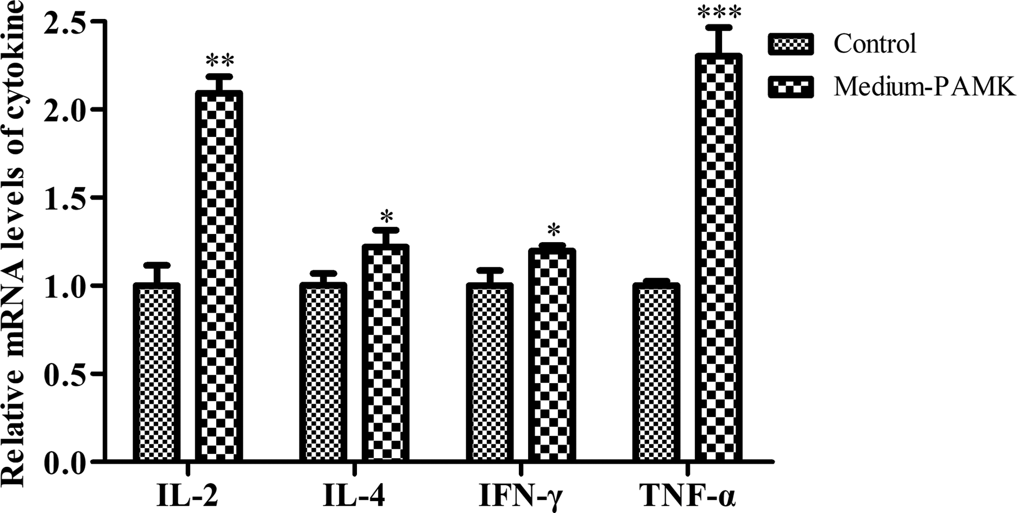
Effects of PAMK on the mRNA levels of IL-2, IL-4, IFN-γ, and TNF-α in the spleen of mice. *P < .05, **P < .01, ***P < .001. Data are means ± SD, n = 6. IL, interleukin; TNF-α, tumor necrosis factor-α.
PAMK regulates the protein expression levels in the TLR4 signaling pathway
Judging by the spleen index and qPCR results, we chose the 200 mg/kg PAMK group (medium-PAMK) for further analyses. The protein expression levels of genes related to the TLR4 signaling pathway are illustrated in Figure 3. The results indicated that PAMK significantly increased the protein levels of TLR4, TRAF6, TRAF3, and NF-κB p65 (P < .05), but had no significant effect on the TBK1 protein amount when compared with the control group (Fig. 3a, A). Furthermore, PAMK markedly raised MyD88 protein levels and decreased those of IκBα; however, RIP1, IKKɛ, and TRIF protein amounts did not change (Fig. 3b, B). The above results suggested that there was some association between the mRNA and protein expression levels of genes related to the TLR4 signaling pathway.

Effects of PAMK on protein levels of related genes in TLR4 pathway in the spleen of mice.
Discussion
A. macrocephala is a traditional Chinese herbal medicine used as an immunopotentiator, PAMK mainly promotes immune organ development, lymphocyte proliferation, and antioxidant and cytokine secretion. 25 Besides, PAMK alleviates immunosuppression in cyclophosphamide-treated geese by maintaining a humoral and cellular immune balance. 9 An increase in the immune organ index is considered one of the characteristics of immunity enhancement. 11 In this study, we found that PAMK promotes spleen development in mice. The spleen index findings indicate that PAMK does affect murine immunity. The raised expression levels of IL-2, IL-4, IFN-γ, and TNF-α can inhibit tumor growth, increase macrophage activity, and enhance both innate and specific immunity. 26 –28 In this study, we found that 200 mg/kg PAMK administered orally markedly increased the expression levels of IL-2, IL-4, IFN-γ, and TNF-α in the spleen of mice. We know that with the development of the spleen, the numbers of T lymphocytes and macrophages increase, thereby leading to upregulation of cytokines and thus enhanced immunity. 9 IL-2, a major factor of T cell proliferation, can promote the production of IFN-γ and IL-4. 29 IFN-γ, a key factor for innate and adaptive immune responses to tumors, can induce macrophage proliferation and differentiation to raise TNF-α production. 30 IL-4 is related to T helper 2 (Th2) cells, and the balance between Th1 and Th2 cells is critical for immune system regulation. 30 The activation of NF-κB is also important for the release of cytokines. 28 One study has revealed that PAMK can promote immune system processes by activating NF-κB to stimulate the release of TNF-α and IFN-γ. 21 As for other traditional Chinese medicine remedies, the polysaccharide from the mycelia of Ganoderma lucidum polysaccharide, Achyranthes bidentata polysaccharide, L. caerulea L. berry polyphenols, and APS can increase the levels of IL-2, IL-4, IL-8, IFN-γ, and TNF-α via the NF-κB pathway. 27,31 –33
TLR4 was the first to be confirmed as a gene upstream of NF-κB among genes of various TLRs and has been well proved to regulate immunity. 34 In the present study, our data indicate that TLR4, MyD88, and NF-κB were positively regulated by PAMK in the spleen of the medium-PAMK group of mice. From these results, we found that expression levels of the genes related to TLR4 signal transduction are consistent with the results on cytokines and the spleen index in this study. It is well known that TLR4's stimulation leads to the activation of a downstream transcription factor, NF-κB, which is linked to TLR4 signals via MyD88. 35 Exogenous agonists can activate inhibitor of kappa B (IκB) kinase to induce IκBα degradation by triggering MyD88-dependent and -independent signaling cascades, 35 which lead to an NF-κB release from the cytoplasmic NF-κB–IκB complex. 36 The activation of NF-κB performs an important function in the secretion of IL-2, IL-4, IFN-γ, and TNF-α, lymphocyte proliferation, and macrophage activation. 26,37 One study suggests that PAMK stimulates macrophages to produce NO and TNF-α through IκBα degradation and NF-κB activation in murine macrophage cell line RAW 264.7. 21 Besides, LPS, APS, and P. grandiflorum have been confirmed as exogenous agonists of TLR4. 15,20 APS 11 and LPS 34 exert immune system regulation and antitumor effects via the TLR4–MyD88–TRAF6–NF-κB pathway in mice and in human cervical cancer cell line SiHa. Moreover, PAMK increased the IκBα mRNA level but reduced its protein level in our study; this effect may be related to IκBα phosphorylation and ubiquitination, which cause degradation of this protein. 37 In addition, TRAF6 and TRAF3 are other adaptor proteins 31 that respond to MyD88 activation. 38 The data presented here show a significant difference in the levels of TRAF3 and TRAF6 between the medium-PAMK group and control group. Nonetheless, the protein expression levels of TBK1 and IKKɛ—genes downstream of TRAF3—were not altered, and the connections among these factors need further research. In addition to MyD88, TRIF is another adaptor protein that responds to the activation of TLR4. 39 Nevertheless, the protein expression data presented here indicate no significant difference in the level of TRIF between the medium-PAMK group and control group. These results imply that PAMK triggers the TLR4–MyD88 pathway rather than the TLR4–TRIF cascade to exert its effects, although further research is necessary to confirm these tentative implications.
In conclusion, these findings indicate that PAMK increases the spleen index and the expression levels of IL-2, IL-4, IFN-γ, and TNF-α in mice possibly by stimulating the TLR4–MyD88–NF-κB signaling pathway. In addition, 200 mg/(kg body weight) is the most suitable dose of PAMK for the immunostimulatory effects in the spleen of mice.
Footnotes
Acknowledgments
This work was supported by National Key Technologies R&D Program of China (No. 2016YFD0501605), Major Research Project in Universities of Guangdong Province (No. 2017KZDXM046), Inter-governmental S & T Exchanges Project between China and Belarus (No. CB01-13), Science & Technology Planning Project of Guangzhou (No. 201604020061). We thank the support of Guangdong Province Key Laboratory of Waterfowl Healthy Breeding, Guangzhou 510225, China.
Author Disclosure Statement
No competing financial interests exist.